Ventral view
Description:
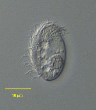
Ventral surface of the dysterine ciliate Orthotrochilia pilula (Deroux, 1976) Song, 2003. The cell is an elongate ellipse in outline. The anterior and posterior ends are broadly rounded. The dorsal surface is arched and the ventral surface flattened. The colorless pellicle is somewhat flexible. The anterior cytostome is inconspicuous and the fine supporting cytopharyngeal rods are seldom visible in vivo even with DIC. The somatic ciliature is restricted to the ventral surface. The two rightmost kineties arch around the cytostome reaching the left anterior side. A small kinetal fragment occupies the left anterior projection of the cell (cilia of this fragment are seen in this image). There are seven postoral kineties, which progressively shorten from right to left. There are two short oblique perioral kineties anterior to and on either side of the cytostome. There is a small posterior podite just to the left of the posterior termination of the right somatic kineties. The podite is often difficult to visualize in vivo. There are two contractile vacuoles just to the right of the midline. There is an ellipsoid heteromerous macronucleus in the midbody. Orthotrochilia pilula is the only species of this genus found in freshwater to date. O. pilula has also been found in a marine habitat providing the basis for reestablishment of the genus by Song (Song, W. Hydrobiologia 499:169-177,2003). Collected from a freshwater pond near Boise, Idaho January, 2005. DIC.
Included On The Following Pages:
- Life (creatures)
- Cellular (cellular organisms)
- Eukaryota (eukaryotes)
- SAR (Stramenopiles, Alveolates, Rhizaria)
- Alveolata (alveolates)
- Ciliophora (ciliates)
- Intramacronucleata
- Phyllopharyngea
- Phyllopharyngia
- Dysteriida
- Dysteriidae
- Orthotrochilia
- Orthotrochilia pilula
This image is not featured in any collections.
Source Information
- license
- cc-by-nc
- author
- William Bourland
- provider
- micro*scope
- original
- original media file
- visit source
- partner site
- micro*scope
- ID