Image of zoanthids
Description:
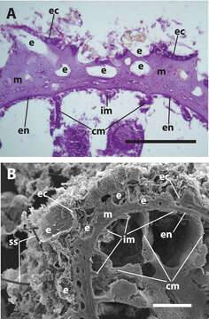
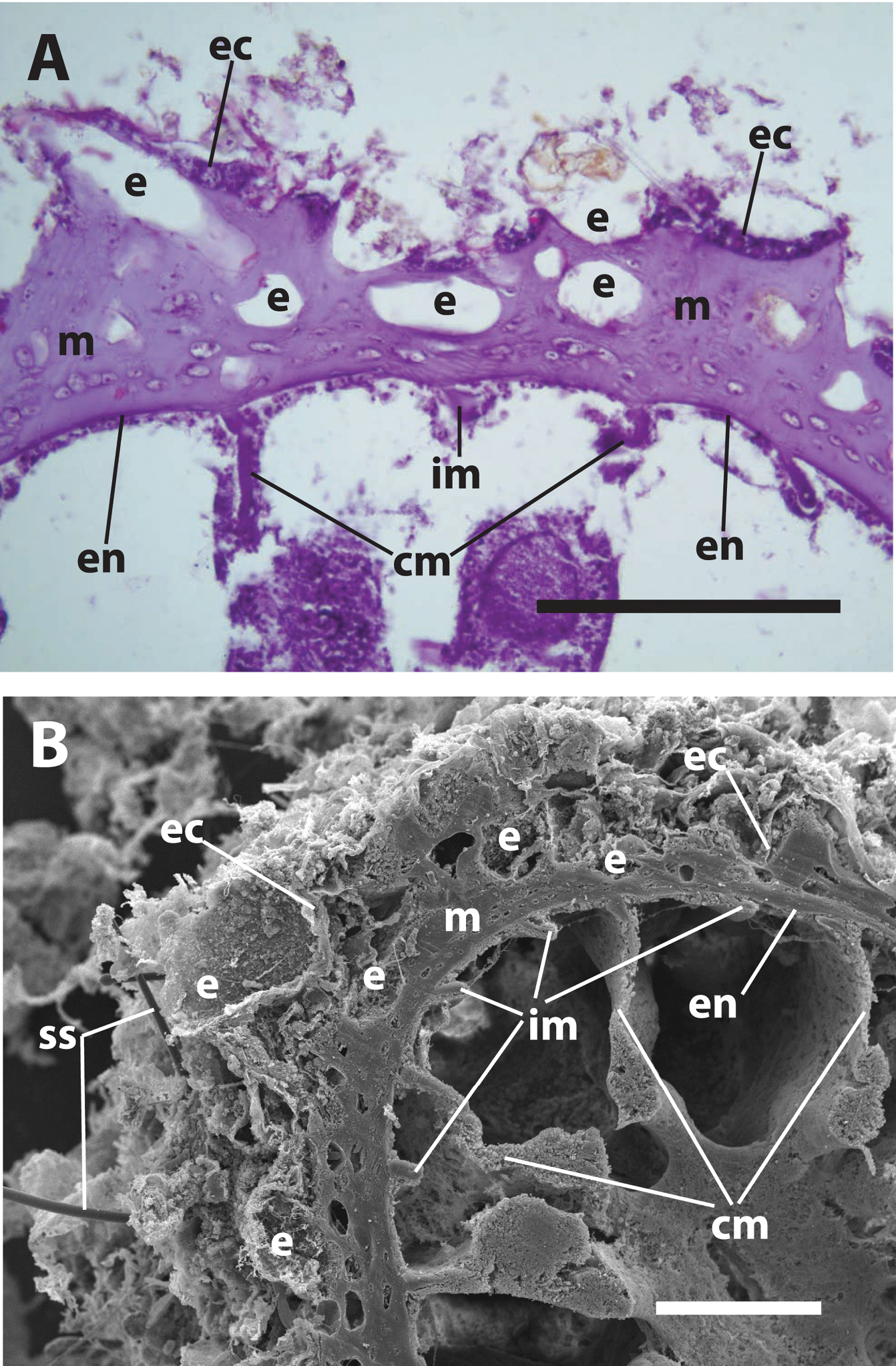
Figure 1.Internal structure of Neozoanthus uchina sp. n. showing encrustation in outer mesoglea and ectoderm, characteristic of Neozoanthus spp., with irregularly-sized encrustation A light microscope histological cross-section, and B scanning electron microscope image. Both images of specimen RMNH Coel 40098 (Table 2). Abbreviations: cm=complete mesenteries, e= sand/detritus encrustation (in B) or where encrustation existed before decalcification (in A), im=incomplete mesenteries, ec=ectoderm, en=endoderm, m=mesoglea, ss=encrusted sponge spicules. Scales: A=100 µm, B=200 µm.
Included On The Following Pages:
- Life (creatures)
- Cellular (cellular organisms)
- Eukaryota (eukaryotes)
- Opisthokonta (opisthokonts)
- Metazoa (Animal)
- Cnidaria (cnidarians)
- Anthozoa (anemones and corals)
- Zoantharia (zoanthids)
- Neozoanthidae
- Neozoanthus
- Neozoanthus uchina
This image is not featured in any collections.
Source Information
- license
- cc-by-3.0
- copyright
- James Davis Reimer, Yuka Irei, Takuma Fujii
- bibliographic citation
- Reimer J, Irei Y, Fujii T (2012) Two new species of Neozoanthus (Cnidaria, Hexacorallia, Zoantharia) from the Pacific ZooKeys 246: 69–87
- original
- original media file
- visit source
- partner site
- Zookeys
- ID