Odontella aurita cells
Description:
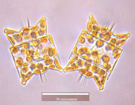
: Quality image This image has been assessed using the Quality image guidelines and is considered a Quality image. العربية | جازايرية | беларуская | беларуская (тарашкевіца) | български | বাংলা | català | čeština | Cymraeg | Deutsch | Schweizer Hochdeutsch | Zazaki | Ελληνικά | English | Esperanto | español | eesti | euskara | فارسی | suomi | français | galego | עברית | हिन्दी | hrvatski | magyar | Հայերեն | italiano | 日本語 | ქართული | 한국어 | Kurdî | Lëtzebuergesch | lietuvių | македонски | മലയാളം | मराठी | Bahasa Melayu | Nederlands | polski | português | português do Brasil | rumantsch | română | русский | sicilianu | slovenčina | slovenščina | српски / srpski | svenska | தமிழ் | తెలుగు | ไทย | Türkçe | українська | vèneto | Tiếng Việt | 中文 | 中文(简体) | 中文(繁體) | +/−. Description: English: The diatom Odontella aurita (400X magnification) The photograph was taken with a Leica DFC 320 microscope camera. Date: 9 May 2007. Source: Own work. Author: Richard A. Ingebrigtsen, Department of Arctic and Marine Biology, University of Tromsø.
Included On The Following Pages:
- Life (creatures)
- Cellular (cellular organisms)
- Eukaryota (eukaryotes)
- SAR (Stramenopiles, Alveolates, Rhizaria)
- Stramenopiles (heterokont)
- Ochrophyta (Ochrophyte)
- Bacillariophyta (diatoms)
- Mediophyceae
- Biddulphiophycidae
- Eupodiscales
- Odontella
This image is not featured in any collections.
Source Information
- license
- cc-by-sa-3.0
- copyright
- Richard A. Ingebrigtsen, Department of Arctic and Marine Biology, University of Tromsø
- creator
- Richard A. Ingebrigtsen, Department of Arctic and Marine Biology, University of Tromsø
- original
- original media file
- visit source
- partner site
- Wikimedia Commons
- ID