Pediculus humanus capitis 2

Description:
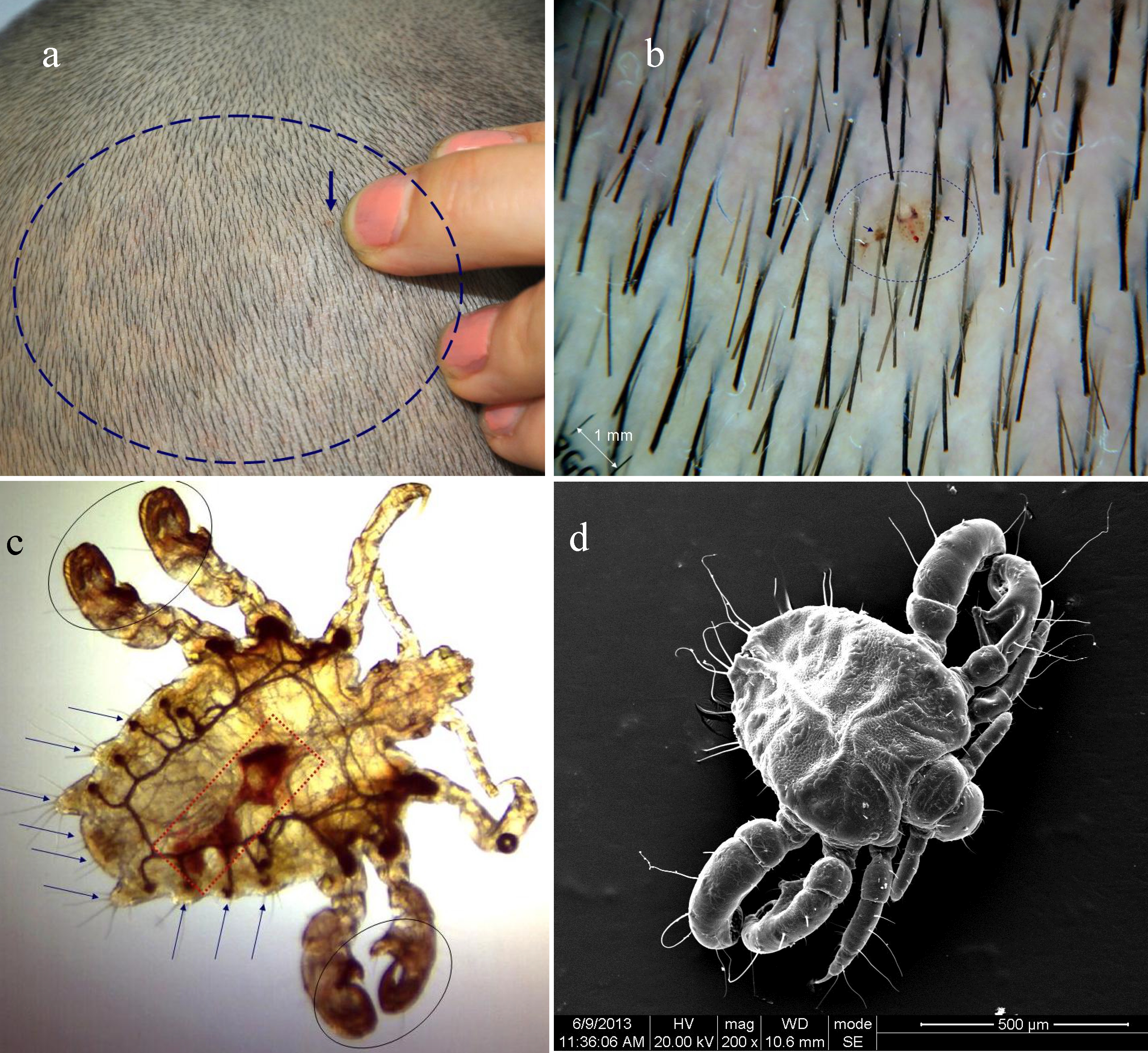
Description: a. There were some small pieces of erythema (in the circle) and a brown dot-like substance on the scalp (arrow). b. The dermoscopy revealed a brown parasite (0.9 mm in horizontal axes and 1.2 mm in vertical axes) with two crab-like feet adhered to the scalp. c. Under the microscope, the parasite was characterized by a flat body like a crab and three pairs of feet in different sizes. There was an area (red box) full of blood in the middle part of the parasite. A large number of short setae (arrow) were noted at the edge of the parasite abdomen. d. The SEM showed a vivid three-dimensional ultrastructure of the parasite: the whole body was composed of three parts including spherical head, chest, and elliptical abdomen; a pair of feelers was noted on the head; the three pairs of feet were in shaped section and curved serrated claws were noted at the end of foot; short setae in different length were not only at the edge of the abdomen but also on the feet. Date: 2016. Source: [1]. Author: Ran Yuping et al.
Included On The Following Pages:
- Life
- Cellular
- Eukaryota
- Opisthokonta
- Metazoa
- Bilateria
- Protostomia
- Ecdysozoa
- Arthropoda (arthropods)
- Pancrustacea
- Hexapoda
- Insecta (insects)
- Pterygota
- Neoptera (neopteran)
- Paraneoptera
- Psocodea
- Troctomorpha (book louse)
- Phthiraptera
- Anoplura (sucking louse)
- Pediculidae
- Pediculus
- Pediculus humanus
- Pediculus humanus capitis (head louse)
This image is not featured in any collections.
Source Information
- license
- cc-by-sa-3.0
- copyright
- Ran Yuping et al.
- creator
- Ran Yuping et al.
- source
- [1]
- original
- original media file
- visit source
- partner site
- Wikimedia Commons
- ID