Fmars-08-706308-g001
Description:
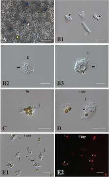
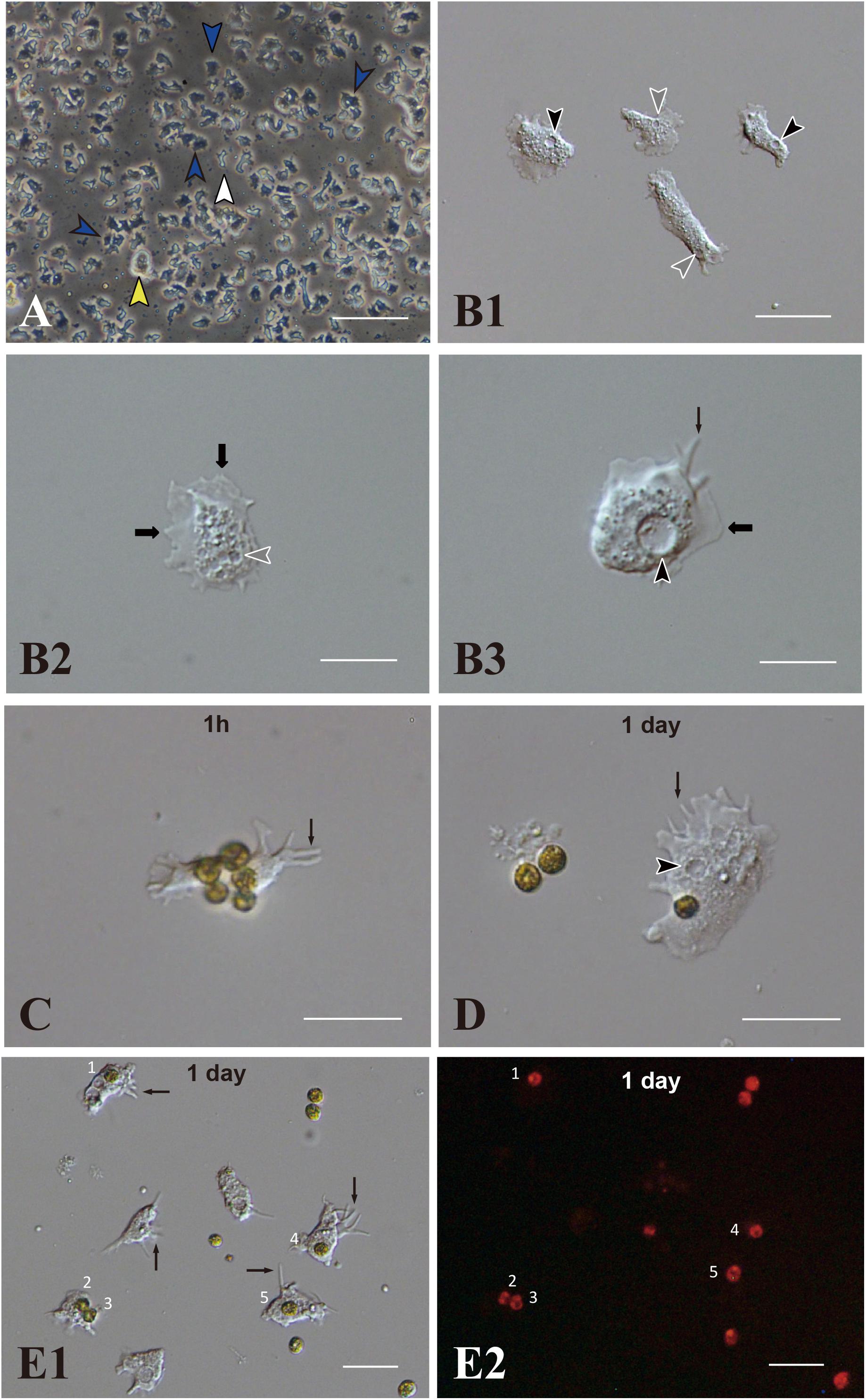
Description: English: In vitro: Acropora tenuis cells of the IVB5 line and symbiosis with the photosynthetic dinoflagellate, Breviolum minutum (Symbiodiniaceae). (A) A vertical microscope image of IVB5 line. Most cells are dark, flattened, amorphous cells (blue arrowheads), while a few brilliant cells (yellow arrowhead) and small cells of different morphology (white arrowhead) are present as well. (B–D) Differential interference microscopy images showing morphological features of flattened, amorphous cells. (B1) Lower and (B2,B3) higher magnification. Flattened amorphous cells contain a large vacuole (black arrowheads) and several small vesicles (white arrowheads) in the cytoplasm. They extend lamellipodia (bold b black arrows) and/or filopodia (thin black arrows) and show locomotor activity. (C–E) Coral cell engulfment of dinoflagellates, (C) 1 hour and (D,E) 1 day after mixturing cells of the two taxa. (E2) is a dark-field image of (E1), showing red auto-fluorescence of dinoflagellates. Cells endocytosed dinoflagellates numbered 1–5. Filopodia are indicated by arrows and vacuoles by black arrowheads. Scale bars, 100 μm in (A), 50 μm in (B1), 20 μm in (B2,B3,C,D), and 50 μm in (E1,E2). Date: 14 July 2021. Source: Fig. 1 at https://www.frontiersin.org/articles/10.3389/fmars.2021.706308/full In vitro Symbiosis of Reef-Building Coral Cells With Photosynthetic Dinoflagellates. In: Front. Mar. Sci., doi:10.3389/fmars.2021.706308 . Author: Kaz Kawamura, Satoko Sekida, Koki Nishitsuji, Eiichi Shoguchi, Kanako Hisata, Shigeki Fujiwara, Noriyuki Satoh. Other versions: This file has an extracted image: File:Fmars-08-706308-g001D.jpg..
Included On The Following Pages:
- Life (creatures)
- Cellular (cellular organisms)
- Eukaryota (eukaryotes)
- Opisthokonta (opisthokonts)
- Metazoa (Animal)
- Cnidaria (cnidarians)
- Anthozoa (anemones and corals)
- Hexacorallia (hexacorallians)
- Scleractinia (stony corals)
- Acroporidae (staghorn corals)
- Acropora (Staghorn corals)
- Acropora tenuis (Staghorn coral)
This image is not featured in any collections.
Source Information
- license
- cc-by-sa-3.0
- copyright
- Kaz Kawamura, Satoko Sekida, Koki Nishitsuji, Eiichi Shoguchi, Kanako Hisata, Shigeki Fujiwara, Noriyuki Satoh
- creator
- Kaz Kawamura, Satoko Sekida, Koki Nishitsuji, Eiichi Shoguchi, Kanako Hisata, Shigeki Fujiwara, Noriyuki Satoh
- source
- Fig. 1 at https://www.frontiersin.org/articles/10.3389/fmars.2021.706308/full In vitro Symbiosis of Reef-Building Coral Cells With Photosynthetic Dinoflagellates. In: Front. Mar. Sci., doi:10.3389/fmars.2021.706308
- original
- original media file
- visit source
- partner site
- Wikimedia Commons
- ID