portrait
Description:
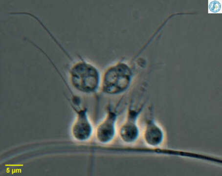
Codosiga - collar flagellate, two cells attached to a common stalk with a collection of Salpingoeca - also a collar flagellate. All these cells have a single apical flagellum that is surrounded with a collar of fine pseudopodia that appears as two dark lines, one to either side of the flagellum in this micrograph. Feed on suspended bacteria. From Lake Donghu, China Phase contrast micrograph.
Included On The Following Pages:
This image is not featured in any collections.
Source Information
- license
- cc-by-nc
- author
- Feng Weisong; Gu Xiwen; Yang Jun; Miao Wei and David Patterson
- provider
- micro*scope
- original
- original media file
- visit source
- partner site
- micro*scope
- ID