Image of Xanthidium antilopaeum var. crameri R. L. Grönblad
Description:
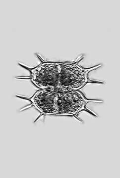
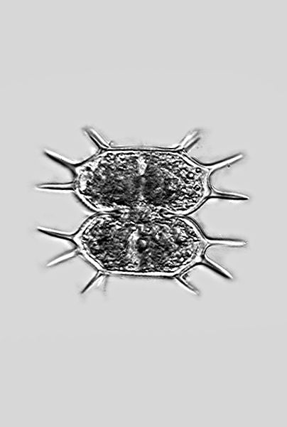
Description: The cells are only slightly wider than they are long, roughly octagonal in outline with straight or slightly concave sides. The central notches are greatly expanded outwards. At each of the side angles there is a pair of staggered spines (note the vertex view!), and the apical angles each have a pair of spines. In the middle of the cell halves there is a flat, hemispherical bulge.
Dimension: Length 45–60 µm, width 58–63 µm (without pricks).
Occurrence: In Central Europe sporadic in moderate acidic waters of fens, siltation zones et cetera.
Left: Original graphic by Prof. Lenzenweger.
Right: Photomicrograph by Prof. Lenzenweger. For microphotography, the microscope illumination was adjusted in such a way that as many features as possible that are important for species identification are prominently displayed.
Copyright by Prof. Rupert Lenzenweger, Ried im Innkreis, Austria.
© Wolfgang Bettighofer,
images under Creative Commons License V 3.0 (CC BY-NC-SA).
For permission to use of (high resolution) images please contact postmaster@protisten.de.
For further information about the image, please click here: Link to protisten.de page
Included On The Following Pages:
- Life (biota)
- Cellular
- Eukaryota (eukaryotes)
- Archaeplastida (plants)
- Chloroplastida
- Streptophyta (streptophytes)
- Zygnemophyceae (zygnemophyceaen algae)
- Zygnematophycidae
- Desmidiales
- Desmidiaceae
- Xanthidium
- Xanthidium antilopaeum
- Xanthidium antilopaeum crameri
This image is not featured in any collections.
Source Information
- license
- cc-by-nc-sa-3.0
- copyright
- Wolfgang Bettighofer
- creator
- Wolfgang Bettighofer [email]
- original
- original media file
- visit source
- partner site
- protisten.de
- ID