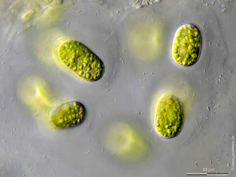
Image of Mesotaenium chlamydosporum
Description:
Sampling date 06/2024. Scale bars indicate 25 µm.
Four images.
Optical journey through a group of cells lying in a common mucilaginous mass. The gelatinous layers around the individual cells are visible; the short sticks in the mucilaginous mass are symbiotic bacteria.
Please click on < or > on the image edges or on the dots at the bottom edge of the images to browse through the slides!
Place name: Wetland Lauchseemoor, Fieberbrunn (Tyrol, Austria)
Latitude: 47.46954439 Longitude: 12.53826499
Microscope Zeiss Universal, camera Olympus OM-D M5 MKII. DOF images.
© Wolfgang Bettighofer,
images under Creative Commons License V 3.0 (CC BY-NC-SA).
For permission to use of (high resolution) images please contact postmaster@protisten.de.
For further information about the image, please click here: Link to protisten.de page
Included On The Following Pages:
- Life (biota)
- Cellular
- Eukaryota (eukaryotes)
- Archaeplastida (plants)
- Chloroplastida
- Streptophyta (streptophytes)
- Zygnemophyceae (zygnemophyceaen algae)
- Zygnematophycidae
- Zygnematales (Desmid)
- Mesotaeniaceae
- Mesotaenium
- Mesotaenium chlamydosporum
This image is not featured in any collections.
Source Information
- license
- cc-by-nc-sa-3.0
- copyright
- Wolfgang Bettighofer
- creator
- Wolfgang Bettighofer [email]
- original
- original media file
- visit source
- partner site
- protisten.de
- ID