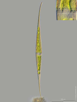
Image of Closterium kuetzingii Brébisson 1856
Description:
Sampling date 06/2024. Scale bars indicate 100 µm.
Two images.
First:Synoptic representation of the cell surface. The bars in the enlarged inset mark 10 µm. The striation has a density of 12 lines/10 µm.Second:Optical cross-section showing cell nucleus and chloroplasts.
Please click on < or > on the image edges or on the dots at the bottom edge of the images to browse through the slides!
Place name: Wetland at the Pillersee (Tyrol, Austria)
Latitude: 47.531785 Longitude: 12.573095
Microscope Zeiss Universal, camera Olympus OM-D M5 MKII. DOF images.
© Wolfgang Bettighofer,
images under Creative Commons License V 3.0 (CC BY-NC-SA).
For permission to use of (high resolution) images please contact postmaster@protisten.de.
For further information about the image, please click here: Link to protisten.de page
Included On The Following Pages:
- Life (biota)
- Cellular
- Eukaryota (eukaryotes)
- Archaeplastida (plants)
- Chloroplastida
- Streptophyta (streptophytes)
- Zygnemophyceae (zygnemophyceaen algae)
- Zygnematophycidae
- Desmidiales
- Closteriaceae
- Closterium
- Closterium kuetzingii
This image is not featured in any collections.
Source Information
- license
- cc-by-nc-sa-3.0
- copyright
- Wolfgang Bettighofer
- creator
- Wolfgang Bettighofer [email]
- original
- original media file
- visit source
- partner site
- protisten.de
- ID