Image of Eastern Carpenter Bee
Description:
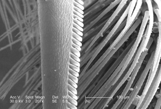
Under the moderate magnification of 207x, this scanning electron micrograph (SEM) revealed the rear leg anatomy of a carpenter bee, Xylocopa virginica, at the region of the "tibial spur". These spurs, which are exoskeletal protuberances, enable the bee to grasp various floral structures, thereby, enhancing its maneuverability inside the flower while it obtains it nectar meal, and pollinates the plant. See PHIL #8828 for a view of the same tibial spur under a lower magnification.
Created: 2006
Included On The Following Pages:
- Life (creatures)
- Cellular (cellular organisms)
- Eukaryota (eukaryotes)
- Opisthokonta (opisthokonts)
- Metazoa (Animal)
- Bilateria
- Protostomia (protostomes)
- Ecdysozoa (ecdysozoans)
- Arthropoda (arthropods)
- Pancrustacea
- Hexapoda (hexapods)
- Insecta (insects)
- Pterygota (winged insects)
- Neoptera (neopteran)
- Endopterygota (endopterygotes)
- Hymenoptera (wasps, bees, and ants)
- Apocrita (wasp)
- Aculeata
- Apoidea (bees & apoid Wasps)
- Apidae (honeybees, bumblebees, and relatives)
- Xylocopa (Carpenter Bees)
- Xylocopa virginica (Eastern Carpenter Bee)
- Panarthropoda
This image is not featured in any collections.
Source Information
- license
- cc-publicdomain
- photographer
- Janice Carr
- provider
- Public Health Image Library
- original
- original media file
- visit source
- partner site
- Public Health Image Library
- ID