Plate 4
Description:
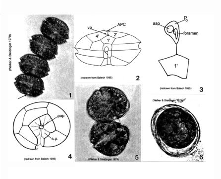
Plate 4. Alexandrium monilatum. Fig. 1. LM: four-cell chain. Cells large, wider than long, flattened anterio-posteriorly. Antapex slightly concave (arrow). Figs. 2-4. Line drawings. Fig. 2. Ventral pore (vp) depicted (Florida specimens) at anterior margin of 1' plate where it comes in contact with plates 2' and 4'. Cingulum (C) deeply excavated, wide, descending; displaced one time its width. Fig. 3. Apical pore plate (Po) does not come in contact with 1' plate. Anterior attachment pore (aap) large, round and dorsally situated in the APC. Foramen comma-shaped. Fig. 4. Antapical view: posterior sulcal plate (sp) large, rhomboid and concave with radial markings. Posterior attachment pore (pap) large and centrally located. Figs. 5-6. LM. Fig. 5. Two isogamous gametes fusing at oblique angles. Fig. 6. Mature resting cysts: dark and round, with a triple layered wall.
Included On The Following Pages:
- Life (creatures)
- Cellular (cellular organisms)
- Eukaryota (eukaryotes)
- SAR (Stramenopiles, Alveolates, Rhizaria)
- Alveolata (alveolates)
- Dinophyceae
- Gonyaulacales
- Gonyaulacaceae
- Alexandrium
- Alexandrium monilatum
- Dinoflagellata (dinoflagellates)
This image is not featured in any collections.
Source Information
- license
- cc-publicdomain
- bibliographic citation
- Faust, Maria A. and Rose A. Gulledge. Identifying Harmful Marine Dinoflagellates. Smithsonian Contributions from the United States National Herbarium, volume 42: 1-144 (including 48 plates, 1 figure and 1 table).
- original
- original media file
- visit source
- partner site
- NMNH Marine Dinoflagellates
- ID