-
-
Lardero, La Rioja, Espaa
-
-
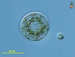
Public Domain, U.S. Government Work 2011 Barry H. Rosen Courtesy of life.nbii.gov
NBII images
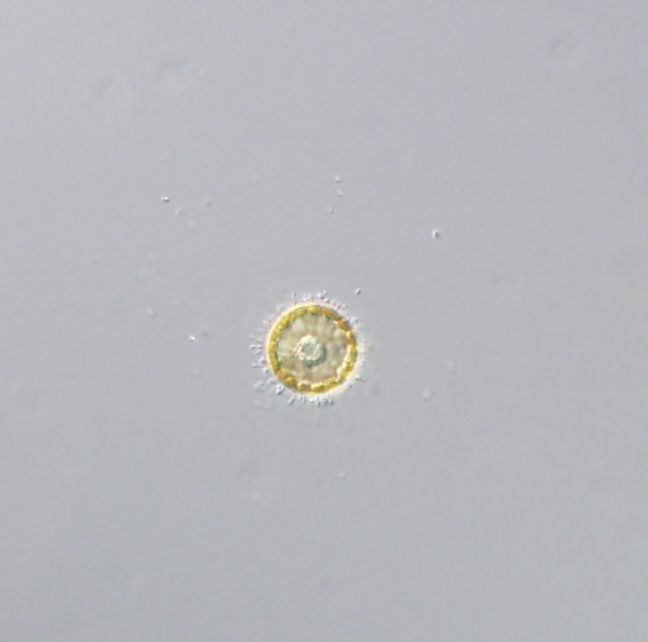
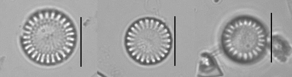
Category hierarchy: Microorganisms | AlgaeDescription: Discoid chromatophores and the nucleus easily observed with epifluorescent illumination. Sample was collected from Raccoon River, Iowa.Capture device: DP71Capture details: 400x with microscopeOriginal date: 20091002|||112306Locality: Latitude: 2.859009900000000e+001; Longitude: -8.119031699999999e+001
-
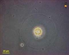
Public Domain, U.S. Government Work 2011 Barry H. Rosen Courtesy of life.nbii.gov
NBII images
Category hierarchy: Microorganisms | MicrofloraDescription: Discoid chromatophores and the nucleus easily observed with normal illumination. Specimen was collected from Raccoon River, Iowa.Capture device: DP71Original date: 20091002|||113054Locality: Latitude: 2.859009900000000e+001; Longitude: -8.119031699999999e+001
-
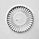
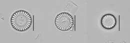
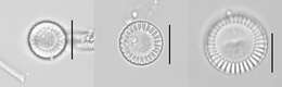
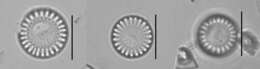
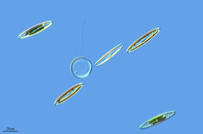
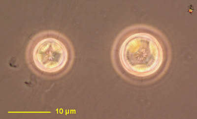
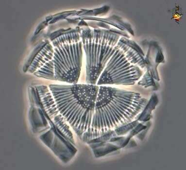
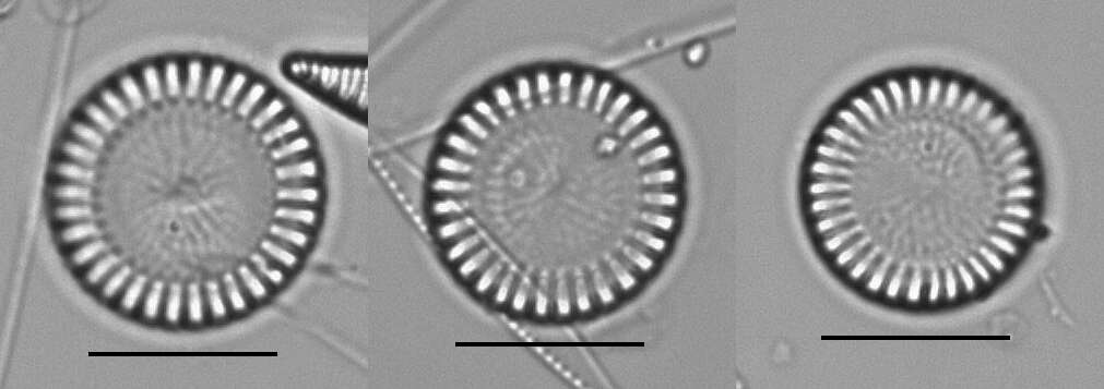
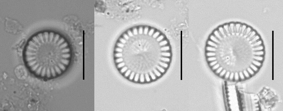
Cyclotella (sike-low-tell-a). Centric diatom, seen from valve view. Three plastid profiles are visible around the periphery of the cell. Long thin organic spines project from the cell - and are believed to have a role in flotation. The pattern of pores in the frustule is used in identification. Marine. Phase contrast.
-
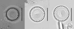
Centric diatom, seen from valve view. The pattern of pores in the frustule is used in identification. Marine. Phase contrast.
-
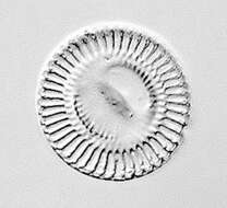
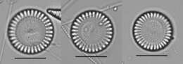
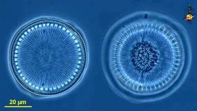
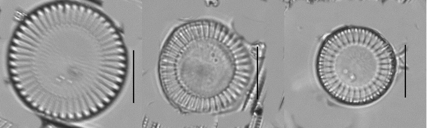
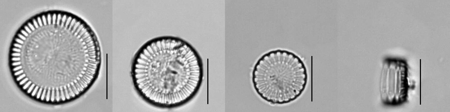
Cyclotella (sike-low-tell-a). Centric diatom, frustule only, seen from valve view, with the two frustules seen at slightly different focal planes. . The pattern of pores in the frustule is used in identification. Phase contrast.
-
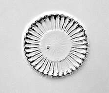
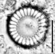
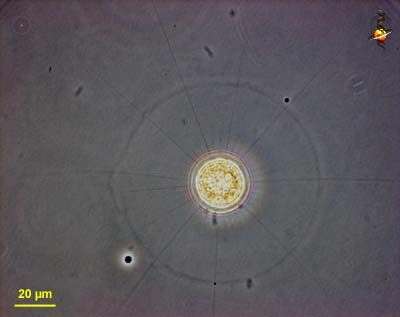
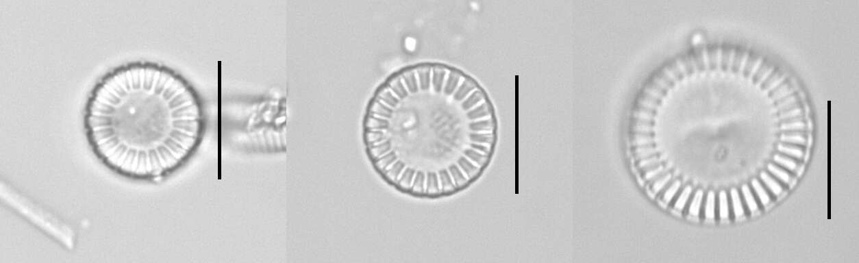
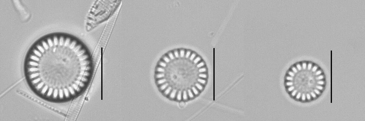
Cyclotella (sike-low-tell-a). Centric diatom, seen from valve view. The cell is surrounded by a sheath of mucus. Long thin organic spines project from the cell - and are believed to have a role in flotation. The pattern of pores in the frustule is used in identification. From a freshwater site. Phase contrast.
-
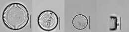
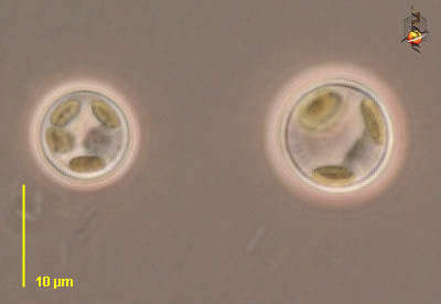
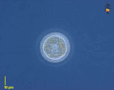
Cyclotella (sike-low-tell-a). Centric diatom, seen from valve view. With many small plastids containing chlorophylls a and c. From a freshwater site. Differential interference contrast.
-
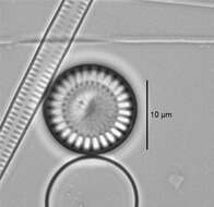
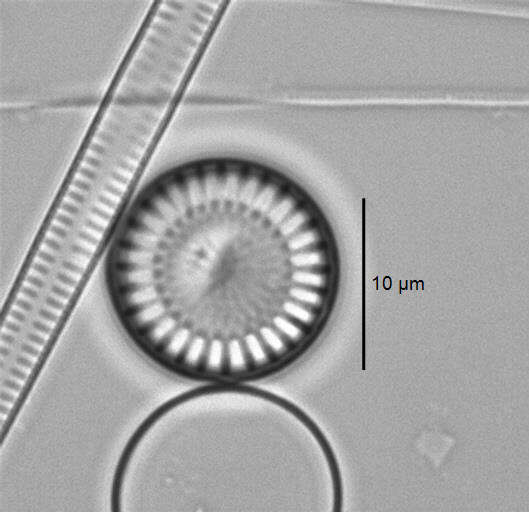
Cyclotella (sike-low-tell-a). Centric diatom, frustule only, seen from valve view, frustule broken from cover-slip pressure to show the brittle nature of the frustule. The pattern of pores in the frustule is used in identification. Phase contrast.
-
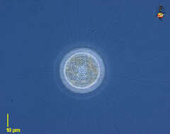
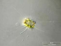
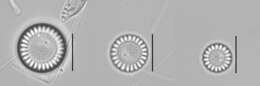
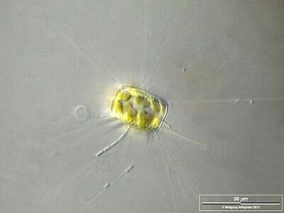
The thin, barely visible floating extensions are made of chitin. Furthermore, filamentous bacteria colonies are attached. Scale bar indicates 50 µm. Sample from the Lake Constance (vicinity of Bodman). The image was built up using several photomicrographic frames with manual stacking technique. Images were taken using Zeiss Universal with Olympus C7070 CCD camera.Image under Creative Commons License V 3.0 (CC BY-NC-SA).
-
Optical tranversal section, showing the nucleus. The thin, barely visible floating extensions are made of chitin. Furthermore, filamentous bacteria colonies are attached. Scale bar indicates 50 µm. Sample from the Lake Constance (vicinity of Bodman). The image was built up using several photomicrographic frames with manual stacking technique. Images were taken using Zeiss Universal with Olympus C7070 CCD camera.Image under Creative Commons License V 3.0 (CC BY-NC-SA).
-
-
-
-
-
-
-
-
-
-