-
Sergey G. Ermilov, Umukusum Ya. Shtanchaeva, Luis S. Subías, Jochen Martens
Zookeys
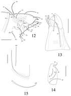
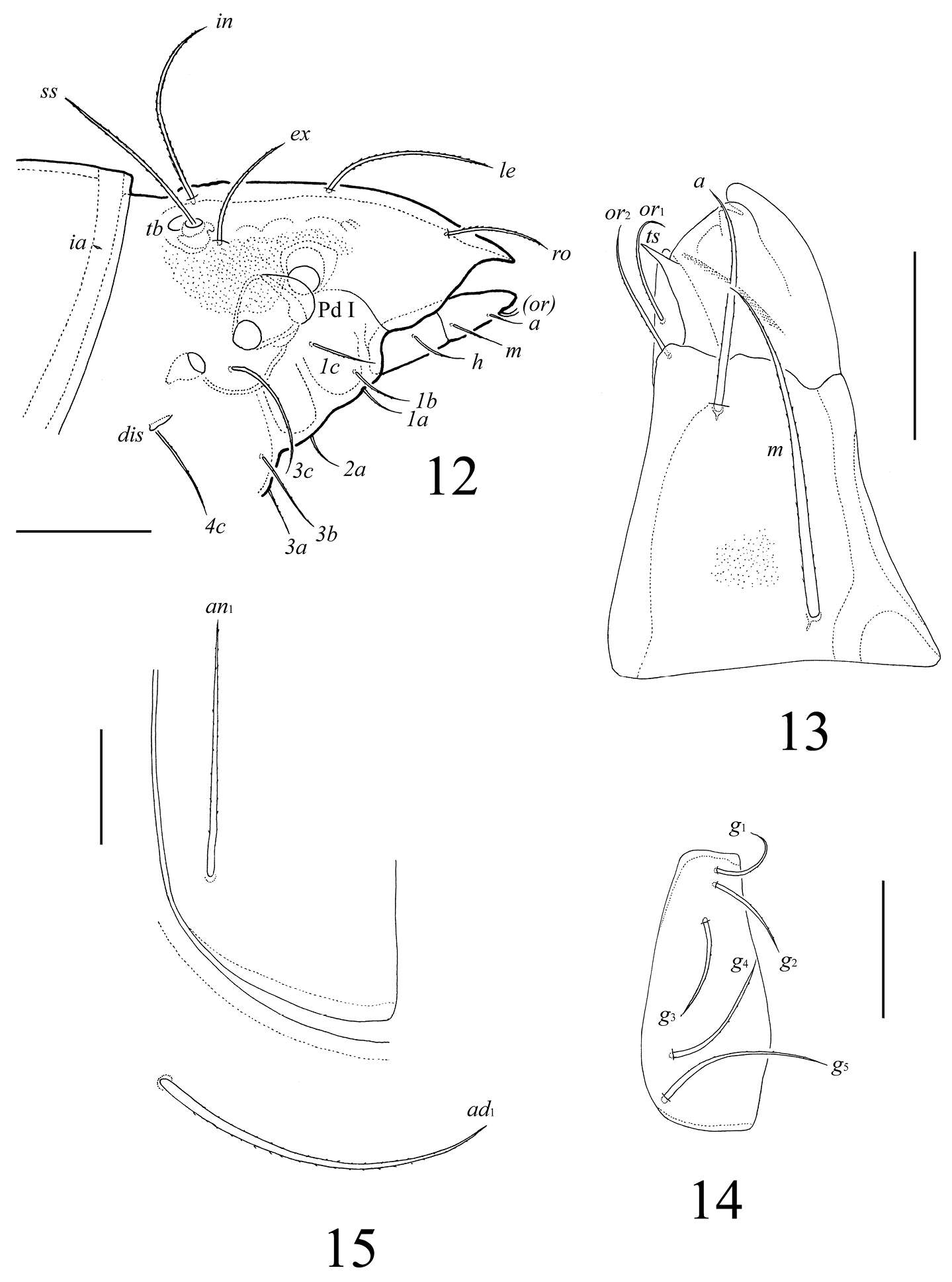
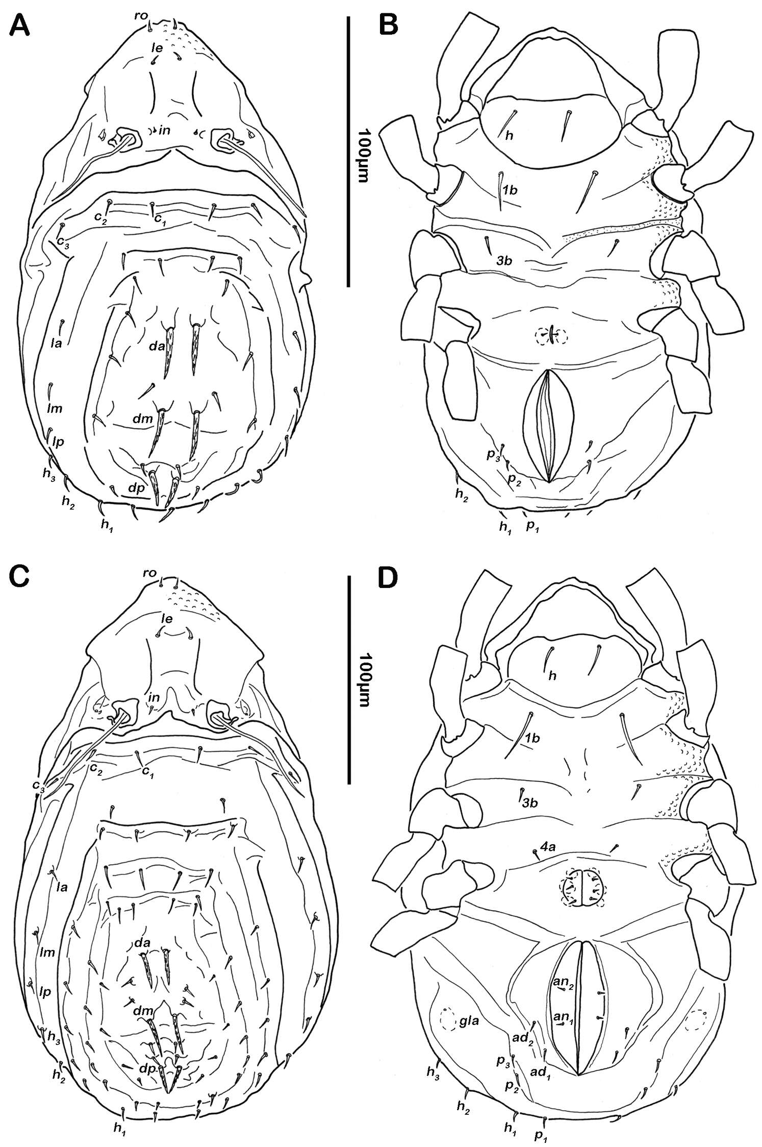
Figures 12–15.Lasiobelba (Antennoppia) nepalica sp. n.: 12 lateral view of prodorsum (legs not illustrated) and anterior part of notogaster 13 left rutellum and gena of subcapitulum, ventral view 14 genital plate, right 15 posterior part of anal plate with seta an1 and adanal seta ad1. Scale bar 50 μm.
-
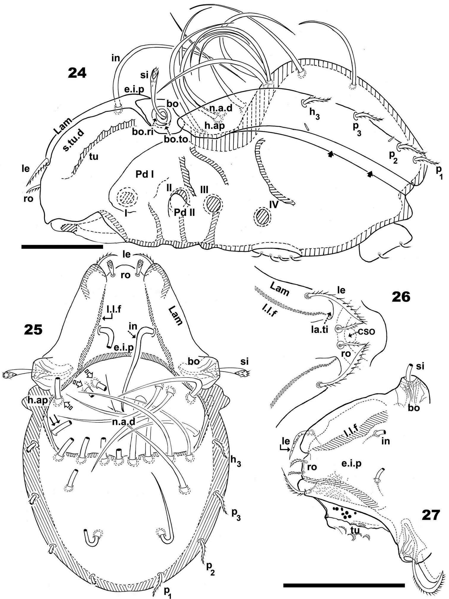
Nestor Fernandez, Pieter Theron, Christine Rollard, Elio Rodrigo Castillo
Zookeys
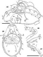
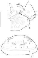
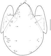
Figures 24–27.Malgasodes hungarorum Mahunka, 2000, adult. Optic observations. 24 lateral, slightly inclined view 25 dorsal view 26 prodorsum anterior part, dorsal view, inclined anteroposterior 27 prodorsum dorsal, inclined laterally. Abbreviations: see “Material and methods”. Scale bar: 24–25 = 60 μm; 26, 27 = 55 μm.
-
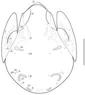
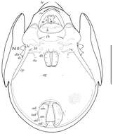
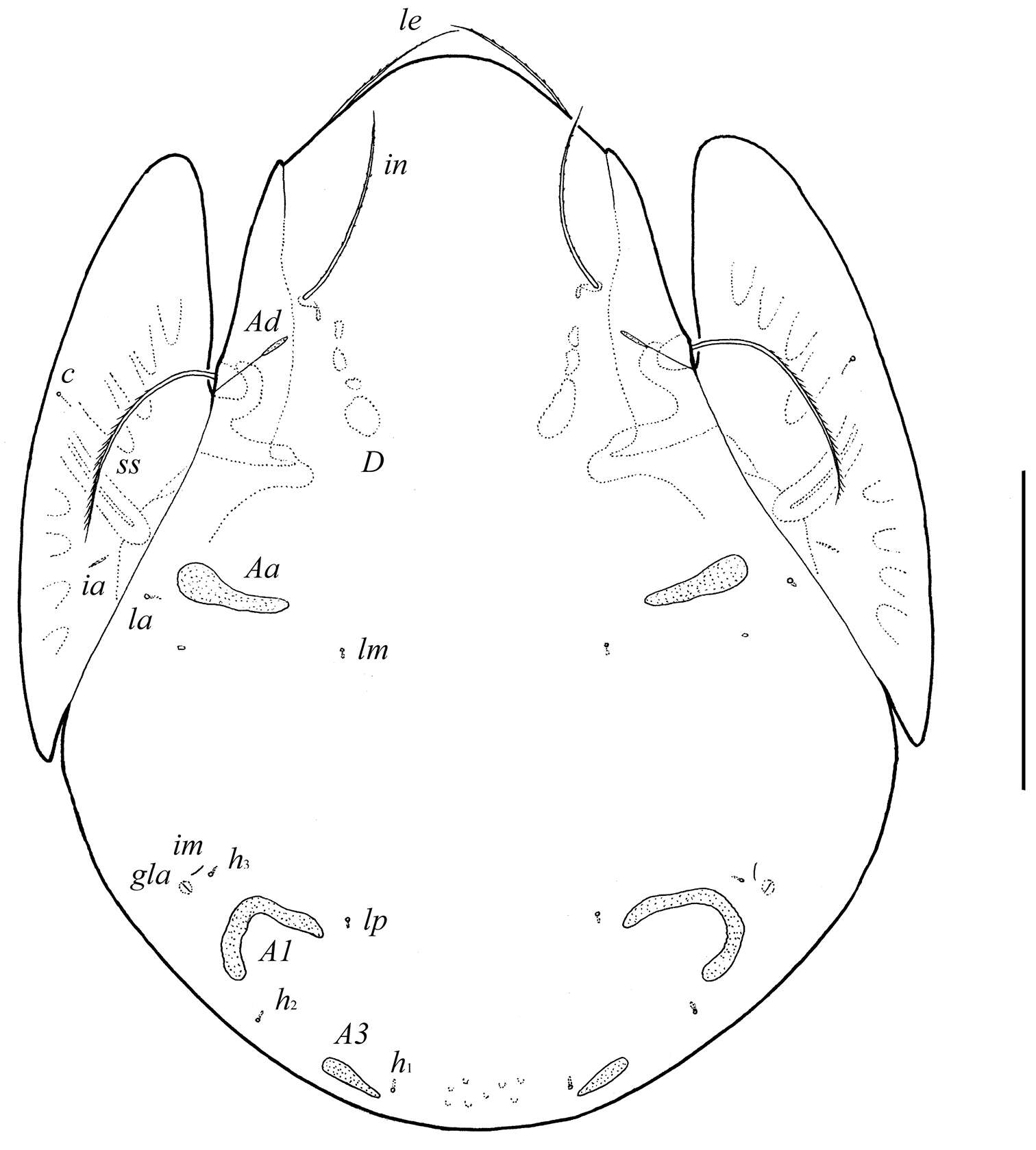
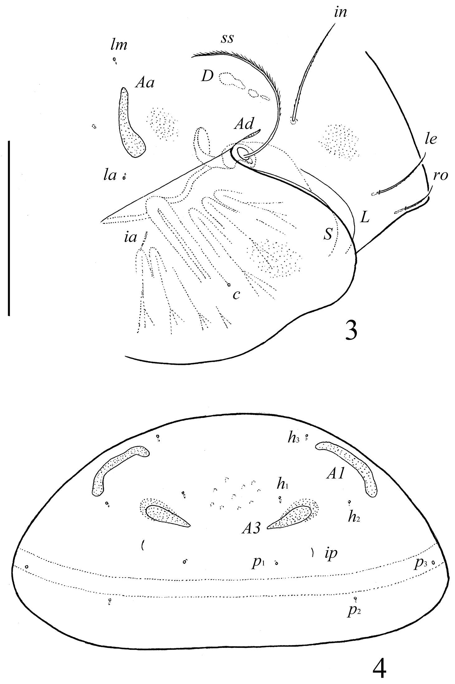
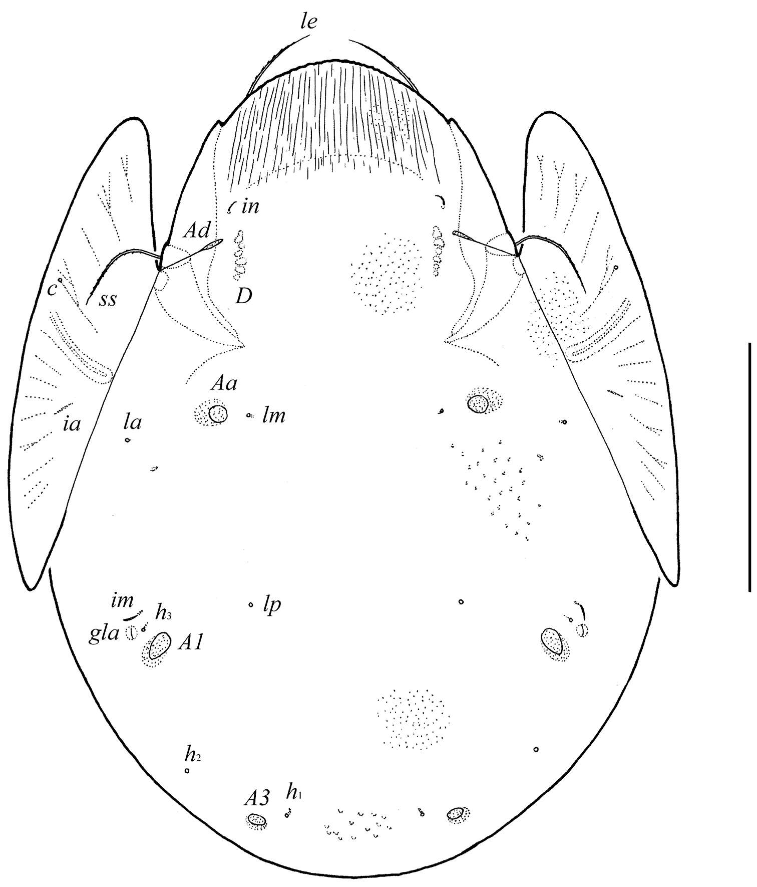
Figure 1.Conchogneta glabrisensillata sp. n. A Dorsal view of idiosoma B Ventral view of idiosoma C Prodorsum D Sensillus and bothridium, lateral view E Slight variation of sensillus, lateral view.
-
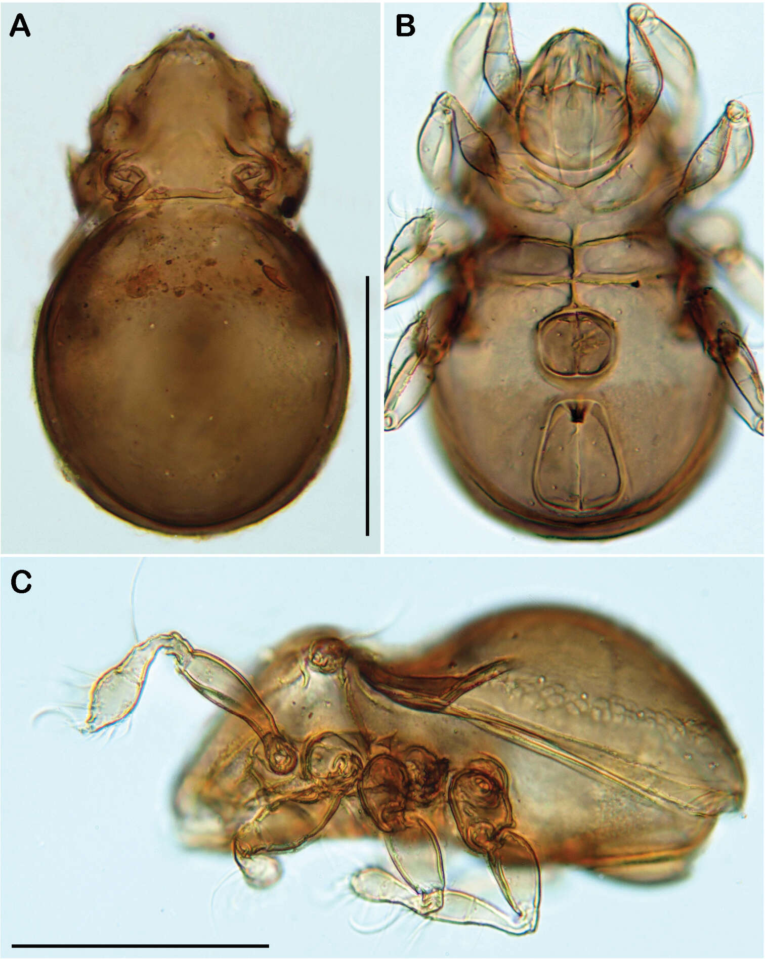
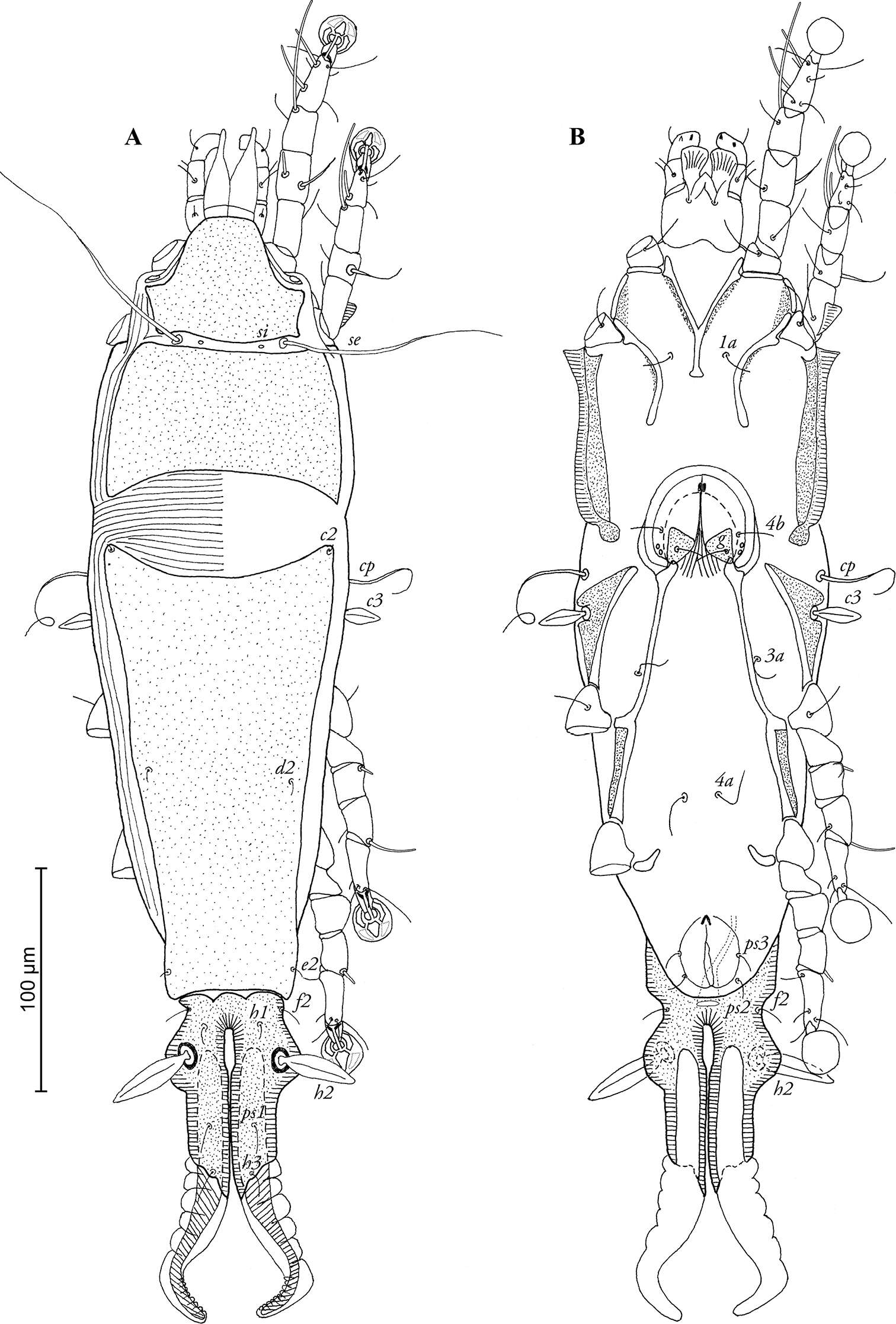
Figure 3.Selenoribates quasimodo sp. n. adult micrographs layered from 5–10 sequentially focused images. A dorsal view B ventral view C lateral view. Scale bars = 100 µm.
-
Ioana Cristina Constantinescu, Gabriel Chişamera, D. Khlur B. Mukhim, Costică Adam
Zookeys
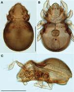
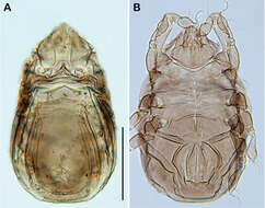
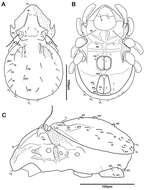
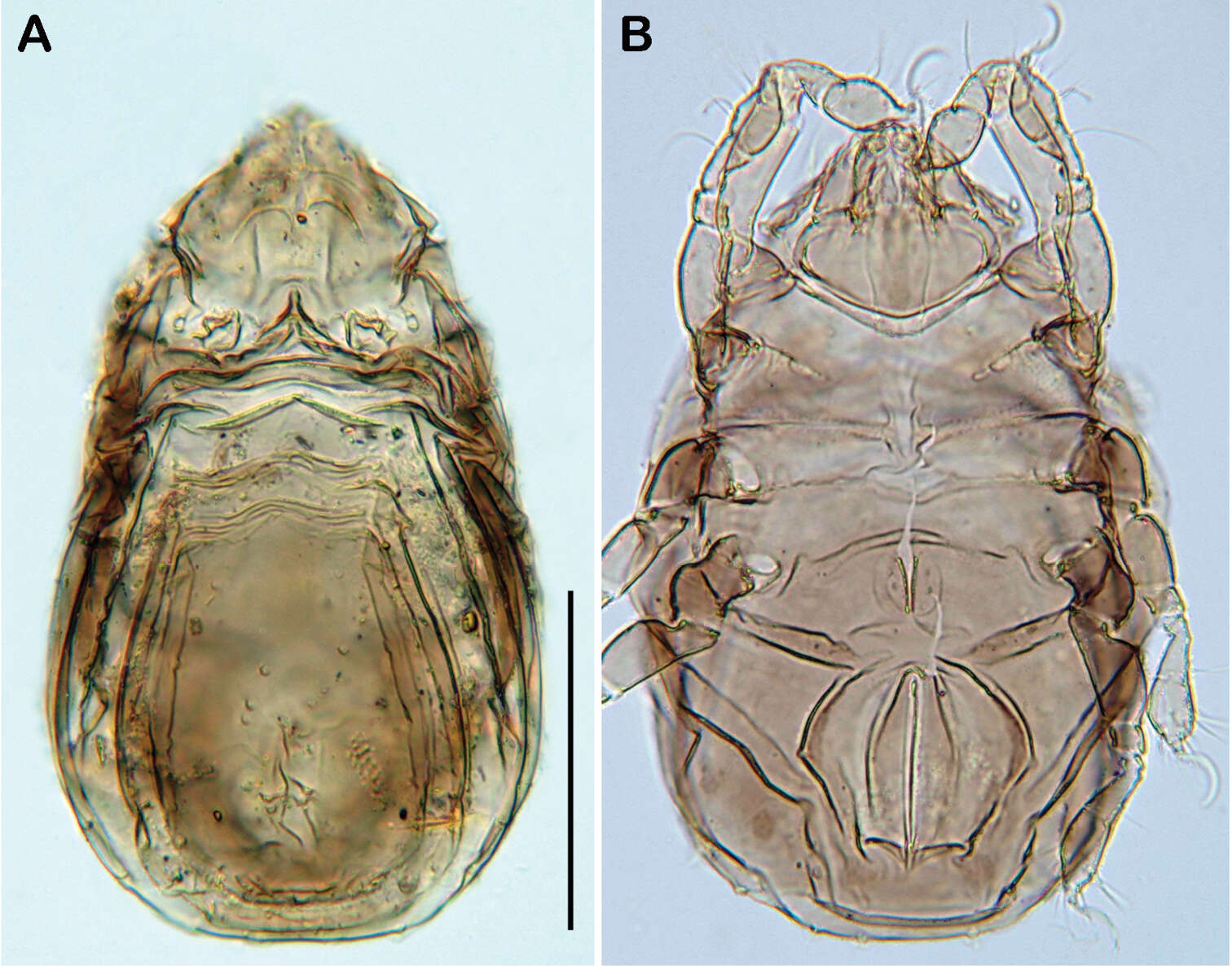
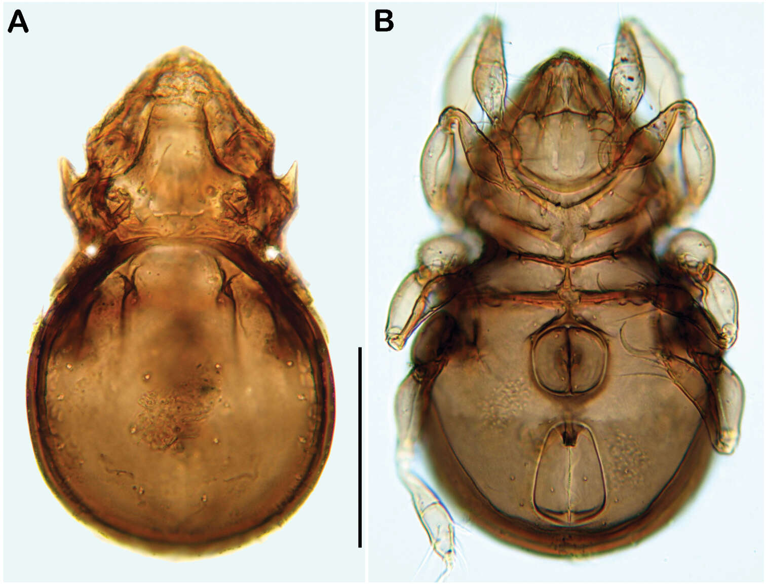
Figure 1.Pedanodectes angustilobus sp. n., male holotype: A dorsal view of idiosoma B ventral view of idiosoma.
-
Sergey G. Ermilov, Olman Alvarado-Rodríguez, Axel P. Retana-Salazar
Zookeys
Figure 1.Pergalumna elongatiporosa sp. n.: dorsal view. Scale bar 100 μm.
-
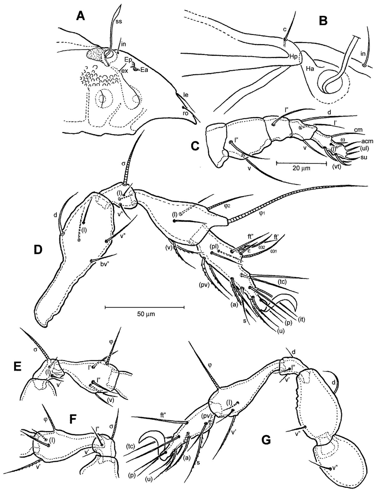
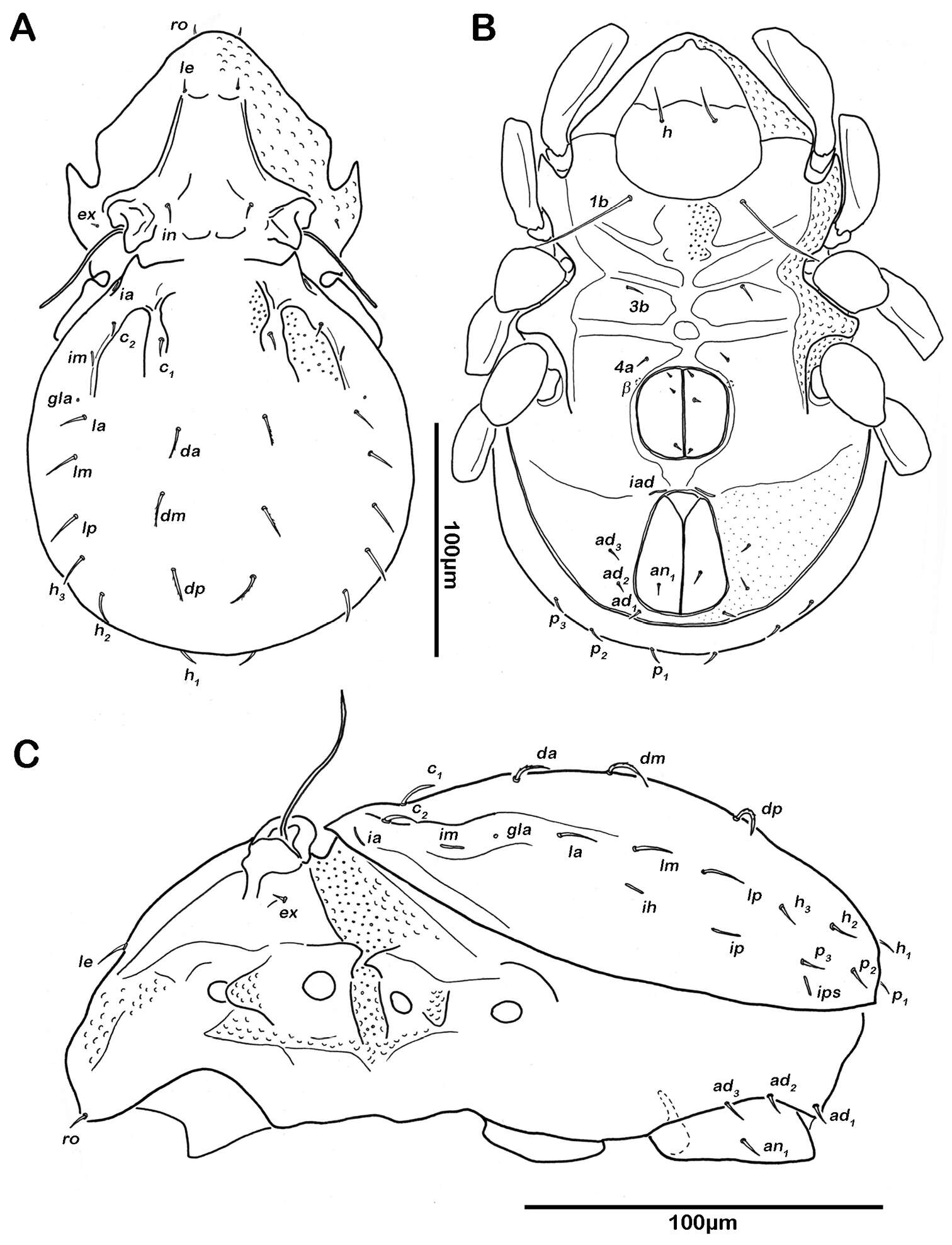
Figure 2.Conchogneta glabrisensillata sp. n. A Lateral view of prodorsum and anterior part of notogaster B Humeral region, showing tubercles Ha and Hp C Palp, right, antiaxial view D Leg I, right, antiaxial view E Genu and tibia of leg II, right, antiaxial view F Genu and tibia of leg III, right, antiaxial view G Leg IV, right, antiaxial view.
-
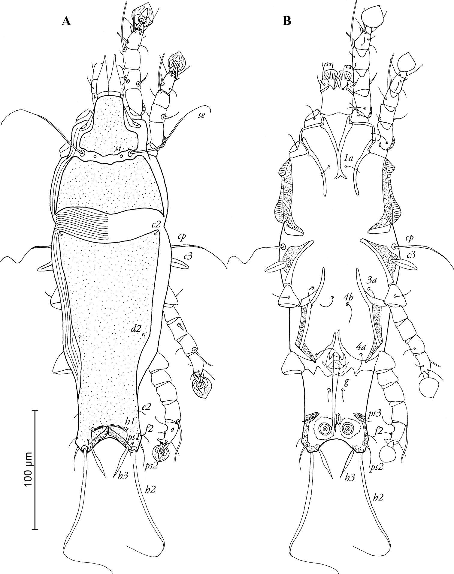
Figure 4.Selenoribates quasimodo sp. n. nymphs. A protonymph dorsal view B protonymph ventral view C tritonymph dorsal view D tritonymph ventral view.
-
Ioana Cristina Constantinescu, Gabriel Chişamera, D. Khlur B. Mukhim, Costică Adam
Zookeys
Figure 2.Pedanodectes angustilobus sp. n., female paratype: A dorsal view of idiosoma B ventral view of idiosoma.
-
Sergey G. Ermilov, Olman Alvarado-Rodríguez, Axel P. Retana-Salazar
Zookeys
Figure 2.Pergalumna elongatiporosa sp. n.: ventral view (legs not illustrated). Scale bar 100 μm.
-
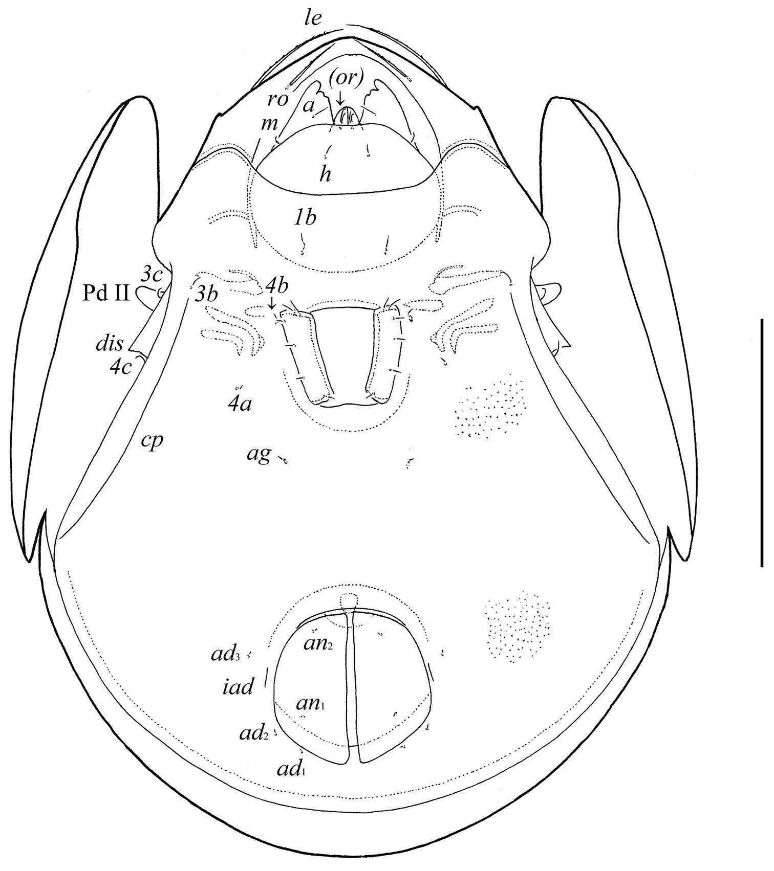
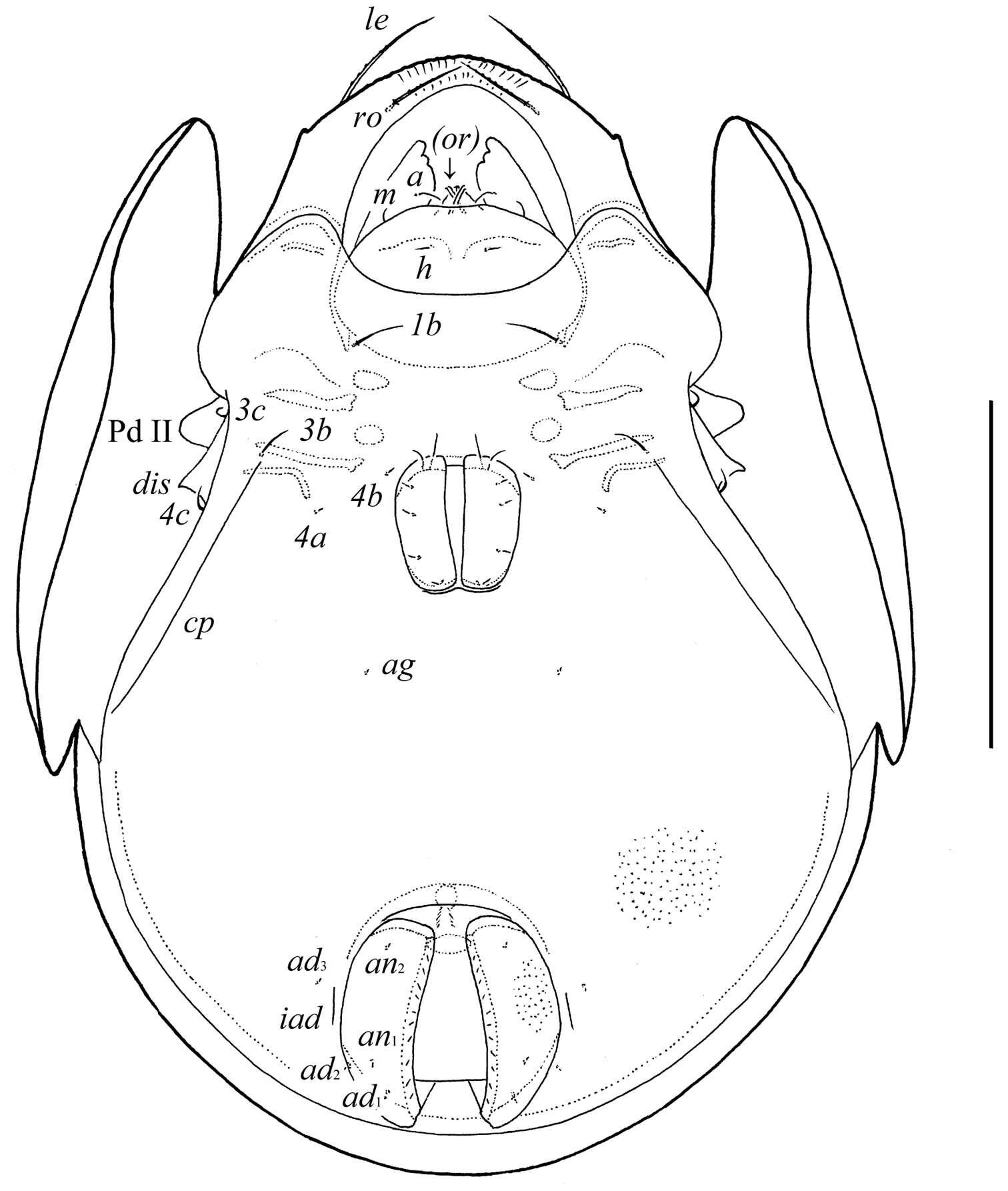
Figure 3.Conchogneta glabrisensillata sp. n. A Prodorsum, showing enantiophysis E, costula and bothridium B Central part of prodorsum, showing alveolus of interlamellar seta and interbothridial tubercle (indicated by arrow) C Part of laterial view of prodorsum, showing sensillus and granular tubercles on humeral region D Lateral view of prodorsal costula E Sensillus, lateral view F Slight variation of sensillus, lateral view G Granular tubercles on lateral part of prodorsum H Humeral region, showing tubercles Ha and Hp.
-
Figure 5.Selenoribates quasimodo sp. n. tritonymph, micrographs layered from 5–10 sequentially focused images. A dorsal view B ventral view. Scale bar = 100 µm.
-
Ioana Cristina Constantinescu, Gabriel Chişamera, D. Khlur B. Mukhim, Costică Adam
Zookeys
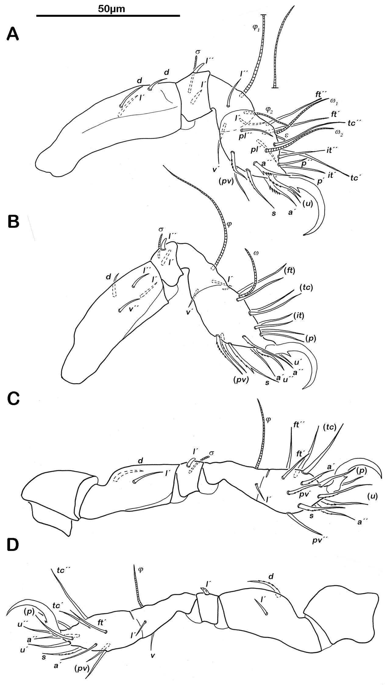
Figure 3.Pedanodectes angustilobus sp. n., details: A–D legs I–IV of male, respectively, dorsal view.
-
Sergey G. Ermilov, Olman Alvarado-Rodríguez, Axel P. Retana-Salazar
Zookeys
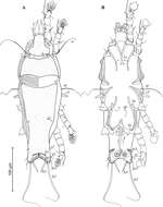
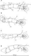
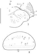
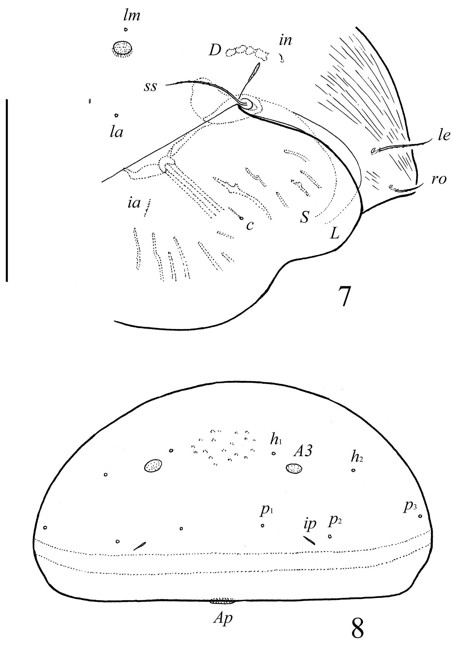
Figures 3–4.Pergalumna elongatiporosa sp. n.: 3 dorso-lateral view of prodorsum and anterior part of notogaster and pteromorph (gnathosoma and legs not illustrated) 4 posterior view of notogaster. Scale bars 100 μm.
-
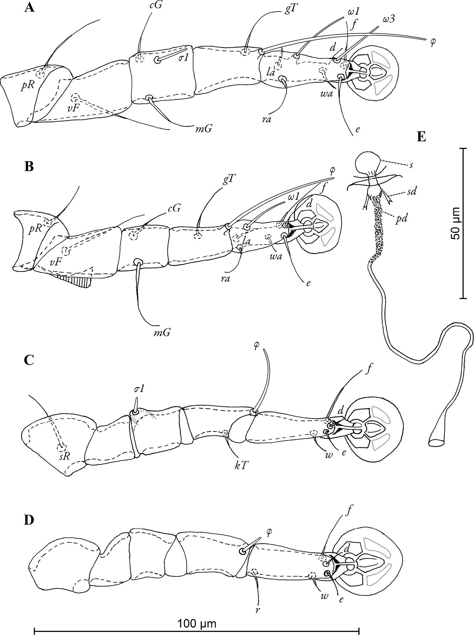
Figure 6.Selenoribates quasimodo sp. n. tritonymph legs. A right leg I antiaxial view B right leg II antiaxial view C left leg III antiaxial view D right leg IV antiaxial view.
-
Ioana Cristina Constantinescu, Gabriel Chişamera, D. Khlur B. Mukhim, Costică Adam
Zookeys
Figure 4.Pedanodectes angustilobus sp. n., details: A–D legs I–IV of female, respectively E spermatheca and spermaducts, dorsal view. Abbreviations: pd - primary spermaduct; s - spermatheca; sd - secondary spermaduct.
-
Sergey G. Ermilov, Olman Alvarado-Rodríguez, Axel P. Retana-Salazar
Zookeys
Figure 5.Pergalumna striatiprodorsum sp. n.: dorsal view. Scale bar 200 μm.
-
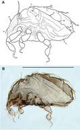
Figure 7.Selenoribates satanicus sp. n. adult. A dorsal view B ventral view C lateral view.
-
Sergey G. Ermilov, Olman Alvarado-Rodríguez, Axel P. Retana-Salazar
Zookeys
Figure 6.Pergalumna striatiprodorsum sp. n.: ventral view (legs not illustrated). Scale bar 200 μm.
-
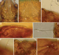
Figure 8.Selenoribates satanicus sp. n. adult micrographs layered from 5–10 sequentially focused images. A dorsal view B ventral view. Scale bar = 100 µm.
-
Sergey G. Ermilov, Olman Alvarado-Rodríguez, Axel P. Retana-Salazar
Zookeys
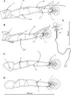
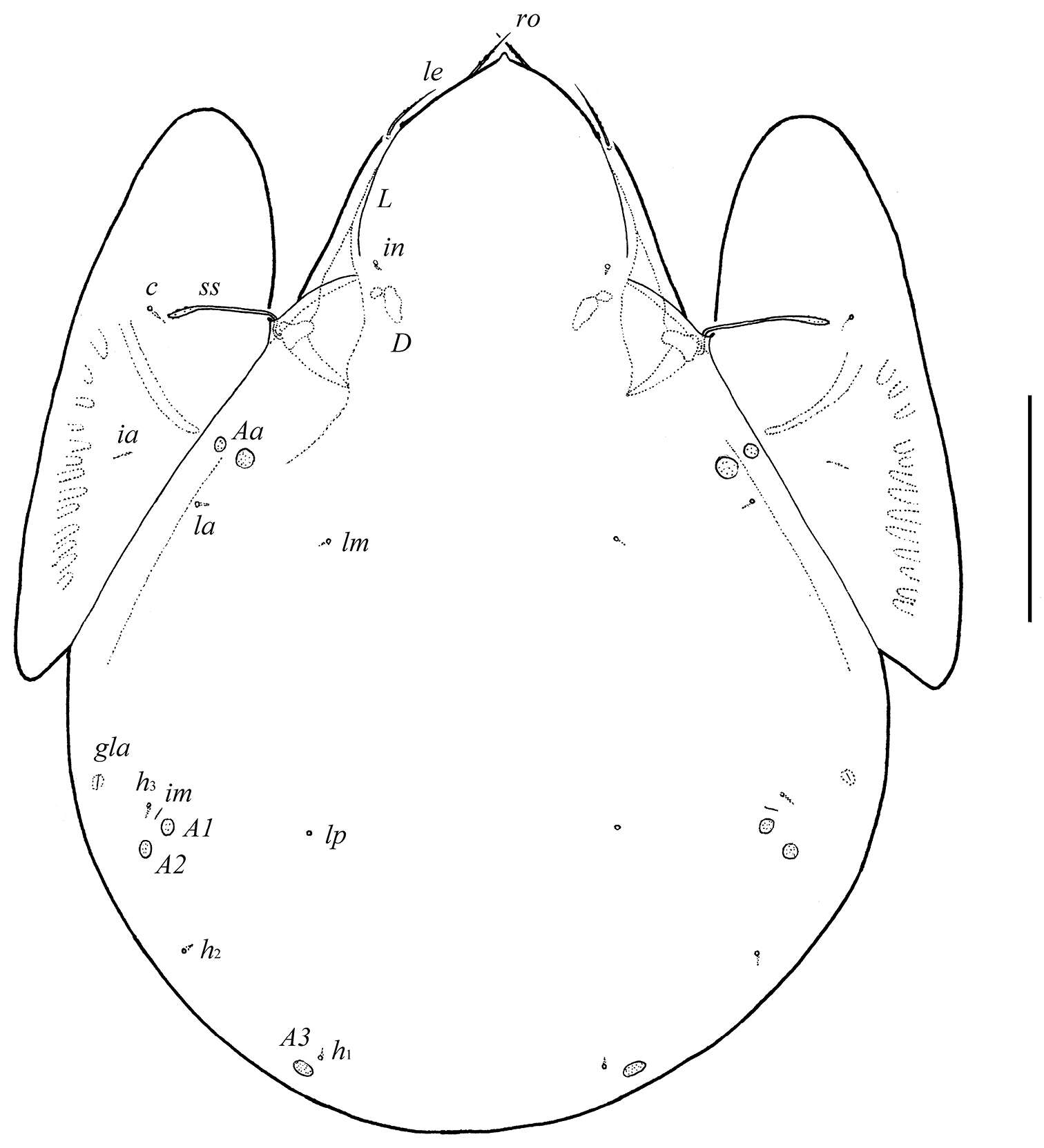
Figures 7–8.Pergalumna striatiprodorsum sp. n.: 7 dorso-lateral view of prodorsum and anterior part of notogaster and pteromorph (gnathosoma and legs not illustrated) 8 posterior view of notogaster. Scale bars 200 μm.
-
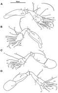
Figure 9.Selenoribates satanicus sp. n. legs. A right leg I antiaxial view B right leg II axial view C right leg III antiaxial view D left leg IV antiaxial view.
-
Sergey G. Ermilov, Jochen Martens, Andrei V. Tolstikov
Zookeys
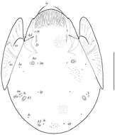
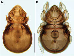
Figure 1.Galumna tetraporosa sp. n., adult: dorsal view. Scale bar 200 μm.
-
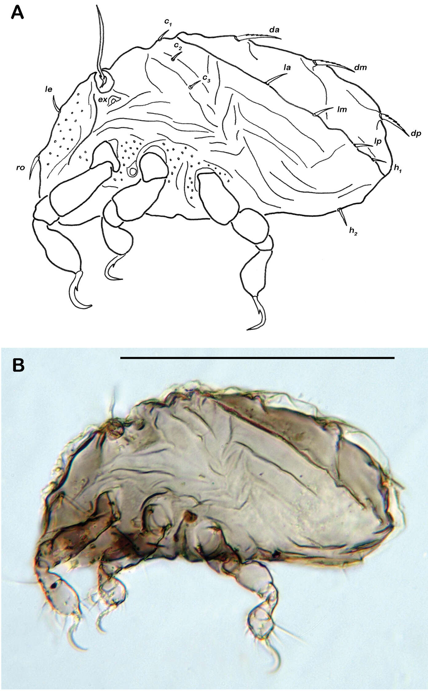
Figure 10.Selenoribates satanicus sp. n. larva. A lateral view B lateral view, micrograph layered from 5–10 sequentially focused images. Scale bar = 100 µm.