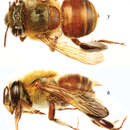
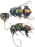
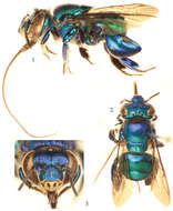
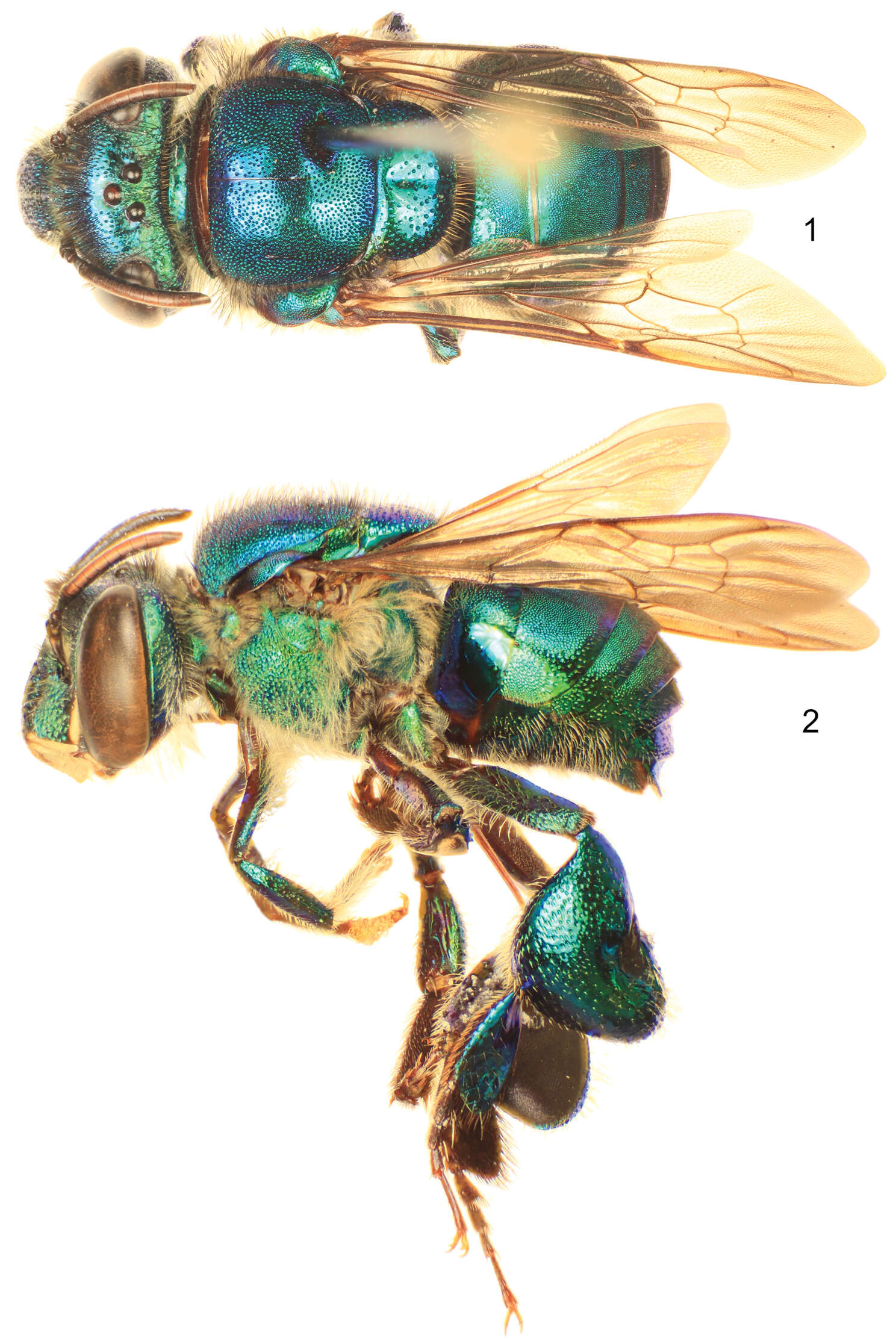
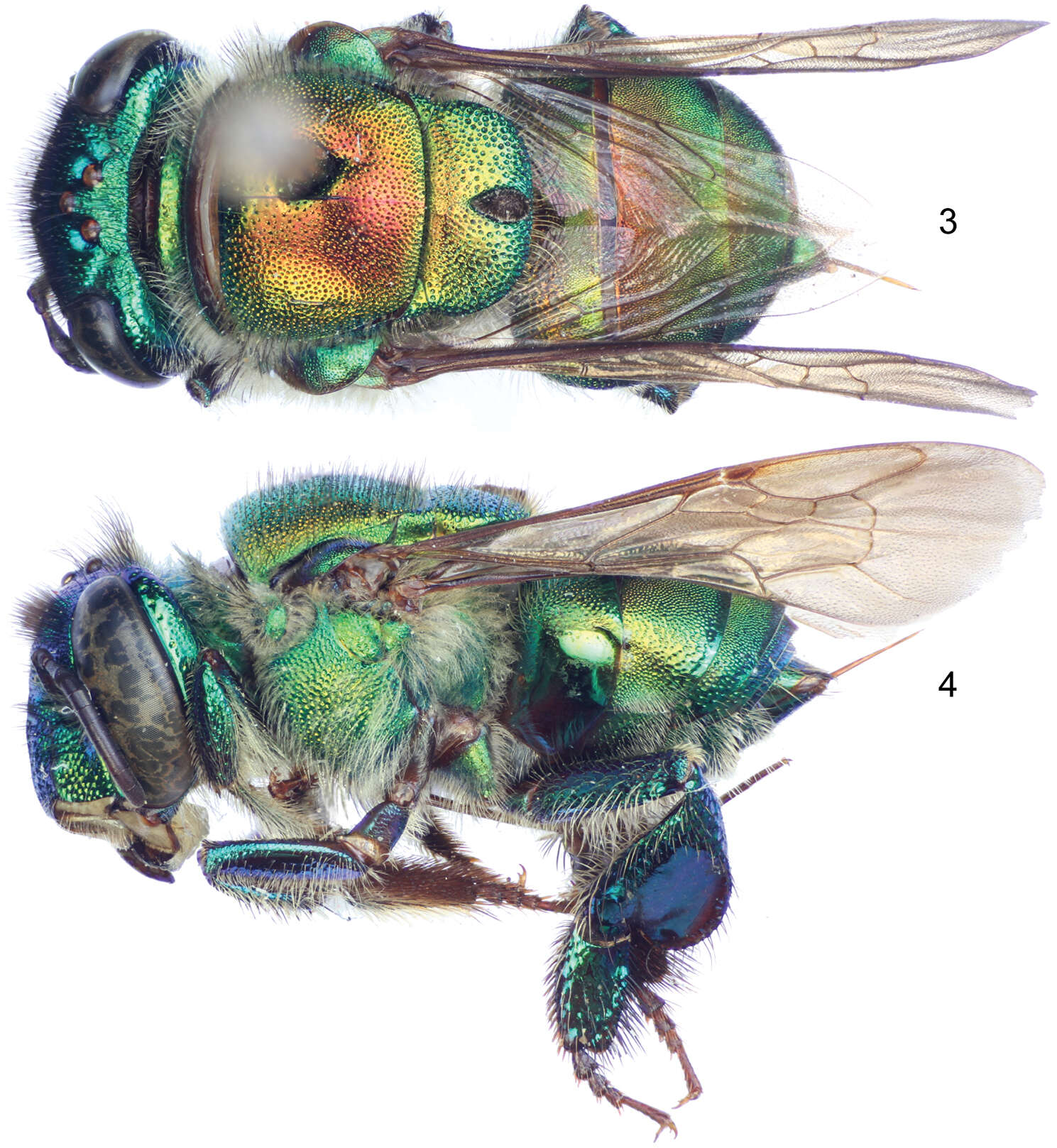
Figures 1–3.Euglossa (Alloglossura) gorgonensis Cheesman, female, red specimen from the Pacific slope of southern Costa Rica. 1 Dorsal habitus 2 Lateral habitus 3 Facial aspect.
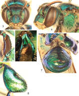
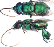
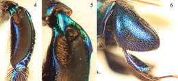
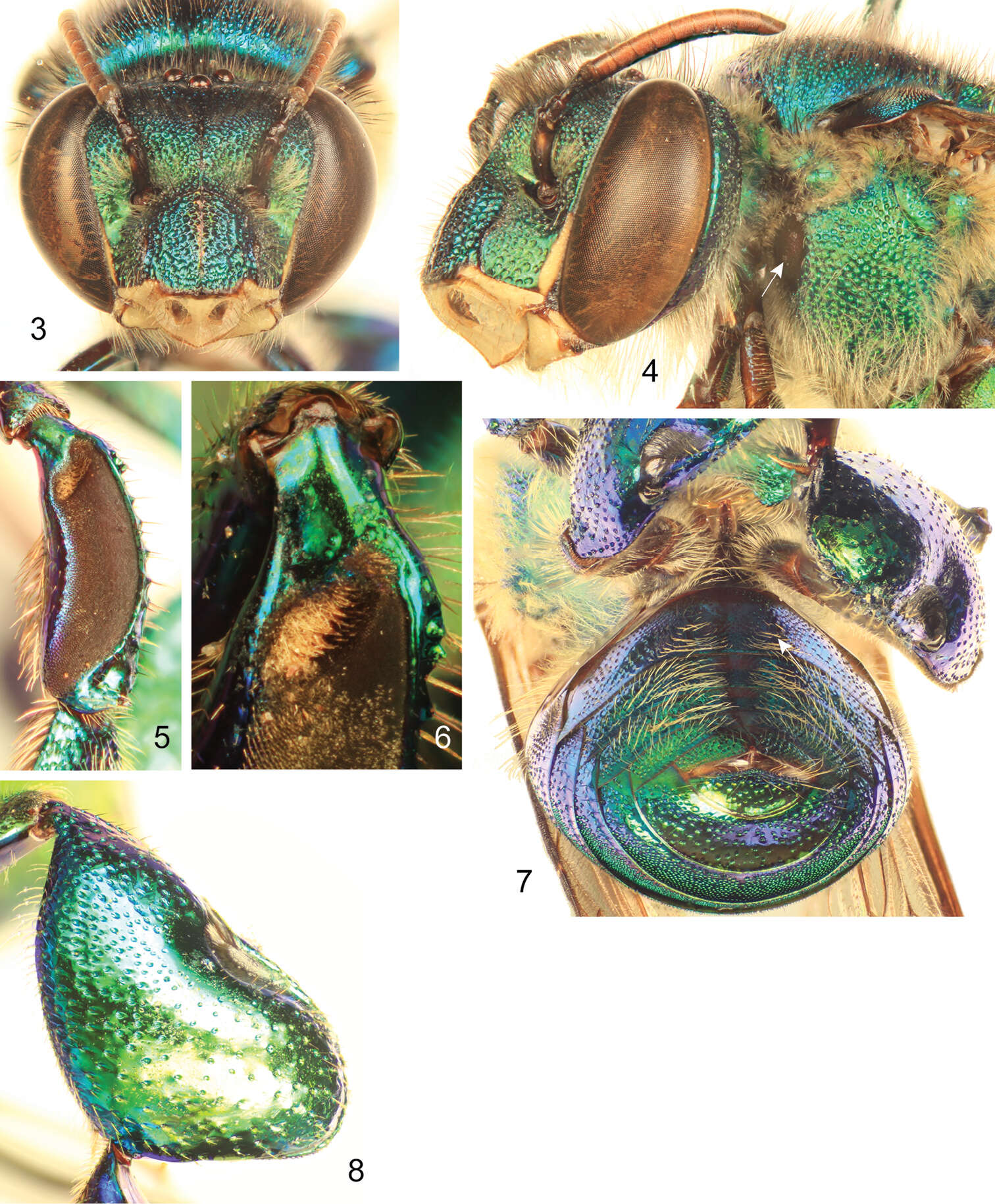
Figures 3–8.Euglossa williamsi Hinojosa-Díaz and Engel sp. n. male holotype 3 Facial aspect 4 Lateral aspect of mesepisterum showing preomaular spot (arrow) 5 Outer surface of mesotibia 6 Mesotibial tufts 7 Ventral view of metasoma showing the absence of integumental modifications on second sternum (arrow) 8 Outer view of metatibia.
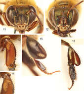
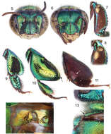
Figures 11–16.Euglossa (Euglossella) aurantia sp. n. 11 Facial aspect of male holotype 12 Facial aspect of female paratype 13 Outer surface of male mesotibia (arrow pointing to anterior surface convexity) 14 Mesotibial tufts 15 Outer view of male metatibia and metatarsus 16 Outer view of female metatibia and metatarsus.
Ismael A. Hinojosa-Díaz, André Nemésio, Michael S. Engel
Zookeys
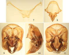
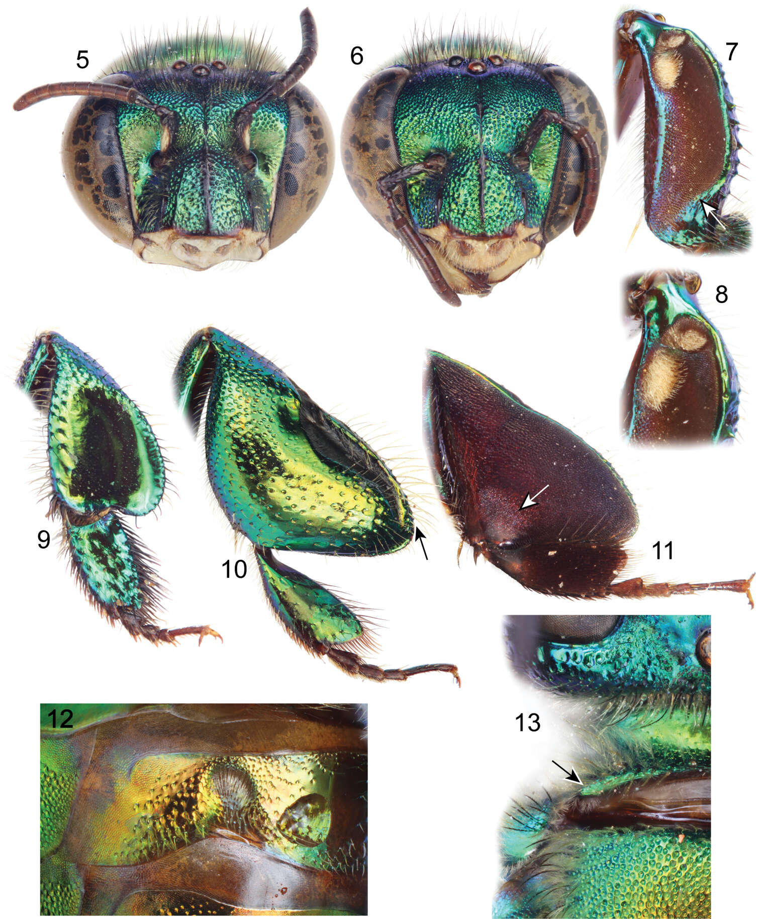
Figures 5–13.Euglossa embera sp. n. 5 Facial aspect of male holotype 6 Facial aspect of female paratype 7 Outer surface of male mesotibia (arrow pointing to oblique truncation of velvety area) 8 Mesotibial tufts of male 9 Outer view of female metatibia and metatarsus 10 Outer view of male metatibia and metatarsus (arrow pointing to distal-most extreme of organ slit) 11Inner view of male metatibia and metatarsus (arrow pointing to circular depression) 12 Section of male second metasomal sternum 13 Dorsal view of pronotal dorso-lateral angle (arrow) of male.
Ismael A. Hinojosa-Díaz, André Nemésio, Michael S. Engel
Zookeys
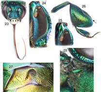
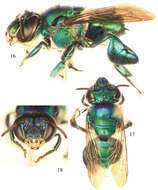
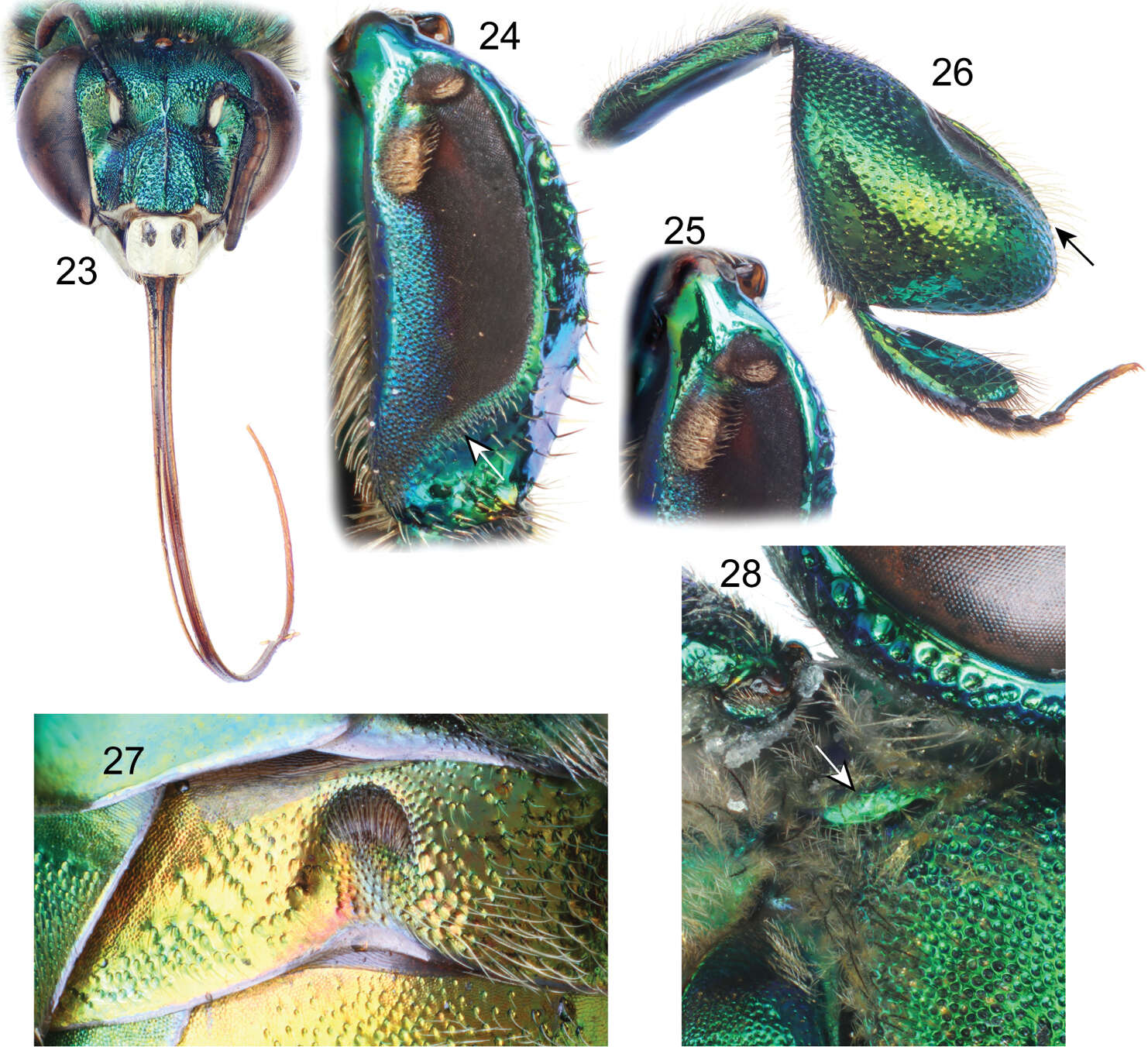
Figures 23–28.Euglossa adiastola sp. n. male holotype. 23 Facial aspect 24 Outer surface of mesotibia (arrow pointing to oblique-concave truncation of velvety area) 25 Mesotibial tufts 26 Outer view of metatibia and metatarsus (arrow pointing to distal-most extreme of organ slit) 27 Section of second metasomal sternum 28 Dorsal view of pronotal dorso-lateral angle (arrow).
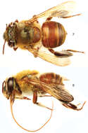
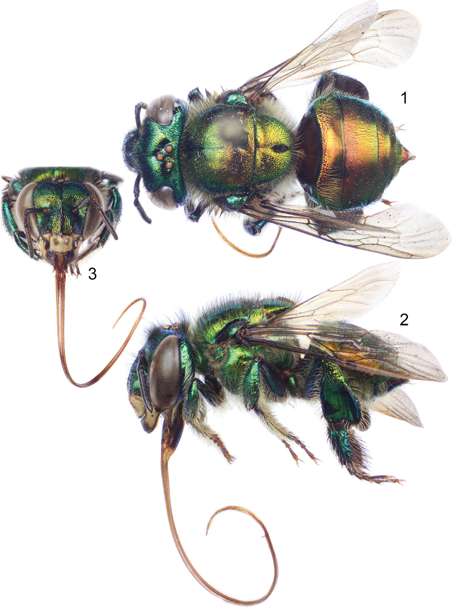
Figures 1–3.Photomicrographs of paratype male of Euglossa clausi Nemésio and Engel, sp. n. 1 Lateral habitus 2 Dorsal habitus (arrow points to rounded pronotal angle) 3 Facial aspect.
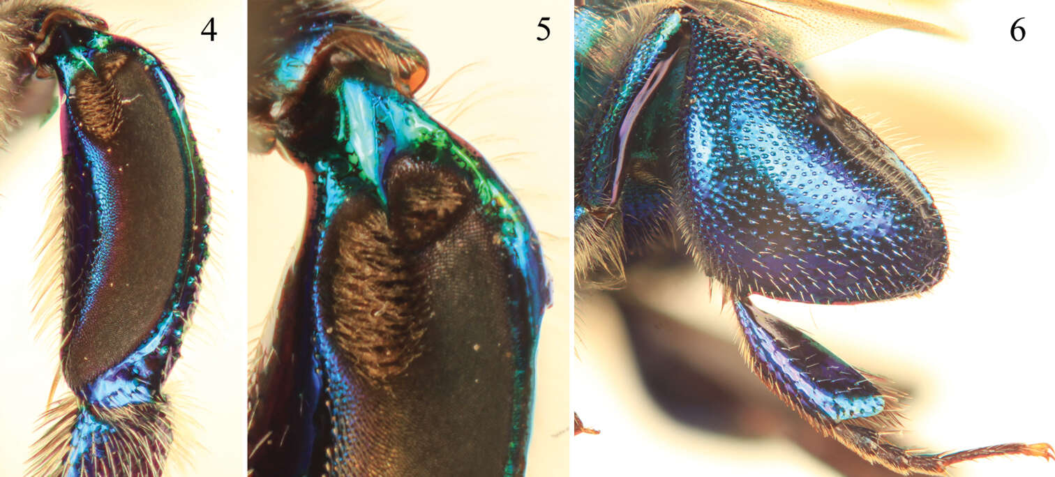
Figures 4–6.Tibial characters of Euglossa clausi Nemésio and Engel, sp. n. 4 Outer surface of mesotibia 5 Detail of mesotibial tufts 6 Outer surface of metatibia.
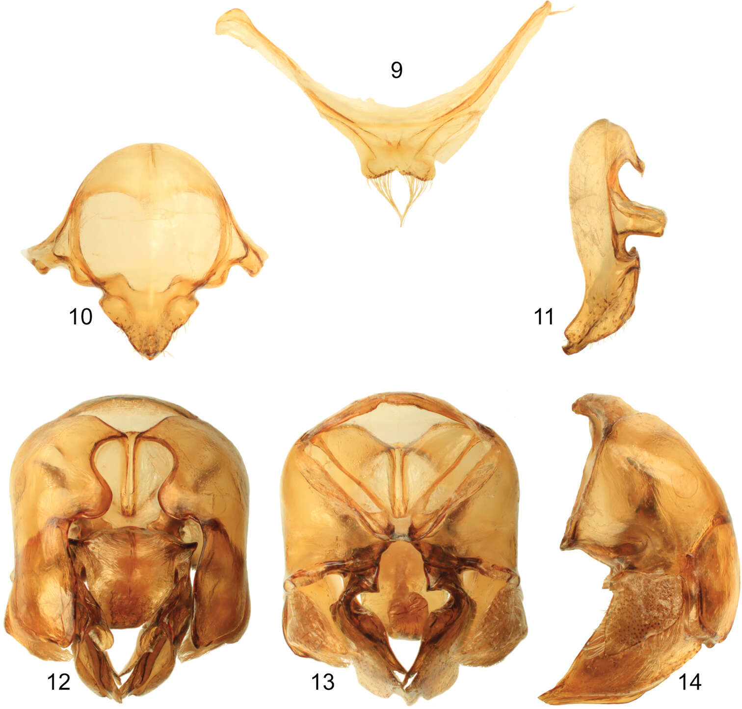
Figures 7–11.Male terminalia of Euglossa clausi Nemésio and Engel, sp. n. 7 Seventh metasomal sternum 8 Eighth sternum (note that relative proportions of the anterior section to the posterior section may be distorted owing to position of sclerite when photographed) 9 Genital capsule, dorsal view 10 Genital capsule, lateral view 11 Genital capsule, ventral view.
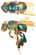
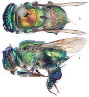
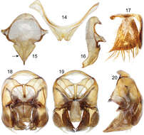
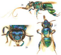
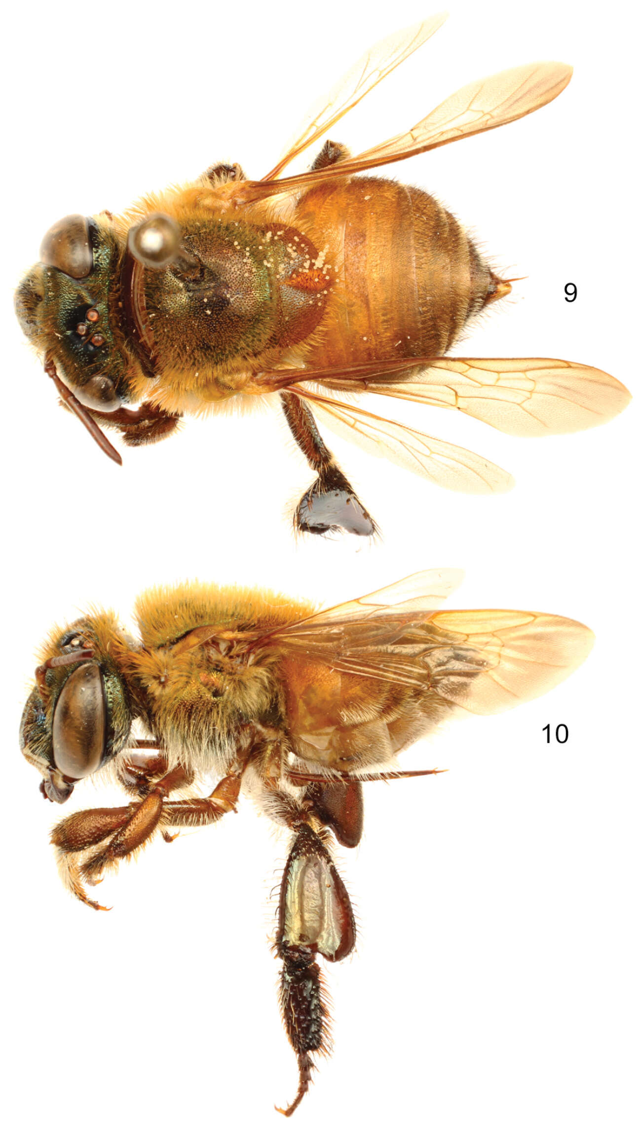
Figures 16–18.Photomicrographs of paratype male of Euglossa moratoi Nemésio and Engel, sp. n. 16 Lateral habitus 17 Dorsal habitus (arrow points to projected pronotal angle) 18 Facial aspect.
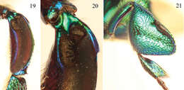
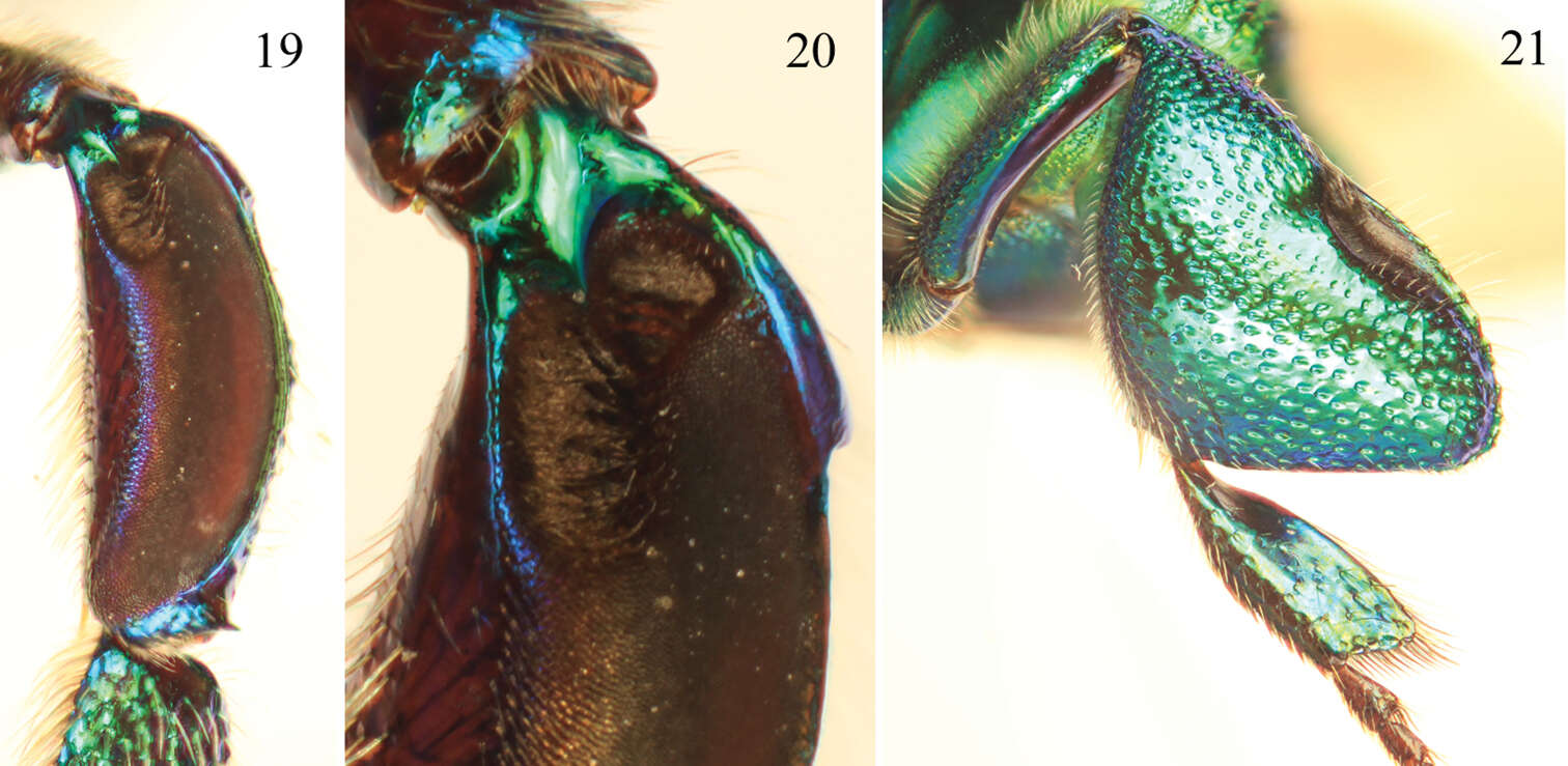
Figures 19–21.Tibial characters of Euglossa moratoi Nemésio and Engel, sp. n. 19 Outer surface of mesotibia 20 Detail of mesotibial tufts 21 Outer surface of metatibia.
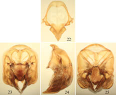
Figures 22–25.Male terminalia of Euglossa moratoi Nemésio and Engel, sp. n. 22 Eighth metasomal sternum (note that relative proportions of the anterior section to the posterior section may be distorted owing to position of sclerite when photographed) 23 Genital capsule, dorsal view 24 Genital capsule, lateral view 25 Genital capsule, ventral view.
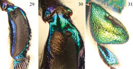
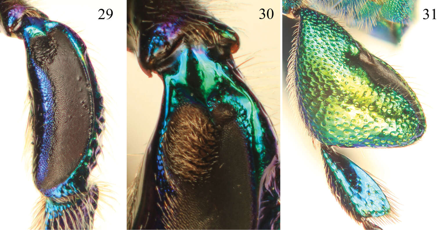
Figures 29–31.Tibial characters of Euglossa pepei Nemésio and Engel, sp. n. 29 Outer surface of mesotibia 30 Detail of mesotibial tufts 31 Outer surface of metatibia.
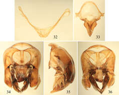
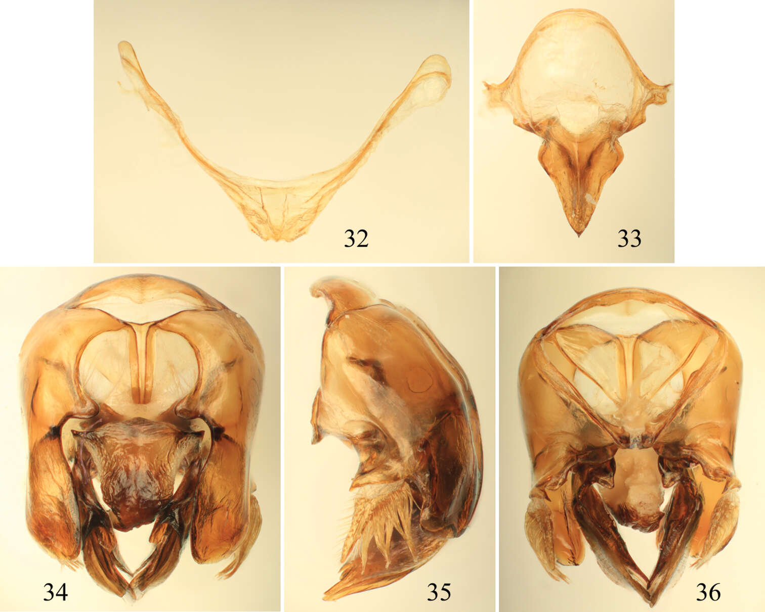
Figures 32–36.Male terminalia of Euglossa pepei Nemésio and Engel, sp. n. 32 Seventh metasomal sternum 33 Eighth sternum (note that relative proportions of the anterior section to the posterior section may be distorted owing to position of sclerite when photographed) 34 Genital capsule, dorsal view 35 Genital capsule, lateral view 36 Genital capsule, ventral view.