-
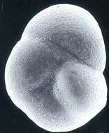
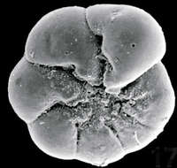
Sample collected at Hamble Estuary, Hampshire, England. Image courtesy of Elisabeth Alve, University of Oslo. Originally published in the Journal of Foraminiferal Research 31:1; used with permission.
-
Collected off the Ile de Porquerroles, France. The test is about 40 microns across. Image courtesy of Jan Pawlowski, University of Geneva. This image first appeared in J. Foram. Res 23:231-237, and is used with permission.
-
From Laguna Madre, Texas. Image courtesy of Pamela Stephens, Midwestern State University.
-
Collected off Capo Colonna, Italy. Image courtesy of Jan Pawlowski, University of Geneva. This image first appeared in J. Foram. Res 23:231-237, and is used with permission.
-
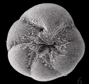
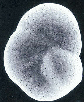
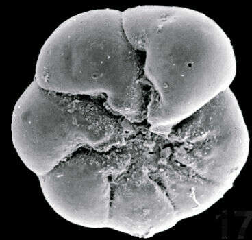
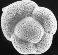
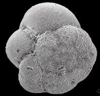
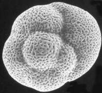
This is the top (spiral) surface of a gamont of Ammonia parkinsoniana. Forams have complex and varied reproductive cycles. Some forms, such as this species, alternate between agamonts/schizonts (which are produced when two gametes fuse) and gamonts (which are produced when an agamont splits up into hundreds of smaller individuals). The two types often look so distinct from each other that they were originally mistaken for different species. Image courtesy of Pamela Stephens, Midwestern State University.
-
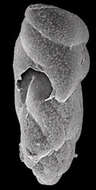
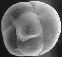
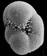
Part of the outer surface of the test is broken, showing the surface of an older chamber below. Image courtesy of Jan Pawlowski, University of Geneva. This image first appeared in J. Foram. Res 23:231-237, and is used with permission.
-
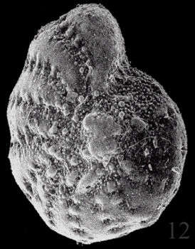
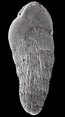
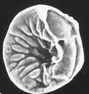
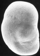
This is the bottom (umbilical) face of an A. parkinsoniana gamont. Image courtesy of Pamela Stephens, Midwestern State University.
-
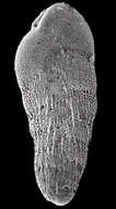
View of the holotype. Image courtesy of Jan Pawlowski, University of Geneva. This image first appeared in J. Foram. Res 23:231-237, and is used with permission.
-
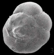
This is the top (spiral) surface of an A. parkinsoniana schizont. Image courtesy of Pamela Stephens, Midwestern State University.
-
Collected off the Ile de Porquerroles, France. The test is about 70 microns across in its long dimension. Image courtesy of Jan Pawlowski, University of Geneva. This image first appeared in J. Foram. Res 23:231-237, and is used with permission.
-
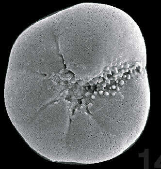
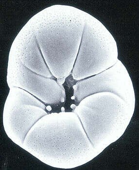
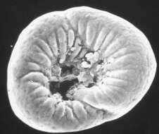
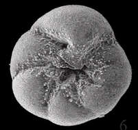
The umbilical surface of an A. parkinsoniana schizont. The "umbilical plug" which is found in many species in the genus Ammonia is prominent here. Image courtesy of Pamela Stephens, Midwestern State University.
-
Collected off the Ile de Porquerroles, France. Individuals from this area have, among other things, fewer and smaller denticles than is considered typical for the species. Image courtesy of Jan Pawlowski, University of Geneva. This image first appeared in J. Foram. Res 23:231-237, and is used with permission.
-
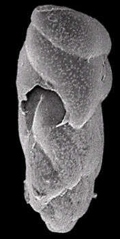
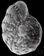
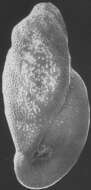
The large hole midway up the test is not formed by the foraminiferan; it is a break in the fragile test wall. Collected at Laguna Madre, Texas. Image courtesy of Pamela Stephens, Midwestern State University.
-
This young foram has only a few chambers, and is 30 microns across. Collected off the Ile de Porquerroles, France. Image courtesy of Jan Pawlowski, University of Geneva. This image first appeared in J. Foram. Res 23:231-237, and is used with permission.
-
Individual collected in Saanich Inlet, Vancouver Island, British Columbia. Image courtesy of R. Timothy Patterson, Carleton University. This image first appeared in J. Foram. Res. 28:201-219 and is used with permission.
-
Collected off the Ile du Levant, France. Image courtesy of Jan Pawlowski, University of Geneva. This image first appeared in J. Foram. Res 23:231-237, and is used with permission.
-
Collected along the South Texas coast. Image courtesy of Pamela Stephens, Midwestern State University.
-
Collected off the Ile de Porquerroles, France. Image courtesy of Jan Pawlowski, University of Geneva. This image first appeared in J. Foram. Res 23:231-237, and is used with permission.
-
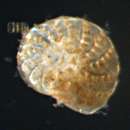
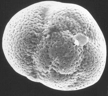
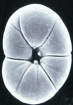
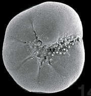
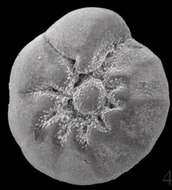
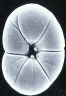
An umbilical (bottom) view of the test. Ammonia species are very tolerant of salinity changes and are common in estuarine environments. Sample collected at Hamble Estuary, Hampshire, England. Image courtesy of Elisabeth Alve, University of Oslo. Originally published in the Journal of Foraminiferal Research 31:1; used with permission.
-
Collected off the Ile du Levant, France. Image courtesy of Jan Pawlowski, University of Geneva. This image first appeared in J. Foram. Res 23:231-237, and is used with permission.
-
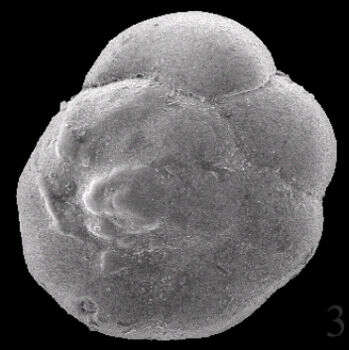
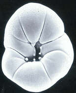
This juvenile has only a few chambers, and is only beginning to show the distinctive coiled pattern of the adult. The genus is named after Ammon, the ram-headed Egyptian god. Sample collected at Hamble Estuary, Hampshire, England. Image courtesy of Elisabeth Alve, University of Oslo. Originally published in the Journal of Foraminiferal Research 31:1; used with permission.
-
Collected off Capo Colonna, Italy. Image courtesy of Jan Pawlowski, University of Geneva. This image first appeared in J. Foram. Res 23:231-237, and is used with permission.
-
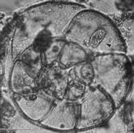
After gametogenesis, there is very little left of the parent cell. In this image, remnant cytoplasm in the empty test (which has been decalcified to make it more transparent) is being scavenged by a ciliate (arrow). Image courtesy of Susan T. Goldstein, University of Georgia. This image first appeared in J. Foram Res. 23:213-220, and is used with permission.
-
This species is unusual for its genus in that the later chambers are inflated. The test is about 60 microns long on its long axis. Image courtesy of Jan Pawlowski, University of Geneva. This image first appeared in J. Foram. Res 23:231-237, and is used with permission.