-
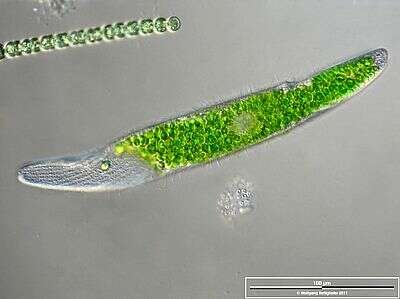
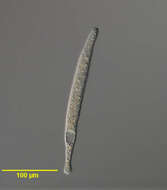
Scale bar indicates 100 µm. Sample from the pond Hegne Moor situated in the vicinity of Lake Constance (Bodensee, Southern Germany). The image was built up using several photomicrographic frames with manual stacking technique. Images were taken using Zeiss Universal with Olympus C7070 CCD camera.
-
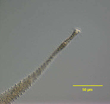
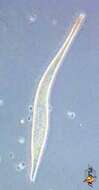
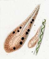
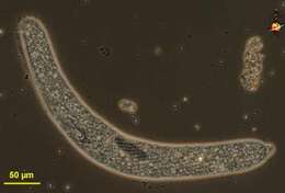
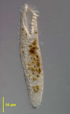
Tracheloraphis (track-ell-owe-ray-fiss) is a karyorelict ciliate. It is one of several genera in which most species have adopted a long thin contractile body form and the identification of this cell is tentative. Mostly found in marine sediments, living within the spaces between sand grains. The anterior end is slightly expanded and thought by most but not all to the site of the mouth. Fairly common. Phase contrast
-
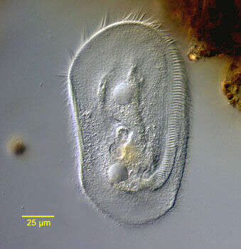
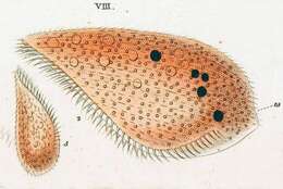
Portrait of the marine heterotrich ciliate, Condylostoma arenarium (Spiegel, 1926). The dorsoventrally flattened elongate cell body is very contractile. Contraction is probably mediated by calcium dependent subcortical myonemes and cell extension by interaction of cortical postciliary microtubular ribbons. The broad anterior V-shaped peristome is bordered on the right by a large undulating membrane. An adoral zone of membranelles (AZM) winds from right anterior clockwise around the left margin of the peristome. Strips of yellowish cortical granules or pigmentocysts separate uniform longitudinal somatic kineties. Pigmentocysts are extrusomes containing toxic substances. They play a role in cell defense against predators and may also function in photoreception. Pigmentocysts are found in other heterotrichs (e.g. Blepharisma and Stentor species). Several cirri are located at the right-most end of the AZM. The long moniliform macronucleus extends along the right cell margin (faintly visible here). No contractile vacuole. Brownish food vacuoles throughout the cytoplasm in this individual contain ingested dinoflagellates (Amphidinium). Collected from a seawater aquarium in Boise, Idaho January 2004. DIC optics.
-
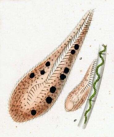
Originally described by Ehrenberg under the name Bursaria lateritia.
-
-
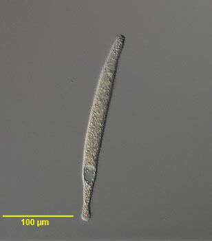
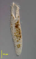
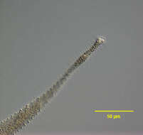
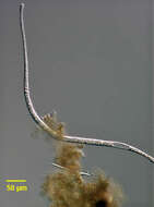
Portrait of the marine interstitial trachelocercid ciliate, Tracheloraphis (Dragesco, 1958). This genus contains many species and identification at the species level is quite difficult. Tracheloraphis is highly contractile. This view shows a contracted individual. Some species are greater than 2 millimeters in length when extended. The left side of the organism Seen here) bears a long unciliated "glabrous stripe". The inconspicuous cytostome is at the anterior apex. There is an inconspicuous cleft on the left margin of the cytostome. Morphology of the macronuclei is highly variable with as few as four to more than 50 macronuclei usually grouped in clusters of two or more. There are multiple micronuclei. Collected from a commercial marine aquarium in Boise, Idaho January 2004. DIC optics.
-
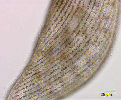
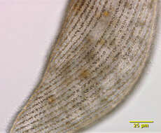
Cortical detail of the marine heterotrich ciliate, Condylostoma arenarium (Spiegel, 1926). The dorsoventrally flattened elongate cell body is very contractile. Contraction is probably mediated by calcium dependent subcortical myonemes and cell extension by interaction of cortical postciliary microtubular ribbons. The broad anterior V-shaped peristome is bordered on the right by a large undulating membrane. An adoral zone of membranelles (AZM) winds from right anterior clockwise around the left margin of the peristome (not visible in this image). Strips of yellowish cortical granules or pigmentocysts separate uniform longitudinal somatic kineties. Pigmentocysts are extrusomes containing toxic substances. They play a role in cell defense against predators and may also function in photoreception. Pigmentocysts are found in other heterotrichs (e.g. Blepharisma and Stentor species). Several cirri are located at the right-most end of the AZM. The long moniliform macronucleus extends along the right cell margin (not visible in this image). No contractile vacuole. Brownish food vacuoles throughout the cytoplasm in this individual contain ingested dinoflagellates (Amphidinium). Collected from a seawater aquarium in Boise, Idaho January 2004. DIC optics.
-
Originally described by Ehrenberg under the name Bursaria lateritia.
-
Ventral view of Condylostomides tardus (Penard,1922) Foissner, 2002. DIC.
-
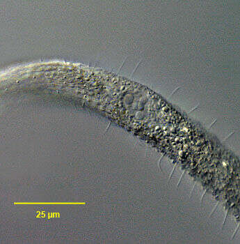
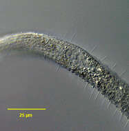
Anterior detail of the marine interstitial trachelocercid ciliate, Tracheloraphis (Dragesco, 1958). This genus contains many species and identification at the species level is quite difficult. Tracheloraphis is highly contractile. This view shows an extended individual. Some species are greater than 2 millimeters in length when extended. The left side of the organism (seen here) bears a long unciliated "glabrous stripe". The inconspicuous cytostome is at the anterior apex. An inconspicuous cleft is seen on the left side of the cytostome. Some authors have suggested that ingestion occurs along the glabrous stripe in Tracheloraphis but this view has been largely discounted. Morphology of the macronuclei is highly variable with as few as four to more than 50 macronuclei usually grouped in clusters of two or more. There are multiple micronuclei. Collected from a commercial marine aquarium in Boise, Idaho January 2004. DIC optics.
-
-
Originally described by Ehrenberg under the name Bursaria lateritia.
-
-
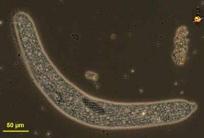
Portrait of the marine interstitial trachelocercid ciliate, Tracheloraphis (Dragesco, 1958). This genus contains many species and identification at the species level is quite difficult. Tracheloraphis is highly contractile. This view shows an extended individual. Some species are greater than 2 millimeters in length when extended. The left side of the organism bears a long unciliated "glabrous stripe". The inconspicuous cytostome is at the anterior apex. Morphology of the macronuclei is highly variable with as few as four to more than 50 macronuclei usually grouped in clusters of two or more. There are multiple micronuclei. Collected from a commercial marine aquarium in Boise, Idaho January 2004. DIC optics.
-
-

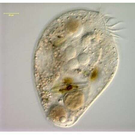
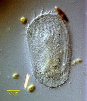
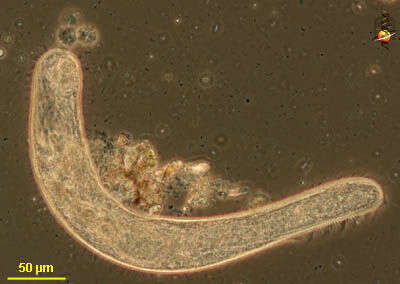
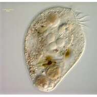
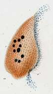
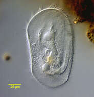
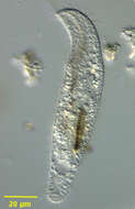
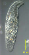
Portrait of Loxodes vorax (left side), a colorless karyorelict ciliate. The cell body is elongate, rounded anteriorly and posteriorly and highly laterally compressed. The anterior is bent ventrally forming a short beak-like rostrum. Very flexible. Somatic ciliature on the left side is restricted to a marginal kinety (seen here). On the right surface there are regular longitudinal kineties. The slit shaped cytostome is located in a ventral concavity posterior to the rostrum. A cone of fibrils forms a primitive cytopharynx at the posterior end of the cytostome. Two small spherical macronuclei flank a micronucleus in the mid body (seen here just anterior to ingested cyanobacteria). Refractile concretions of barium sulfate occupy several Müller's vesicles on the dorsal side. These probably act as statoreceptors, orienting the organism in the gravitational field. L. vorax is very similar to L. rostrum, the type species, but lacks zoochlorellae and also has a prominent posterior vacuole of uncertain function (seen here). Several food vacuoles are visible. From organically enriched freshwater pond sediment near Boise, Idaho. DIC optics.
-
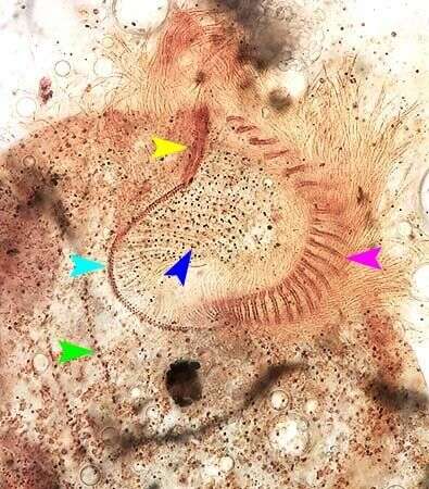
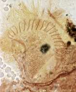
Oral infraciliature of Condylostomides tardus (PENARD,1922). The yellow arrowhead indicates the frontal membranelles along the anterior portion of the paraoral membrane (light blue arrowhead). There is a prominent adoral zone of membranelles on the left side of the buccal cavity (pink arrowhead).A system of fibrils arises from the basal bodies of the paraoral membrane (dark blue arrowhead). The green arrowhead indicates a somatic kinety.From non-flooded Petri dish culture of soil from the intermittently flooded grass lawn of a park in Boise,Idaho. Stained by the silver carbonate technique (Foissner,W. Europ. J. Protistol.27:313-330;1991).Brightfield.
-
Detail view of the marine interstitial trachelocercid ciliate, Tracheloraphis (Dragesco, 1958)showing the macronuclei. This genus contains many species and identification at the species level is quite difficult. Tracheloraphis is highly contractile. Some species are greater than 2 millimeters in length when extended. The left side of the organism bears a long unciliated "glabrous stripe". The inconspicuous cytostome is at the anterior apex. Morphology of the macronuclei is highly variable with as few as four to more than 50 macronuclei usually grouped in clusters of two or more.This species has a central cluster of at least five macronuclei. There are multiple micronuclei. Collected from a commercial marine aquarium in Boise, Idaho January 2004. DIC optics.
-
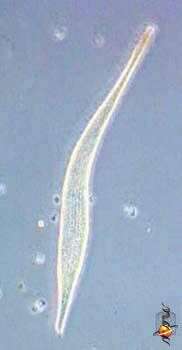
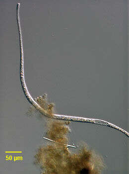
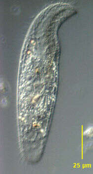
Spirostomum (spire-owe-stow-mum) is a genus of heterotrich ciliates, and some species in this genus may extend up to 1 mm in length. With a line of membranelles leading from the anterior of the cell to the cytostome which is located about half way down the cell. Contractile. Contractile vacuole at posterior end of cell. In habitats with low oxygen. Phase contrast.
-
Portrait of Loxodes vorax (left side), a colorless karyorelict ciliate. The cell body is elongate, rounded anteriorly and posteriorly and highly laterally compressed. The anterior is bent ventrally forming a short beak-like rostrum. Very flexible. Somatic ciliature on the left side is restricted to a marginal kinety (seen here). On the right surface there are regular longitudinal kineties. The slit shaped cytostome is located in a ventral concavity posterior to the rostrum. A cone of fibrils forms a primitive cytopharynx at the posterior end of the cytostome. Two small spherical macronuclei flank a micronucleus in the mid body dorsally. Refractile concretions of barium sulfate occupy several Müller's vesicles on the dorsal side. These probably act as statoreceptors, orienting the organism in the gravitational field. L. vorax is very similar to L. rostrum, the type species, but lacks zoochlorellae. Several food vacuoles are visible. From organically enriched freshwater pond sediment near Boise, Idaho. DIC optics.
-
Ventral view of the infraciliature of Condylostomides tardus (PENARD,1922). From non-flooded Petri dish culture of soil from the intermittently flooded grass lawn of a park in Boise,Idaho. Stained by the silver carbonate technique (Foissner,W. Europ. J. Protistol.27:313-330;1991).Brightfield.
-
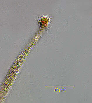
Detail view of the marine trachelocercid ciliate, Tracheloraphis (Dragesco, 1958). This genus contains many species and identification at the species level is quite difficult. This view shows the completely ciliated right side. The left side of the organism bears a long unciliated "glabrous stripe". This view shows ingestion of a dinoflagellate at the expansible anterior apical cytostome. Some authors have suggested that ingestion occurs along the glabrous stripe in Tracheloraphis but this view has been largely discounted. Collected from a commercial marine aquarium in Boise, Idaho January 2004. DIC optics.
-
Spirostomum (spire-owe-stow-mum) is a genus of heterotrich ciliates, and some species in this genus may extend up to 1 mm in length. With a line of membranelles leading from the anterior of the cell to the cytostome which is located about half way down the cell. Contractile. The dark central structure is the macronucleus Contractile vacuole at posterior end of cell. In habitats with low oxygen. Phase contrast.
-
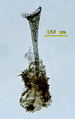
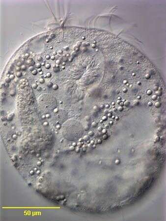
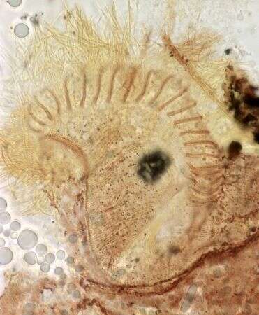
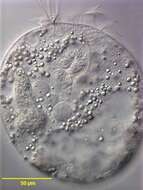
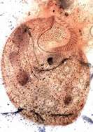
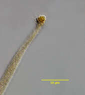
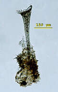
Stentor (sten-torr) is a genus of large, trumpet-shaped ciliates, commonly found in freshwater ponds, usually attached with the posterior end to vegetation or other surfaces. Detached individuals may swim freely and adopt an oval to pyriform shape. At the anterior end of the cells a conspicuous peristomial field with a system of adoral membranelles spiralling clockwise to the cytostome. The cilia of the membranelles are much longer than the somatic cilia. The macronuclei of Stentor may be spherical, elongate to a long strand or formed like a string of pearls. There is a single contractile vacuole with two collecting canals near the cytostome. Some species build transparent loricas of secreted mucus. The cells can be intensive coloured by pigmentation granules located in the pellicula (green, pink, blue, orange or violet). This specimen of Stentor roeselii was collected in freshwater ponds near Konstanz, Germany. Stentor roeselii is colourless and the macronucleus is band-shaped. This specimen built up a pyriform lorica covered with foreign bodies. Bright field.