-
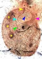
Ventral view of the infraciliature of Condylostomides tardus (PENARD,1922). The yellow arrowhead indicates the frontal membranelles along the anterior portion of the paraoral membrane (light blue arrowhead). There is a prominent adoral zone of membranelles on the left side of the buccal cavity (pink arrowhead).A system of fibrils arises from the basal bodies of the paraoral membrane (dark blue arrowhead). The green arrowhead indicates a somatic kinety.The three macronuclear nodules are indicated by yellow asterisks.From non-flooded Petri dish culture of soil from the intermittently flooded grass lawn of a park in Boise,Idaho. Stained by the silver carbonate technique (Foissner,W. Europ. J. Protistol.27:313-330;1991).Brightfield.
-
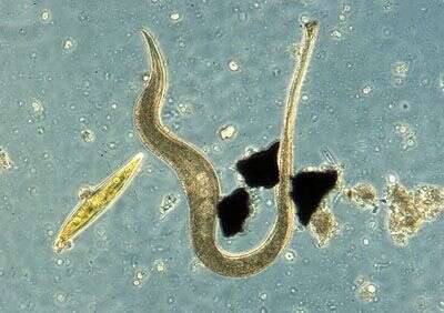
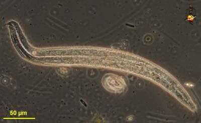
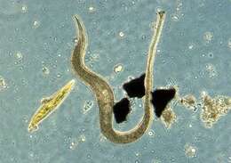
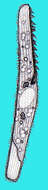
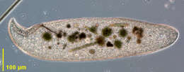
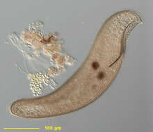
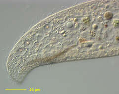
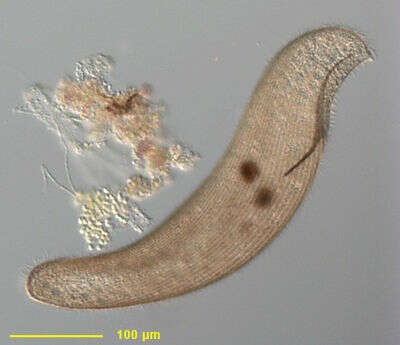
This is one of the many species of karyorelict ciliates that are long, thin, contractile and live between the grains of sediment of fine sandy beaches.
-
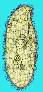
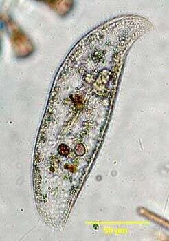
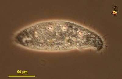
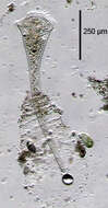
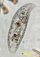
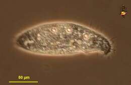
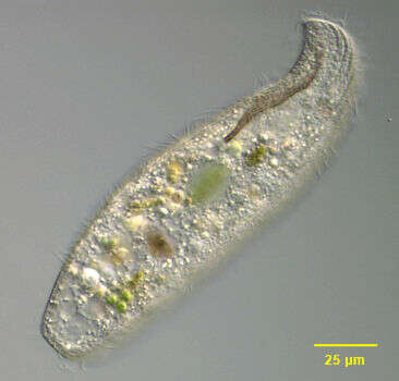
Spirostomum (spire-owe-stow-mum) is a genus of heterotrich ciliates, and some species in this genus may extend up to 1 mm in length. With a line of membranelles leading from the anterior of the cell to the cytostome which is located about half way down the cell. Contractile. The dark central structure is the macronucleus Contractile vacuole at posterior end of cell. In habitats with low oxygen. Phase contrast.
-
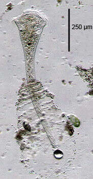
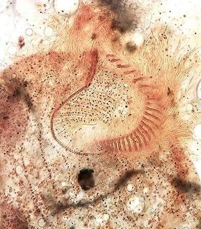
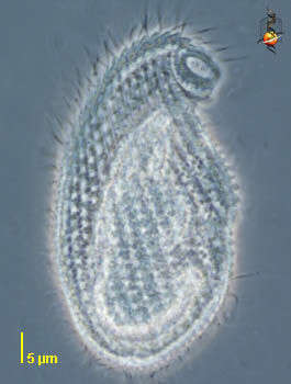
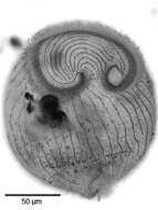
Portrait of the spirotrich ciliate, Stentor roeselii (Ehrenberg, 1835).This extended individual, slightly over 800 µm long, protrudes from a hyaline mucous lorica to which some debris adheres.The posterior terminal holdfast structure is seen here adhering to detritus near a gas bubble in this image.The bandform macronucleus extends nearly the length of the cell.There are approximately 80 longitudinal somatic kineties although this number is quite variable between individuals. Longer stiff thigmotactic bristles are interspersed with the shorter somatic cilia.An adoral zone of membranelles borders the flared anterior end and spirals into the buccal cavity (anteriorly to viewer's right).An undulating membrane runs parallel to the inner aspect of the AZM. The broad surface of the anterior (peristomial bottom)bears concentric curved kineties.The contractile vacuole is seen here adjacent to the buccal cavity (anterior to viewer's right). A long connecting canal extends posteriorly along the lateral edge of the cell.Collected from a freshwater pond near Boise, Idaho.Oblique illumination.
-
-
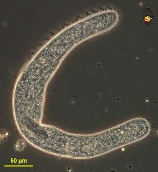
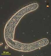
Karyorelectid ciliate collected at Chappaquoit marsh, MA. Cell is highly contracted. Photo by Becky Zufall.
-
-
Portrait of the spirotrich ciliate, Stentor roeselii (Ehrenberg, 1835).This specimen has contracted with fixation.There are usually approximately 80 longitudinal somatic kineties although this number is quite variable between individuals.This individual has about 40-50 somatic kineties. Longer stiff thigmotactic bristles are interspersed with the shorter somatic cilia.An adoral zone of membranelles borders the flared anterior end and spirals into the buccal cavity (anteriorly to viewer's right here).An undulating membrane runs parallel to the inner aspect of the AZM (seen well here). The broad surface of the anterior end (peristomial bottom)bears concentric curved kineties.Collected from a freshwater pond near Boise, Idaho.Stained by the silver carbonate technique (see Foissner, W. Europ. J. Protistol., 27:313-330;1991).Brightfield.
-
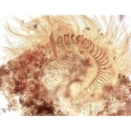
Oral infraciliature of Condylostomides tardus (PENARD,1922).From non-flooded Petri dish culture of soil from the intermittently flooded grass lawn of a park in Boise,Idaho. Stained by the silver carbonate technique (Foissner,W. Europ. J. Protistol.27:313-330;1991).Brightfield.
-
-
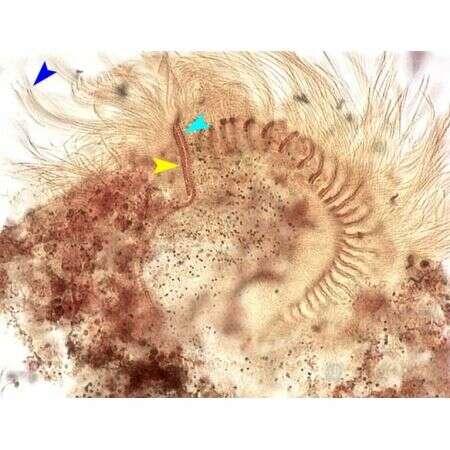
Oral infraciliature of Condylostomides tardus (PENARD,1922). The yellow arrowhead indicates the frontal membranelles along the anterior portion of the paraoral membrane (light blue arrowhead). There is a prominent adoral zone of membranelles on the left side of the buccal cavity.A system of fibrils arises from the basal bodies of the paraoral membrane. The dark blue arrowhead indicates the longer cilia comprising the frontal membranelles.From non-flooded Petri dish culture of soil from the intermittently flooded grass lawn of a park in Boise,Idaho. Stained by the silver carbonate technique (Foissner,W. Europ. J. Protistol.27:313-330;1991).Brightfield.
-
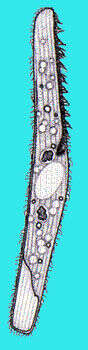
Loxodes, a compressed (flattened side-to-side) karyolectid ciliate. Detail of anterior with slit-like curved cytostome just posterior to hooked "rostrum". Right surface is densely ciliated while left side has only marginal kineties. Numerous Müller's vesicles with refractile inclusions are seen at intervals along the dorsal margin. The function of these is unknown. They contain concretions of barium salts. Body flexible. Several freshwater species. From standing freshwater with abundant decomposing leaves near Boise, Idaho. Brightfield.
-
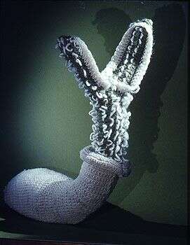
The enthusiasm that many protistologists have for their organisms can be impressive. This crocheted representation of the ciliate was created by Maria Mulisch. I think the living populations of Euglena deses in ear-rings, migrating up and down through glass beads in response to long distant tidal and solar cycles was my favorite. There were of course the knitwares, the songs, even the website (damit).
-
Portrait of Loxodes. Karyolectid ciliate. Laterally compressed. Slit-like curved cytostome just posterior to hooked "rostrum". Right surface is densely ciliated while left side has only marginal kineties. Numerous Müller's vesicles with refractile inclusions are seen at intervals along the dorsal margin. The function of these is unknown. They contain concretions of barium salts. Body flexible. Several freshwater species. From standing freshwater with abundant decomposing leaves near Boise, Idaho. Brightfield.
-

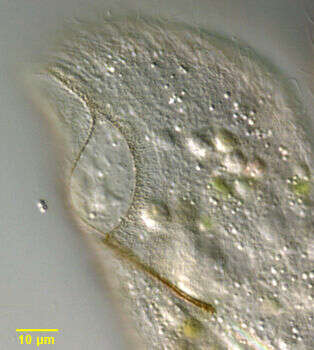
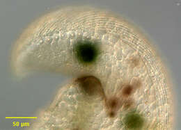
Portrait (right side) of Loxodes magnus, the largest species in the karyorelict ciliate genus, Loxodes. This individual is 755? long. Loxodes magnus is brown to orange. The cell body is elongate, rounded anteriorly and posteriorly and highly laterally compressed. The anterior is bent ventrally forming a short beak-like rostrum. Very flexible. Somatic ciliature on the left side is restricted to a marginal kinety. On the right surface there are regular longitudinal kineties. The slit shaped cytostome is located in a ventral concavity posterior to the rostrum (seen here). A thin cone of dark fibrils forms a primitive cytopharynx at the posterior end of the cytostome (seen here). There are from 3- 31 small macronuclei and a similar number of micronuclei scattered through the cell. Refractile concretions of barium sulfate occupy several Müller's vesicles on the dorsal side. These probably act as statoreceptors, orienting the organism in the gravitational field. There are also subcortical pigment granules, which may have chemo- and phototactic functions. L. magnus lacks contractile vacuoles. Found in polysaprobic habitats. Feeds on cyanobacteria, algae, flagellates and other ciliates. From organically enriched freshwater pond sediment near Boise, Idaho. Phase contrast.
-
Detail view of dorsum left side of Loxodes magnus, the largest species in the karyorelict ciliate genus, Loxodes. The termination of the right side kineties is seen here. Large orange refractile concretions of barium sulfate occupy several Müller's vesicles on the dorsal side in this image. These probably act as statoreceptors, orienting the organism in the gravitational field. There are also smaller subcortical pigment granules, which may have chemo- and phototactic functions (seen here). Several food vacuoles are visible. From organically enriched freshwater pond sediment near Boise, Idaho. DIC optics
-
Portrait (right side) of Loxodes magnus, the largest species in the karyorelict ciliate genus, Loxodes. This individual is 500? long. Loxodes magnus is brown to orange. The cell body is elongate, rounded anteriorly and posteriorly and highly laterally compressed. The anterior is bent ventrally forming a short beak-like rostrum. Very flexible. Somatic ciliature on the left side is restricted to a marginal kinety. On the right surface there are regular longitudinal kineties (seen here). The slit shaped cytostome is located in a ventral concavity posterior to the rostrum (seen here). A thin cone of dark fibrils forms a primitive cytopharynx at the posterior end of the cytostome (seen here). There are from 3- 31 small macronuclei and a similar number of micronuclei scattered through the cell. Refractile concretions of barium sulfate occupy several Müller's vesicles on the dorsal side. These probably act as statoreceptors, orienting the organism in the gravitational field. There are also subcortical pigment granules, which may have chemo- and phototactic functions. L. magnus lacks contractile vacuoles. Found in polysaprobic habitats. Feeds on cyanobacteria, algae, flagellates and other ciliates. From organically enriched freshwater pond sediment near Boise, Idaho. DIC optics.
-
Portrait (left side) of Loxodes striatus, a medium-size karyorelict ciliate. Loxodes striatus is colorless to slightly brown. The cell body is elongate, rounded anteriorly and posteriorly and highly laterally compressed. The anterior is bent ventrally forming a short beak-like rostrum. Very flexible. Somatic ciliature on the left side is restricted to a marginal kinety. On the right surface there are regular longitudinal kineties. In this species there are longitudinal pellicular striations on the left surface. The slit shaped cytostome is located in a ventral concavity posterior to the rostrum. A thin cone of dark fibrils forms a primitive cytopharynx at the posterior end of the cytostome (seen here). There are two spheroid macronuclei, one anterior and one in the mid body, each with an adherent micronucleus (neither seen well in this image). The arrangement of the macronuclei and the pellicular striations distinguish L. striatus from the similar L. vorax and L. rostrum. Refractile concretions of barium sulfate occupy several Müller's vesicles on the dorsal side. These probably act as statoreceptors, orienting the organism in the gravitational field. There are also subcortical pigment granules, which may have chemo- and phototactic functions. Found in polysaprobic habitats. Feeds on cyanobacteria, algae, flagellates and other ciliates. From organically enriched freshwater pond sediment near Boise, Idaho. DIC optics.
-
Detail of the oral aperture and cytopharynx of Loxodes striatus (left surface), a medium-size karyorelict ciliate. Loxodes striatus is colorless to slightly brown. The cell body is elongate, rounded anteriorly and posteriorly and highly laterally compressed. The anterior is bent ventrally forming a short beak-like rostrum. Very flexible. Somatic ciliature on the left side is restricted to a marginal kinety. On the right surface there are regular longitudinal kineties. In this species there are longitudinal pellicular striations on the left surface. The slit shaped cytostome is located in a ventral concavity posterior to the rostrum. A thin cone of dark fibrils forms a primitive cytopharynx at the posterior end of the cytostome (seen here). Many food vacuoles are seen. Found in polysaprobic habitats. Feeds on cyanobacteria, algae, flagellates and other ciliates. From organically enriched freshwater pond sediment near Boise, Idaho. DIC optics.
-
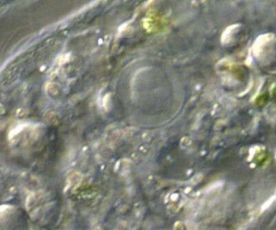
Detail of one of the two macronuclei of Loxodes striatus with its adherent micronucleus. There are two spheroid macronuclei, one anterior and one in the mid body, each with an adherent micronucleus. The arrangement of the macronuclei and the pellicular striations distinguish L. striatus from the similar L. vorax and L. rostrum. Many food vacuoles are seen. Found in polysaprobic habitats. Feeds on cyanobacteria, algae, flagellates and other ciliates. From organically enriched freshwater pond sediment near Boise, Idaho. DIC optics.
-
Detail of Loxodes striatus, a medium-size karyorelict ciliate. Loxodes striatus is colorless to slightly brown. The cell body is elongate, rounded anteriorly and posteriorly and highly laterally compressed. The anterior is bent ventrally forming a short beak-like rostrum. Very flexible. Somatic ciliature on the left side is restricted to a marginal kinety. On the right surface there are regular longitudinal kineties. In this species there are longitudinal pellicular striations on the left surface. The slit shaped cytostome is located in a ventral concavity posterior to the rostrum. A thin cone of dark fibrils forms a primitive cytopharynx at the posterior end of the cytostome (seen here). There are two spheroid macronuclei, one anterior and one in the mid body, each with an adherent micronucleus (both seen well in this image). The arrangement of the macronuclei and the pellicular striations distinguish L. striatus from the similar L. vorax and L. rostrum. Refractile concretions of barium sulfate occupy several Müller's vesicles on the dorsal side. These probably act as statoreceptors, orienting the organism in the gravitational field. There are also subcortical pigment granules, which may have chemo- and phototactic functions. Both the vesicles and pigment granules are seen here. Found in polysaprobic habitats. Feeds on cyanobacteria, algae, flagellates and other ciliates. From organically enriched freshwater pond sediment near Boise, Idaho. DIC optics.
-
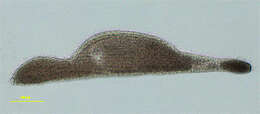
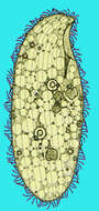
Remanella (rem-ann-ell-a) is karyorelictid ciliate - so called because they have macronuclei which do not divide - and this is thought to be an ancestral trait. Remanella is common in marine sediments or associated with detritus, especially in areas with little oxygen. With a hook-shaped anterior end and a line of Muellers bodies along the convex dorsal side of the body. The Muellers bodies are globular inorganic deposits (usually pink or orange in colour) and are organelles which provide information on the orientation of the body. Phase contrast.
-
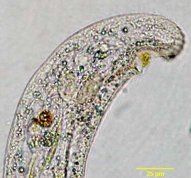
Remanella (re-man-ell-a) a karyorelict ciliate found in marine sediments, seems to prefer slightly anoxic zones. With the mouth located at the base of a hooked anterior end, dorsal side of body with granules in vesicles acting to determine the direction of gravitational pull. This species has an elongate body, Phase contrast micrograph.
-
Cryptopharynx (crypt-oh-far-inks), one of the cuter little ciliates. Small protruberant mouth. A karyorelict? Phase contrast.