-
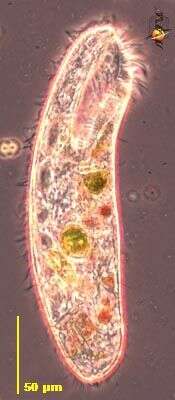
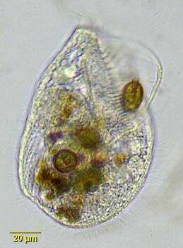
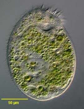
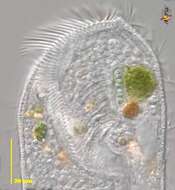
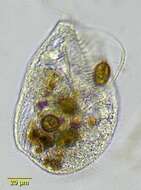
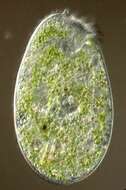
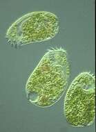
Condylostoma (con-dee-lost-oh-ma) is a medium to large ciliate (usually over 100 microns long). Commonly associated with surfaces, and slightly enriched habitats. The large anterior mouth can pick up relatively large particles of food. It is a heterotrich ciliate, with conventional cilia over most of the body and used to propel the cell, but with larger cilia around the mouth. This cell has obviously been consuming a variety of algae. This Differential Interference Contrast (DIC = Nomarski) image shows the cilia of the Adoral Zone of Membranelles (AZM) around the front of the cell and leading to the mouth at the end of the groove slightly to the right of centre near the bottom of the picture.
-
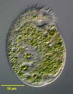
Condylostoma (con-dee-lost-oh-ma) is a medium to large ciliate (usually over 100 microns long). Commonly associated with surfaces, and slightly enriched habitats. The large anterior mouth can pick up relatively large particles of food . It is a heterotrich ciliate, with conventional cilia over most of the body and used to propel the cell, but with larger cilia around the mouth. This cell has obviously been consuming a variety of algae. Phase contrast.
-
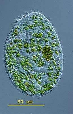
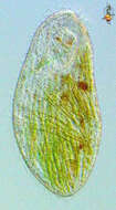
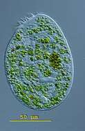
Condylostoma (con-dee-lost-oh-ma) is a medium to large ciliate (usually over 100 microns long). Commonly associated with surfaces, and slightly enriched habitats. The large anterior mouth can pick up relatively large particles of food . It is a heterotrich ciliate, with conventional cilia over most of the body and used to propel the cell, but with larger cilia around the mouth. This cell has obviously been consuming lots of diatoms. Differential interference contrast.
-
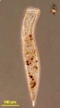
Condylostoma (con-dee-lost-oh-ma) is a medium to large ciliate (usually over 100 microns long). The large anterior mouth can pick up relatively large particles of food. It is a heterotrich ciliate, with conventional cilia over most of the body and used to propel the cell, but with larger cilia around the mouth. Phase contrast.
-
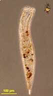
Condylostoma (con-dee-lost-oh-ma) is a medium to large ciliate (usually over 100 microns long). Commonly associated with surfaces, and slightly enriched habitats. The large anterior mouth can pick up relatively large particles of food . It is a heterotrich ciliate, with conventional cilia over most of the body and used to propel the cell, but with larger cilia around the mouth. Phase contrast.
-
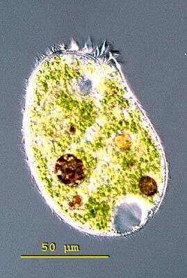
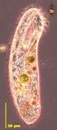
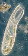
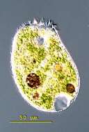
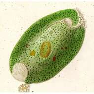
Condylostoma vastum. Brightfield portrait of Condylostoma, a large heterotrich ciliate. The anterior is truncate, and the cell is tapered posteriorly. The posterior contractile vacuole is served by fine collecting canals which are difficult to visualize by light microscopy. There is a well-developed adoral zone of membranelles on the left margin of the V-shaped peristomal region and a large undulating membrane on the right margin, well seen here. This individual is consuming Trachelomonas. From a freshwater aquaculture pond near Boise, Idaho.
-
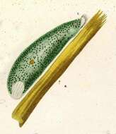
COndylostoma from marine sediments, anterior oral area includes adoral zone of membranelles. Vody coated with cilia. Consumes diatoms.
-
-
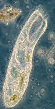
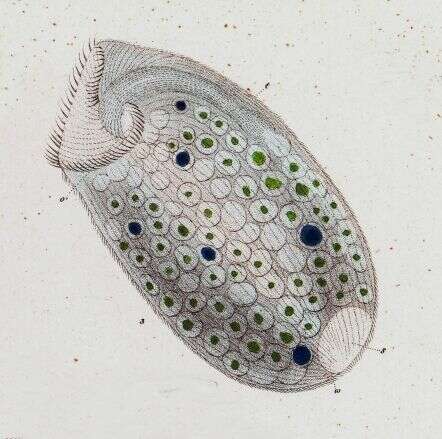
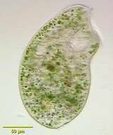
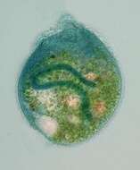
Portrait of Climacostomum virens, a large spirotrich ciliate containing zoochlorellae. Body flattened, truncate anteriorly and rounded posteriorly. Uniform ciliation. Prominent adoral zone of membranelles along left border of peristome. Macronucleus ribbon-like (not well seen in this image). Posterior contractile vacuole has two long collecting canals. From freshwater pond near Boise, Idaho. Oblique illumination.
-
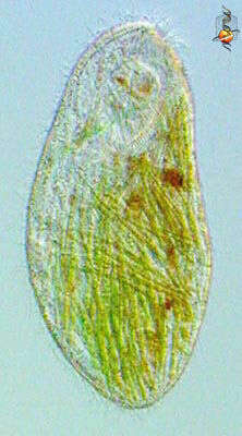
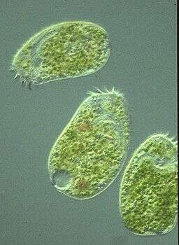
Climacostomum is a ciliate that feeds on detritus and algae using the specialized feeding cilia at the front of the cell (top of image). This species also contains symbiotic algae.
-
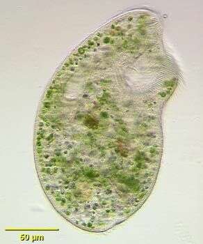
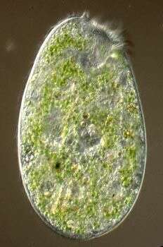
Climacostomum (clee-mack-ost-owe-mum) virens is coloured green by endosymbiotic algae. The contractile vacuole is located in the posterior end of the cell. The buccal cavitiy with the adorale membranelle is visible at the anterior. Differential interference contrast.
-
Climacostomum (clee-mack-ost-owe-mum) virens is coloured green by endosymbiotic algae. The contractile vacuole is located in the posterior end of the cell. Squashed specimen, a long meandering macronucleus is visible. Differential interference contrast.
-
Ventral view of the heterotrich ciliate, Climacostomum virens (Ehrenberg, 1838) Stein, 1859. Collected from a slow-flowing temporary freshwater stream near Boise, Idaho. May 2005. DIC.
-
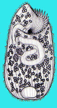
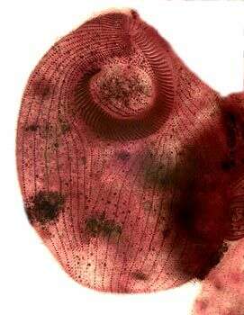
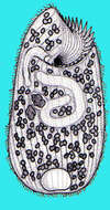
Ventral infraciliature of the heterotrich ciliate, Climacostomum virens (Ehrenberg, 1838) Stein, 1859. Silver carbonate stain (see Foissner, W. Europ. J. Protistol., 27:313-330;1991). Brightfield.
-
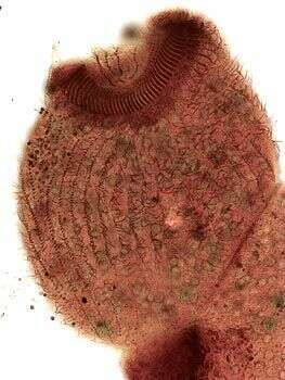
Lateral view of infraciliature of the heterotrich ciliate, Climacostomum virens (Ehrenberg, 1838) Stein, 1859. Silver carbonate stain (see Foissner, W. Europ. J. Protistol., 27:313-330;1991). Brightfield.
-
Vermiform macronucleus of the heterotrich ciliate, Climacostomum virens (Ehrenberg, 1838) Stein, 1859. Collected from a slow-flowing temporary freshwater stream near Boise, Idaho. Methyl green pyronin stain (see Foissner, W. Europ. J. Protistol., 27:313-330;1991). Brightfield
-
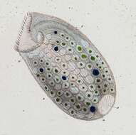
Heterotriuch ciliate with anterior adoral zone of membranelles leading into the curving buccal cavity. Cytoplasm with endosymbiotic eukaryotic algae. Differential interference contrast.
-
Originally described by Ehrenberg under the name Spirostomum virens.
-
Originally described by Ehrenberg under the name Spirostomum virens.
-
Originally described by Ehrenberg under the name Spirostomum virens.