-
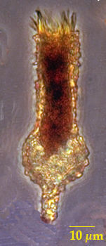
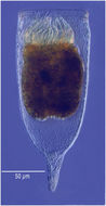
Specimen from the Scripps Canyon area in July 2009
-
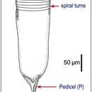
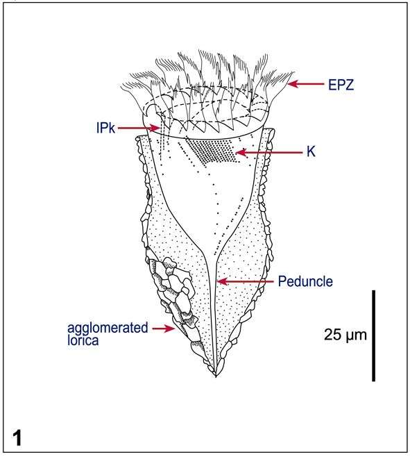
Fig 1: Tintinnopsis baltica Schematic drawings of lorica morphologie: After Laval-Peuto & Brownlee 1986;
-
-
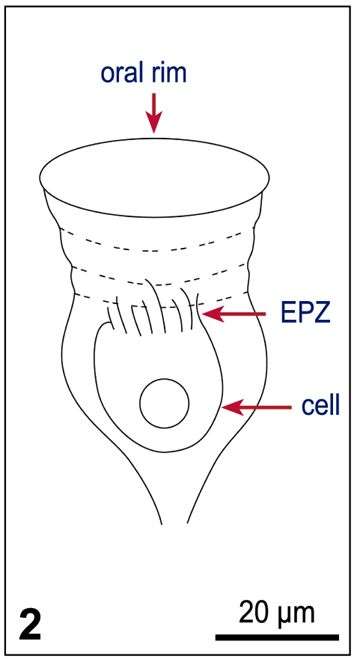
Fig 2 Original drawing of Tintinnopsis baltica (after Möbius, 1887);
-
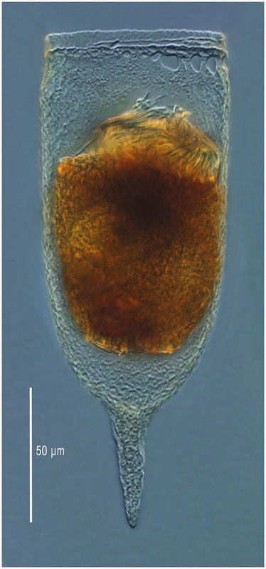
Specimen from the Etang de Thau (Sète, France). Imaged using a 20x objective, lugol's-fixed sample.
-
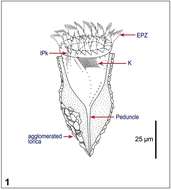
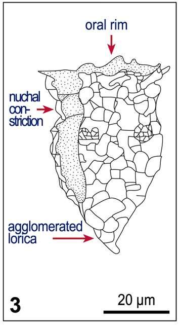
Fig 3 After Kofoid & Campbell 1929.
-
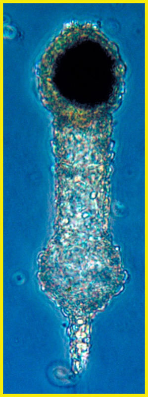
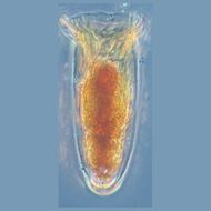
From the Etang de Thau (Sète, France) in May 2012.
-
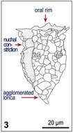
Fig 1: Leegaardiella ovalis Line drawings of protargol stained cells, showing kineties, oral structures and nucleus
-
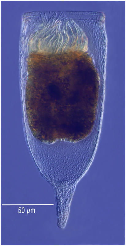
From the Etang de Thau (Sète, France) in May 2012.
-
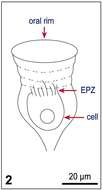
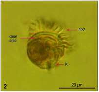
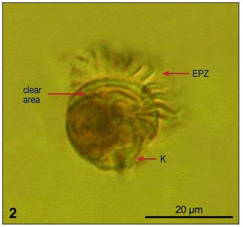
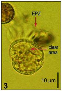
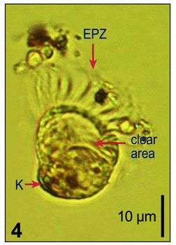
Fig 2: Leegaardiella ovalis Lugol?s fixed cell, showing the clear area (corresponds to oral cavity), the somatic kinety, and the EPZ: Aboral view
-
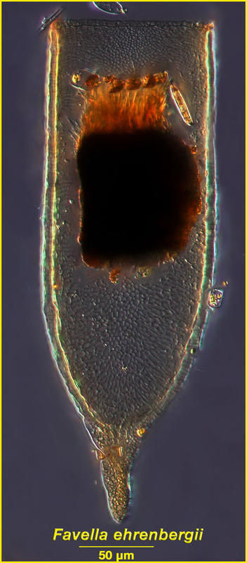
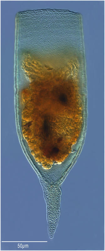
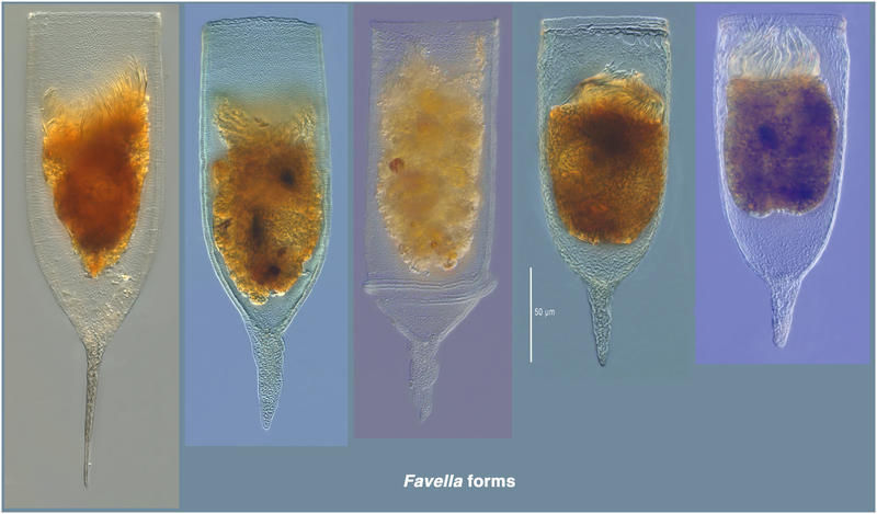
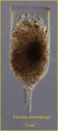
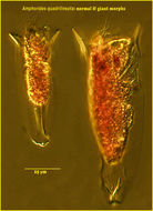
The specimens were all found in samples from the mesocosm experiment WarmAcid. They appear to be variants of a single species, Favella ehrenbergii.
-
Fig 3: Leegaardiella ovalis Lugol?s fixed cell, showing the clear area (corresponds to oral cavity), the somatic kinety, and the EPZ: Lateral view
-
-
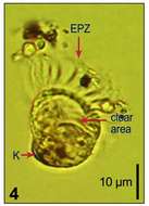
Fig 4: Leegaardiella ovalis Lugol?s fixed cell, showing the clear area (corresponds to oral cavity), the somatic kinety, and the EPZ: Lateral view, cell slightly deformed
-
Specimen from the Chesapeake Bay.
-
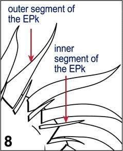
Fig 8: Leegaardiella ovalis Detail of the oral ciliature, showing the inner and outer segments of the EPks and their different ciliation
-
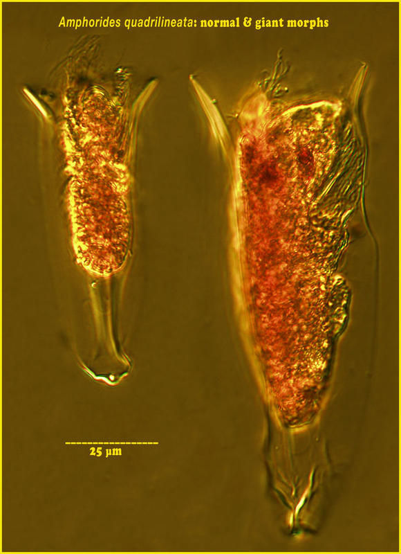
In a sample from the Tara expedition (Station 68) there were 30 large cells for about 1000 'normal-size' cells.
-
-
-
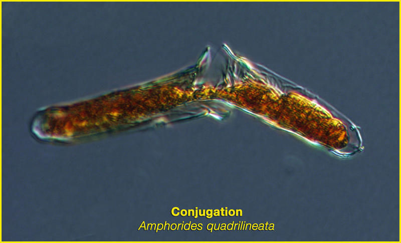
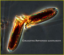
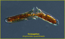
Genetic material is shared between two individuals by the formation of a cytoplasmic bridge
-
A pair of tintinnid ciliates caught in the act of exchanging genetic material.
-
Specimen from the Scripps Canyon area in July 2009
-
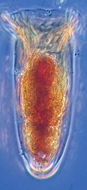
Lugol's-fixed specimen from the Bay of Villefranche in October 2010.
-
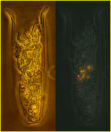
This specimen is shown as viewed with transmitted light (left) and epifluoresence (visible light emitted when subjected to ultraviolet light) showing the algae it ate (right panel).