-
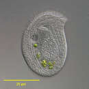
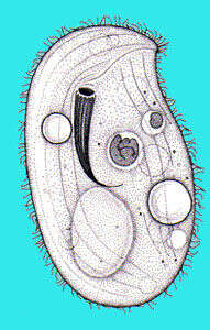
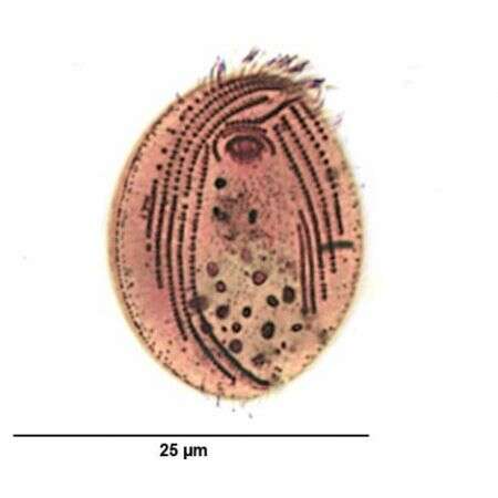
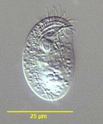
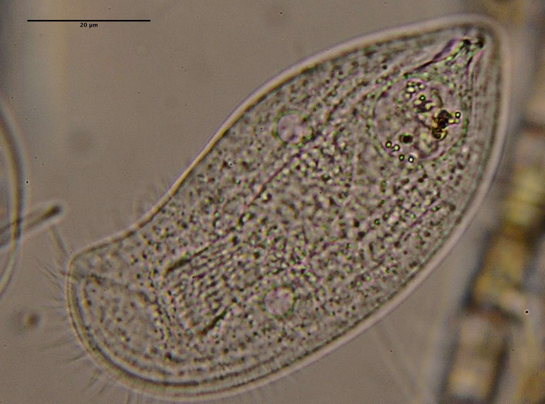
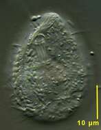
Chilodonella (kai-low-don-ella) is a hypostome ciliate with a mouth stiffened by a palisade of microtubular rods protruding from the ventral surface of the cell. The mouth is used to pick up bacteria and small pieces of detritus and manipulate them into the body. This individual has been eating diatoms. Common in freshwater and marine habitats. Differential interference contrast.
-
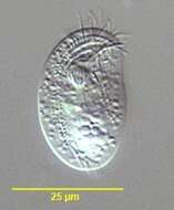
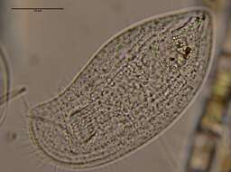
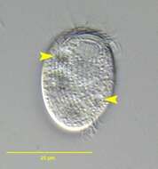
Chilodonella (kai-low-don-ella) is a hypostome ciliate with a mouth stiffened by a palisade of microtubular rods protruding from the ventral surface of the cell. The mouth is used to pick up bacteria and small pieces of detritus and manipulate them into the body. This image shows the ciliature on the ventral surface of the cell. Phase contrast.
-
-
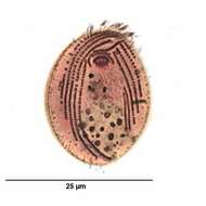
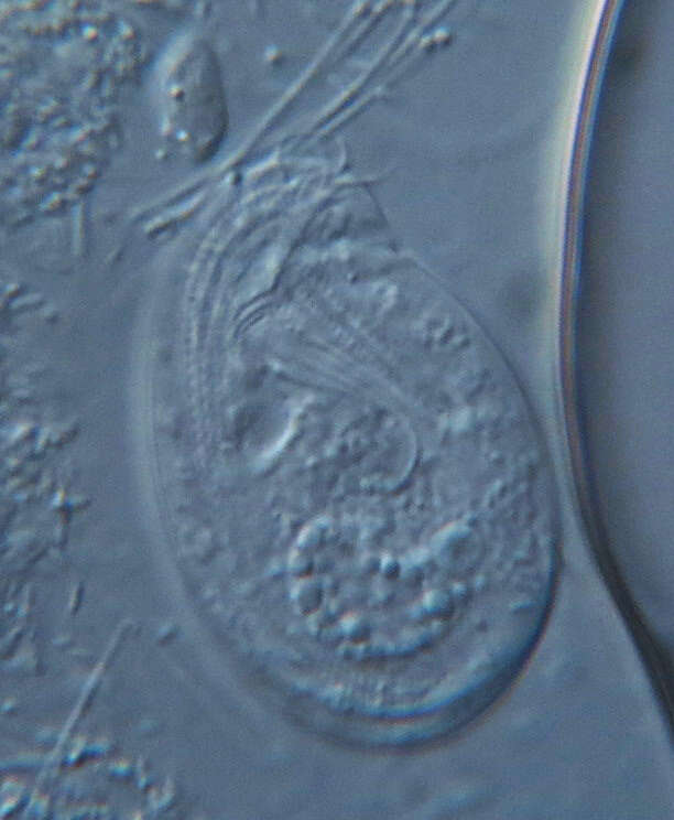
Chilodonella, hypostome ciliate. Macronucleus in posterior part of cell. Mouth with supporting rods (nemadesmata) cruves into the cell. With a few marginal kineties of cilia. Differential interference contrast.
-
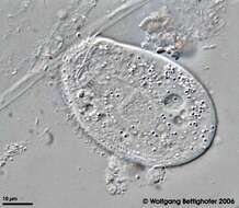
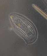
Browsing ciliate, from an immersed slide. Phase contrast micrograph.
-
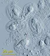
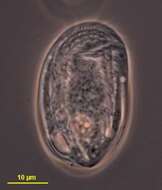
Resting cyst of Chilodonella (STRAND,1928) species (surface view on left;optical section on right). DIC.
-
Differential interference microscopy of Chilodonella cell showing mouth, cilia, and sausage-shaped bacteria adhering to the cell surface.
-
Differential interference microscopy of Chilodonella cell showing mouth, cilia, and sausage-shaped bacteria adhering to the cell surface.
-
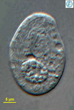
Ventral infraciliature of Chilodonella uncinata (EHRENBERG,1838) STRAND, 1928. Stained by the silver carbonate technique (Foissner,W. Europ. J. Protistol.27:313-330;1991).Brightfield.
-
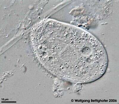
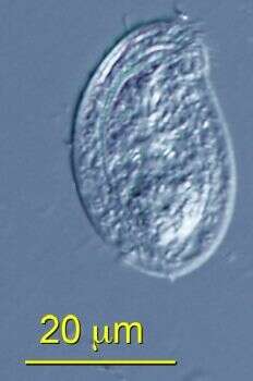
Ventral view of Chilodonella uncinata (EHRENBERG,1838) STRAND,1928. DIC
-
Chilodonella belongs to a morphological group called cyrtophorids which is characterized by special basket formed oral region. It was collected from littoral region (stand of Phragmites) of a rain storage reservoir in Kiel (Schleswig-Holstein, Germany). Images were taken using Zeiss Universal with Olympus C7070 CCD camera.
-
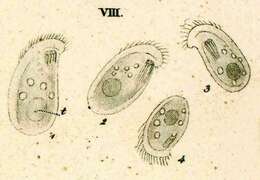
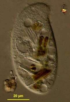
This image was taken with DIC of ATCC 50194. Chlamydodontid ciliates, with distinct postoral break or unciliated field in ventral somatic kineties; preoral kinety complete. The cell has a flat ventral surface and an arched dorsal surface. The cytostome is anterio-ventrally situated and is supported by a protrusible basket of nemadesmata. The dorsal surface is unciliated, except subapically near the left margin there is a single kinety consisting of 5-20 ciliated kinetosomes. Three kineties are associated with the oral region: two circumoral kineties and one preoral kinety above the cytostome; the preoral kinety extends obliquely to the left of the ciliate. There are several longitudinal somatic kineties on the ventral surface; those on the right curve around the anterior end while those on the left extend only to the preoral kinety. The central postoral region lacks cilia. The macronucleus is spheroid to ellipsoidal and located near the posterior. Two contractile vacuoles are usually located on the upper right and lower left of the cell. This ciliate glides quickly over surfaces.
-
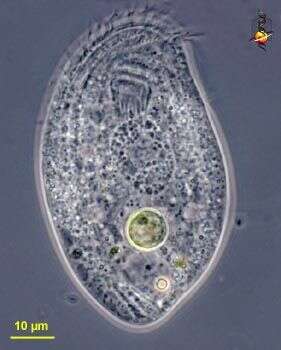
Group of Chilodonella feeding on bacteria. Macronuclei are prominent. ATCC 50194.
-
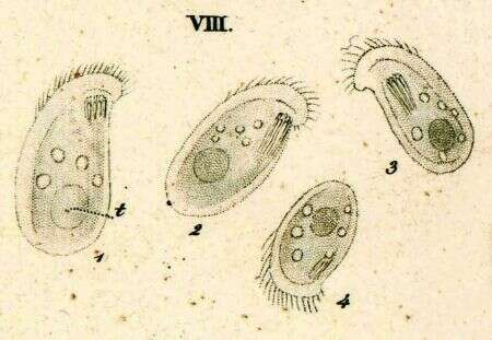
Originally described by Ehrenberg under the name Chilodon uncinata.
-
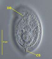
Portrait of Chilodonella caudata (Stokes, 1885) , a hypostome ciliate. This species is distinguished by the sharply pointed caudal spine (CS) arising from the dorsal surface and a sharply notched anterolateral rostrum. The dorsal brush of cilia (DB) is visible here. Collected from a fresh water pond near Boise, Idaho.DIC.
-
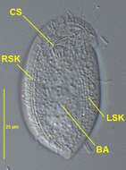
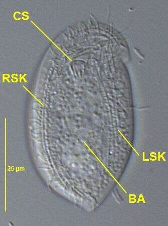
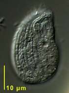
Ventral view of Chilodonella caudata (Stokes, 1885) , a hypostome ciliate. This species is distinguished by the sharply pointed caudal spine (not seen from the ventral aspect) arising from the dorsal surface and a sharply notched anterolateral rostrum. The cytostome (CS), right and left somatic kineties (RSK,LSK) and postoral bare area (BA) are seen well here.Collected from a fresh water pond near Boise, Idaho.DIC.
-
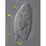
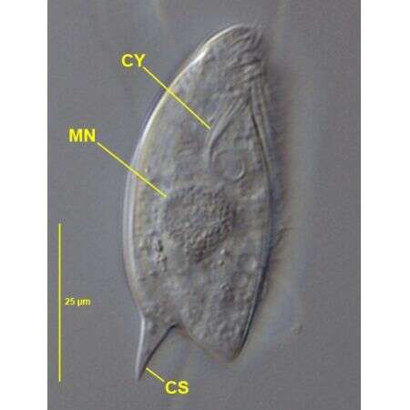
Lateral view of Chilodonella caudata (STOKES, 1885) , a hypostome ciliate. This species is distinguished by the sharply pointed caudal spine (CS) arising from the dorsal surface and a sharply notched anterolateral rostrum. The cytopharyngeal basket of the cyrtos type (CY) and the macronucleus (MN) are seen well here.Collected from a fresh water pond near Boise, Idaho.DIC.
-
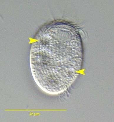
in vivo image of undescribed species of Chlamydonellopsis (BLATTERER & FOISSNER,1990).The yellow arrowheads indicate the two diagonally opposed contractile vacuoles. Collected from a slow-moving freshwater stream near Boise,Idaho.March 2007.DIC.
-
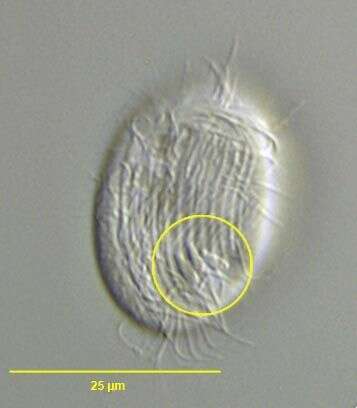
in vivo image of undescribed species of Chlamydonellopsis (BLATTERER & FOISSNER,1990).The yellow circle marks three nonmotile ventral protoplasmic protuberances. At first glance these can be mistaken for adherent bacteria.Usually visible only with DIC. This species differs from the other freshwater species ( C. pleurivacuolata and C. polonica) both of which have more numerous somatic kineties. C. pleurivacuolata has >2 contractile vacuoles and more numerous ventral protuberances. The ventral protuberances of C. polonica have probably been overlooked. Collected from a slow-moving freshwater stream near Boise,Idaho.March 2007.DIC.
-
-
-
-

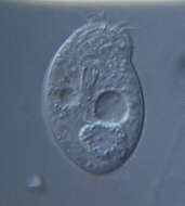
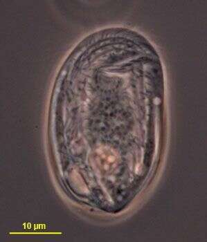
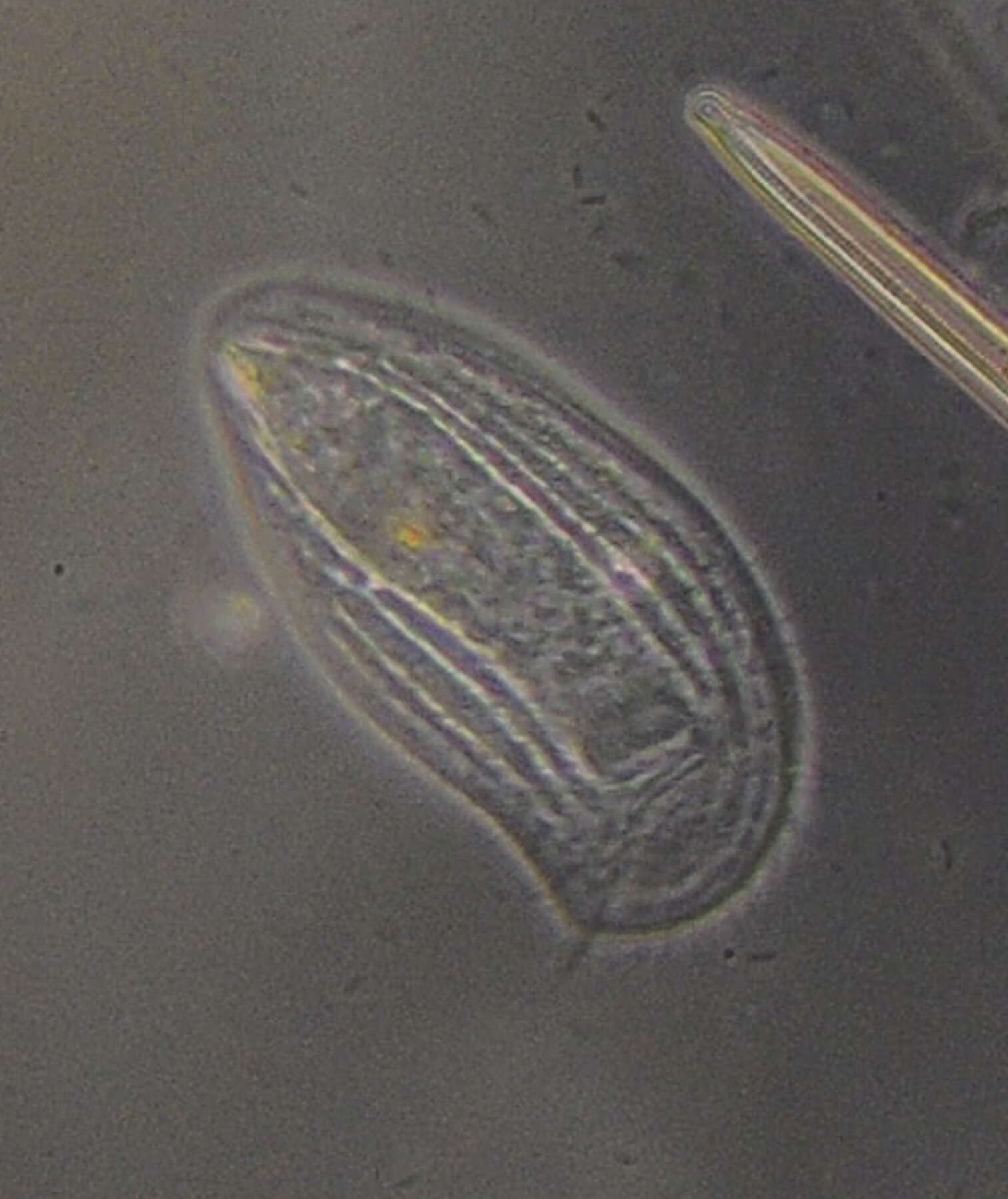
Summary.mw-parser-output table.commons-file-information-table,.mw-parser-output.fileinfotpl-type-information{border:1px solid #a2a9b1;background-color:#f8f9fa;padding:5px;font-size:95%;border-spacing:2px;box-sizing:border-box;margin:0;width:100%}.mw-parser-output table.commons-file-information-table>tbody>tr,.mw-parser-output.fileinfotpl-type-information>tbody>tr{vertical-align:top}.mw-parser-output table.commons-file-information-table>tbody>tr>td,.mw-parser-output table.commons-file-information-table>tbody>tr>th,.mw-parser-output.fileinfotpl-type-information>tbody>tr>td,.mw-parser-output.fileinfotpl-type-information>tbody>tr>th{padding:4px}.mw-parser-output.fileinfo-paramfield{background:#ccf;text-align:right;padding-right:0.4em;width:15%;font-weight:bold}.mw-parser-output.commons-file-information-table+table.commons-file-information-table,.mw-parser-output.commons-file-information-table+div.commons-file-information-table>table{border-top:0;padding-top:0;margin-top:-8px}@media only screen and (max-width:719px){.mw-parser-output table.commons-file-information-table,.mw-parser-output.commons-file-information-table.fileinfotpl-type-information{border-spacing:0;padding:0;word-break:break-word;width:100%!important}.mw-parser-output.commons-file-information-table>tbody,.mw-parser-output.fileinfotpl-type-information>tbody{display:block}.mw-parser-output.commons-file-information-table>tbody>tr>td,.mw-parser-output.commons-file-information-table>tbody>tr>th,.mw-parser-output.fileinfotpl-type-information>tbody>tr>td,.mw-parser-output.fileinfotpl-type-information>tbody>tr>th{padding:0.2em 0.4em;text-align:left;text-align:start}.mw-parser-output.commons-file-information-table>tbody>tr,.mw-parser-output.fileinfotpl-type-information>tbody>tr{display:flex;flex-direction:column}.mw-parser-output.commons-file-information-table+table.commons-file-information-table,.mw-parser-output.commons-file-information-table+div.commons-file-information-table>table{margin-top:-1px}.mw-parser-output.fileinfo-paramfield{box-sizing:border-box;flex:1 0 100%;width:100%}} Description: Chilodonella uncinata - 400x. Date: 18 May 2013, 21:38. Source:
Chilodonella uncinata - 400x. Author:
Picturepest.
-
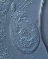
Summary.mw-parser-output table.commons-file-information-table,.mw-parser-output.fileinfotpl-type-information{border:1px solid #a2a9b1;background-color:#f8f9fa;padding:5px;font-size:95%;border-spacing:2px;box-sizing:border-box;margin:0;width:100%}.mw-parser-output table.commons-file-information-table>tbody>tr,.mw-parser-output.fileinfotpl-type-information>tbody>tr{vertical-align:top}.mw-parser-output table.commons-file-information-table>tbody>tr>td,.mw-parser-output table.commons-file-information-table>tbody>tr>th,.mw-parser-output.fileinfotpl-type-information>tbody>tr>td,.mw-parser-output.fileinfotpl-type-information>tbody>tr>th{padding:4px}.mw-parser-output.fileinfo-paramfield{background:#ccf;text-align:right;padding-right:0.4em;width:15%;font-weight:bold}.mw-parser-output.commons-file-information-table+table.commons-file-information-table,.mw-parser-output.commons-file-information-table+div.commons-file-information-table>table{border-top:0;padding-top:0;margin-top:-8px}@media only screen and (max-width:719px){.mw-parser-output table.commons-file-information-table,.mw-parser-output.commons-file-information-table.fileinfotpl-type-information{border-spacing:0;padding:0;word-break:break-word;width:100%!important}.mw-parser-output.commons-file-information-table>tbody,.mw-parser-output.fileinfotpl-type-information>tbody{display:block}.mw-parser-output.commons-file-information-table>tbody>tr>td,.mw-parser-output.commons-file-information-table>tbody>tr>th,.mw-parser-output.fileinfotpl-type-information>tbody>tr>td,.mw-parser-output.fileinfotpl-type-information>tbody>tr>th{padding:0.2em 0.4em;text-align:left;text-align:start}.mw-parser-output.commons-file-information-table>tbody>tr,.mw-parser-output.fileinfotpl-type-information>tbody>tr{display:flex;flex-direction:column}.mw-parser-output.commons-file-information-table+table.commons-file-information-table,.mw-parser-output.commons-file-information-table+div.commons-file-information-table>table{margin-top:-1px}.mw-parser-output.fileinfo-paramfield{box-sizing:border-box;flex:1 0 100%;width:100%}} Description: Chilodonella uncinata - 630x. Date: 18 May 2013, 21:40. Source:
Chilodonella uncinata - 630x. Author:
Picturepest.