-
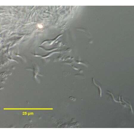
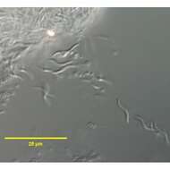
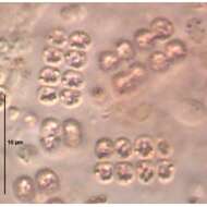
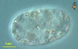
These cells of the endosymbiont Holospora undulata (ex Hafkine, 1890) Gromov and Ossipov,1981are escaping from the micronucleus of a ruptured cell of the host, Paramecium caudatum (Ehrenberg,1833).
-
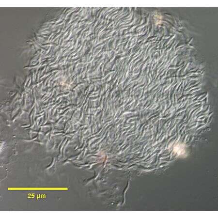
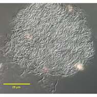
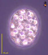
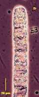
This is a detail of the grossly swollen micronucleus of a ruptured Paramecium caudatum (Ehrenberg,1833) infected with Holospora undulata (ex Hafkine, 1890) Gromov and Ossipov. The micronucleus has burst releasing individual cells of H. undulata..DIC.
-
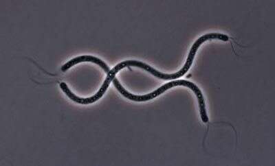
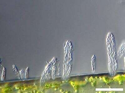
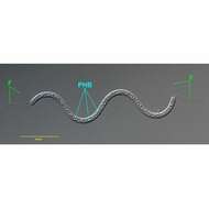
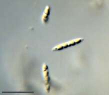
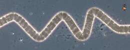
in vivo view of the chemoheterotrophic bacterium Spirillum volutans (EHRENBERG,1832). Tufts of flagella (F) occur at both poles. The species name derives from the term "volutin" or metachromatic granules composed of polyphosphates.However, the granules (PHB) of S. volutans are,in fact,composed of the energy reserve compound,poly-β-hydroxybutyrate and do not contain polyphosphates.Collected from a putifying raw culture from a freshwater pond near Boise,Idaho.DIC.
-
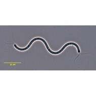
in vivo view of the chemoheterotrophic bacterium Spirillum volutans (EHRENBERG,1832). Tufts of flagella occur at both poles. The species name derives from the term "volutin" or metachromatic granules composed of polyphosphates.However, the granules of S. volutans are,in fact,composed of the energy reserve compound,poly-β-hydroxybutyrate and do not contain polyphosphates.Collected from a putifying raw culture from a freshwater pond near Boise,Idaho.Phase contrast.
-
-
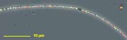
Macromonas contains calcium carbonate bodies and is motile using a single flagellum (not visible here). The scale bar indicates 10 µm. Sample from sphagnum pond Dosenmoor near Neumünster (Schleswig-Holstein, Germany). Images were taken using Zeiss Universal with Olympus C7070 CCD camera.
-
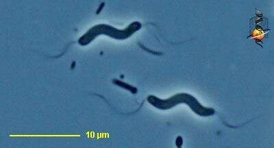
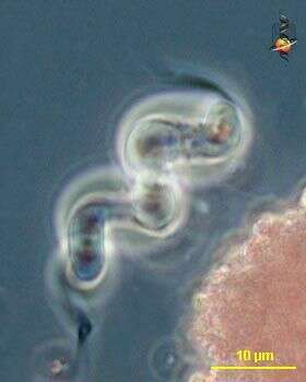
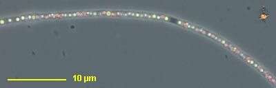
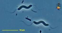
Spirillum (spire-ill-um), a motile bacterium, twisted body form, with (polar) flagella at both ends of the cell. Don t believe any microscopists who pontificate that you cannot see bacterial flagella with the light microscope. Phase contrast.
-
These two Spirillum cells have stiff, corkscrew-like flagella that they use like propellers to move through the environment.
-
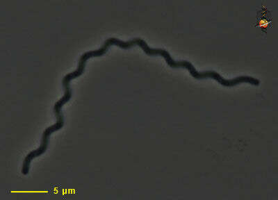
Motile spiral bacterium. No flagella are evident. Bacteria with this shape are often found in sediments and in gelatinous environments. Phase contrast micrograph.
-
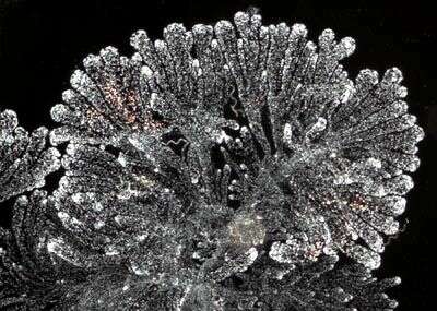
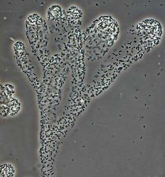
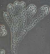
Zoogloeal bacteria grow within a soft mucus material and form slimy filaments in irregular flower-like structures. They are usually common in environments with lots of organic matter.
-
Detail of a zoogloeal aggregate. The individual bacterial cells are short rods and bound together in a delicate mucous
-
Scale bar indicates 25 µm. Sample from the Lake Constance (vicinity of Bodman). The image was built up using several photomicrographic frames with manual stacking technique. Images were taken using Zeiss Universal with Olympus C7070 CCD camera.Image under Creative Commons License V 3.0 (CC BY-NC-SA).
-
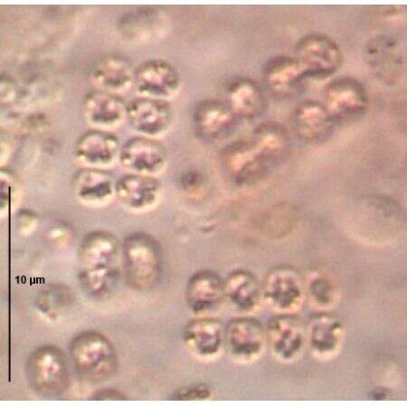
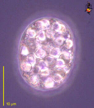
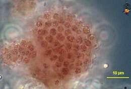
Thiocystis (thigh-owe-cyst-is) is a red sulphur bacterium (or purple sulphur bacterium). It is found in sediments above the reduced zone. It oxidizes hydrogen sulphide producing elemental sulphur which is deposited within the bacterial cell as granules of sulphur. Forms aggregates and individual cells are often hard to distinguish. The cellular nature of these aggregates is hard to determine at first glance. This detail shows that the mass is made of individual cells and the cells have granular inclusions. Phase contrast.
-
Thiocystis (thigh-owe-cyst-is) is a red sulphur bacterium (or purple sulphur bacterium). It is found in sediments above the reduced zone. It oxidizes hydrogen sulphide producing elemental sulphur which is deposited within the bacterial cell as granules of sulphur. Forms aggregates and individual cells are often hard to distinguish. Phase contrast.
-
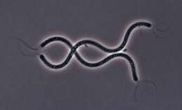
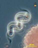
Thiospirillum (thigh-ow-spire-ill-um) is one of the sulphur bacterium. Rather like Spirillum, cells take the form of short-cork-screw and have flagella at both ends of the cells. The flagella occur in very substantial tufts so can be seen easily seen with the light microscope. This cell was rather large. Phase contrast.
-
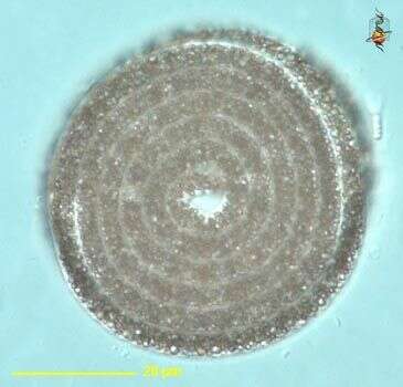
Achromatium (a-chrome-ace-ee-um) one of the larger bacteria, relatively common, and usually distinguished by the presence of large sulphur deposits inside the cell. Phase contrast.
-
Achromatium (a-chrome-ace-ee-um) is one of the non-photosynthetic sulphur bacteria. It is a heterotrophic bacterium which relies on a supply of organic matter to assist the degradation of reduced sulphur to elemental sulphur - which is then deposited as slightly pink granules inside the cell. Large (for a bacterium). Differential interference contrast.
-
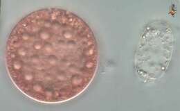
This picture compares Achromatium (a-chrome-ace-ee-um) (right) - a heterotrophic sulphur bacterium, from the red or purple sulphur bacterium, Chromatium. Differential interference contrast.
-
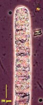
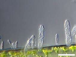
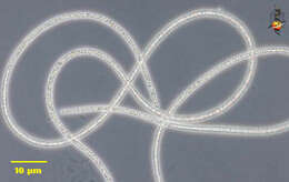
Beggiatoa (beg-ee-a-toe-a) is a colourless sulphur bacterium which occurs as filaments of various widths. Beggiatoa is found in sediments above the reduced zone. It oxidizes hydrogen sulphide, producing elemental sulphur which is deposited within the bacterial cell as sulphur granules and give this filament its opaque appearance. Very long. These cells can glide, a good trait for organisms which live in a habitat the characteristics of which are changing depending on whether there is or is not overlying water or if or if not there is sunlight. Phase contrast.
-
Beggiatoa (beg-ee-a-toe-a), commonly seen on the surface of very rich sediments - such as those of salt marshes. The bacteria metabolise reduced hydrogen sulphide and produce granules of sulphur as a by product. They can be seen as refractile elements within the cells which make up this filament. Phase contrast.
-
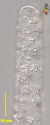
Beggiatoa (beg-ee-a-toe-a), commonly seen on the surface of very rich sediments - such as those of salt marshes. The bacteria metabolise reduced hydrogen sulphide and produce granules of sulphur as a by product. They can be seen within the cells which make up this filament. Differential interference contrast.
-
Beggiatoa (beg-ee-a-toe-a) is a colourless sulphur bacterium which occurs as filaments of various widths. Beggiatoa is found in sediments above the reduced zone. It oxidizes hydrogen sulphide, producing elemental sulphur which is deposited within the bacterial cell as sulphur granules and give this filament its opaque appearance. Layers of Beggiatoa are often seen on the surface of muds or other places where there is a lot of decaying organic matter (around dead dogs is a good place) and looks like white tissue paper lying over the sediment. These cells can glide, a good trait for organisms which live in a habitat the characteristics of which are changing depending on whether there is or is not overlying water or if or if not there is sunlight. Phase contrast.
-
Beggiatoa (beg-ee-a-toe-a) is a colourless sulphur bacterium which occurs as filaments of various widths. Beggiatoa is found in sediments above the reduced zone. It oxidizes hydrogen sulphide, producing elemental sulphur which is deposited within the bacterial cell as sulphur granules. In this delicate filament, the individual sulphur grains can be seen as pink refractile inclusions. Phase contrast.
-
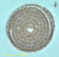
Beggiatoa (beg-ee-a-toe-a) is a colourless sulphur bacterium which occurs as filaments of various widths. Beggiatoa is found in sediments above the reduced zone. It oxidizes hydrogen sulphide, producing elemental sulphur which is deposited within the bacterial cell as sulphur granules visible inside this filament . May be very long. These cells can glide, a good trait for organisms which live in a habitat the characteristics of which are changing depending on whether there is or is not overlying water or if or if not there is sunlight. They don t usually make these cute spirals. Differential interference contrast.