-
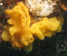
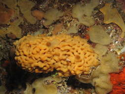
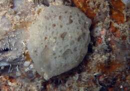
Clathrina lutea in Abrolhos Archipelago, Bahia, Brazil
-
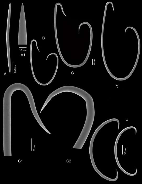
Flagellosigma of subgenus Flagellia
-
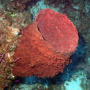
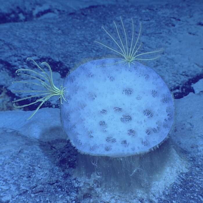
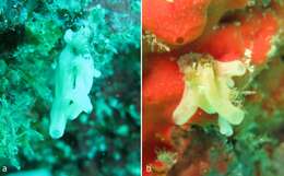
In situ photo of holotype (courtesy N.J. de Voogd)
-
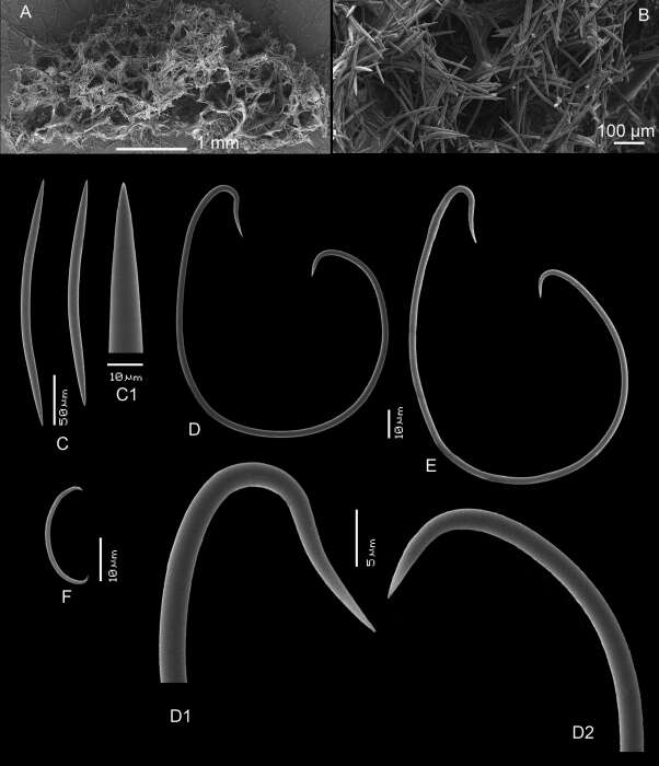
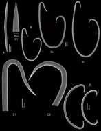
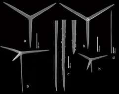
Skeleton and spicules of the holotype RMNH Por. 2326
-
Photo of ZMA Por. 09285 from Sumba, Indonesia
-
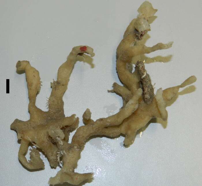
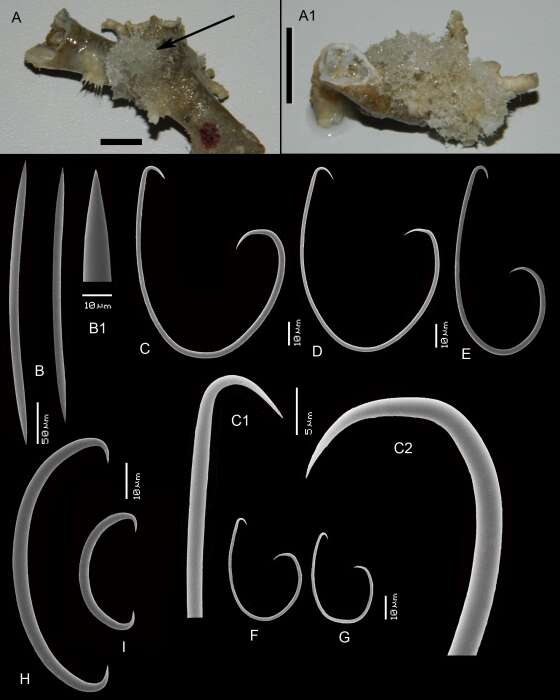
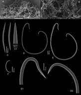
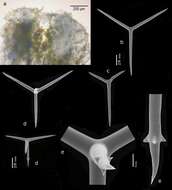
Habitus of holotype (upper left), paratype (upper right), and spicules.
-
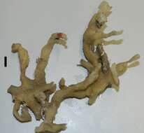
Holotype RMNH Por. 9921.
-
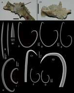
SEM images of spicules of holotype RMNH Por. 9921.
-
-
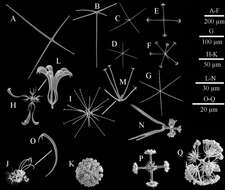
A, the centre part of choanosomal pentactin; B–C, atrial pinular pentactin; D, dermal pinular pentactin; E–F, choanosomal pinular pentactin; G, pinular hexactin; H, stauractin; I, spiny microdiactin; J, smooth middle shaft of marginalia; K, details of the middle shaft of mesouncinate; L, details of the middle shaft of macrouncinate; M, micramphidisc.
-
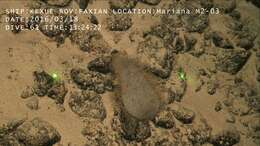
rocher du Diamant, La Martinique, 20 m , Eastern Caribbean
-
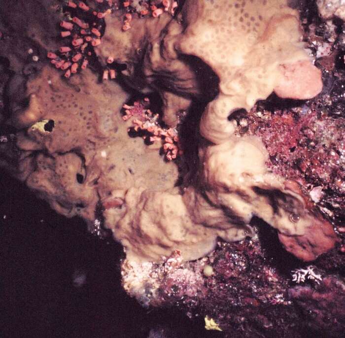
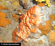
Plakina hellenica in a marine cave (Aegean Sea, Greece).
-
-
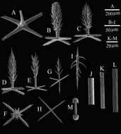
A) choanosomal stauractin; (B, C) atrial pentactins; (D) dermal hexactin; (E) discostauractin; (F) hemi-discohexaster; (G) microhexactin; (H) floricome I; (I) onychohexaster; (J) floricome II; (K) discaster; (L) detail of primary rays of floricome I; (M) detail of primary rays of onychohexaster; (N) regular hexaster; (O) detail of second ray of floricome II; (P) detail of primary rosette of graphiocome; (Q) anchorate discohexaster.
-
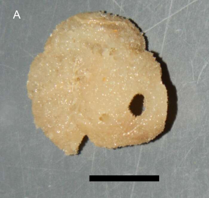
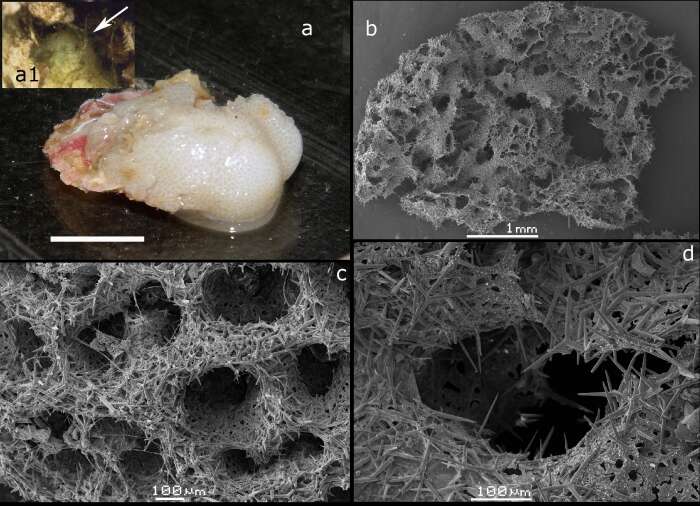
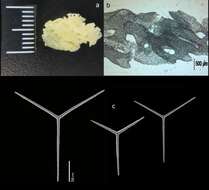
Habitus (a) and SEM cross sections (b-d)
-
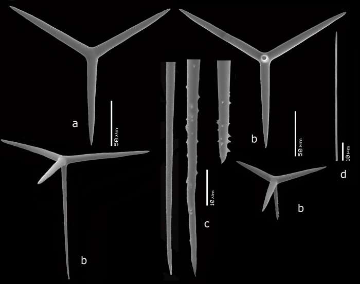
SEM images of spicules
-
In situ photos made by Nicole J. de Voogd
-
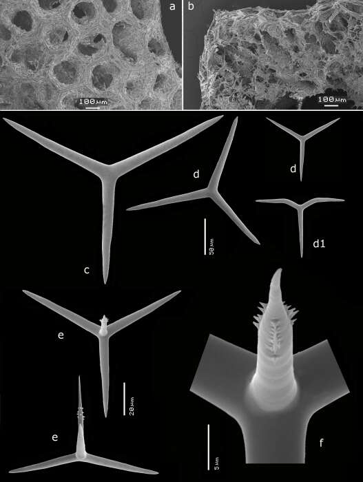
SEM images of surface, cross section and spicules
-
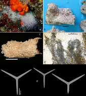
Habitus in situ, photo N.J. de Voogd
-
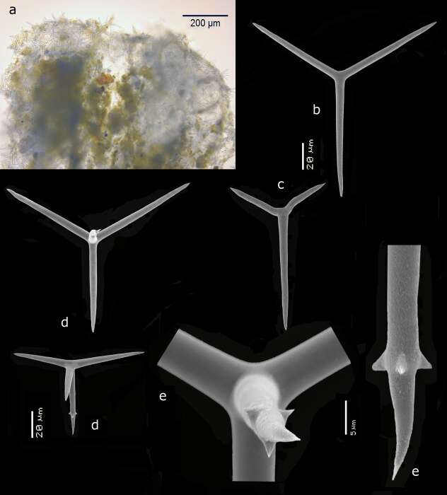
Light microscopy cross section and SEM images of spicules
-
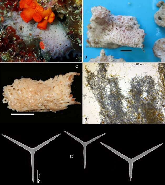
In situ and on deck image by N.J. de Voogd, SEM images of spicules, light microscopic virew of cormus, preserved holotype (c)
-
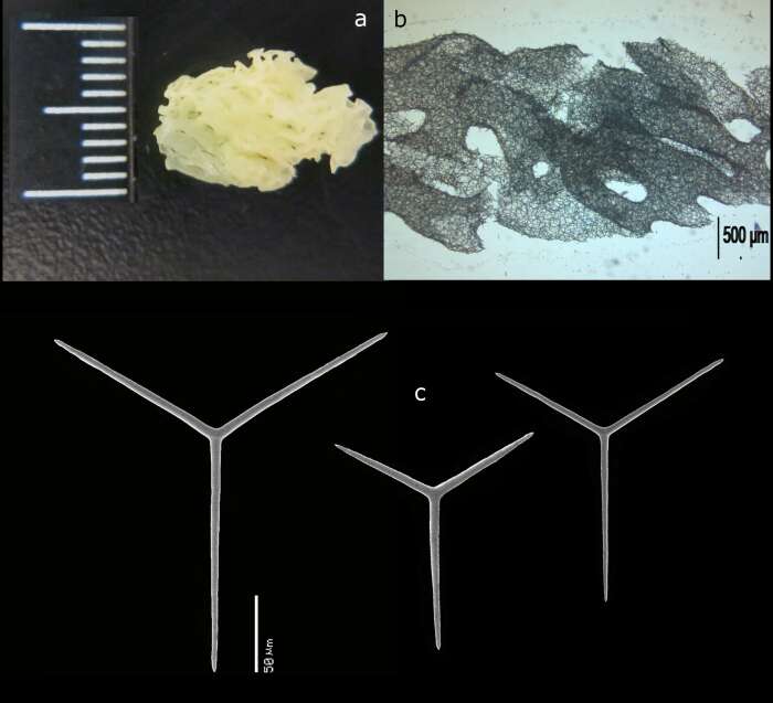
Preserved holotype, light microscopic view of cormus, SEM images of spicules
-
In situ images (photo N.J. de Voogd), preserved holotype, light microscopic view of tubule, SEM images of spicules
-
In situ, preserved and SEM cross sections