-
Longuera-Toscal, Canary Islands, Spain
-
Arredondo, Cantabria, Spain
-
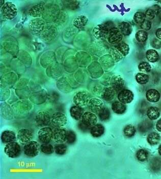
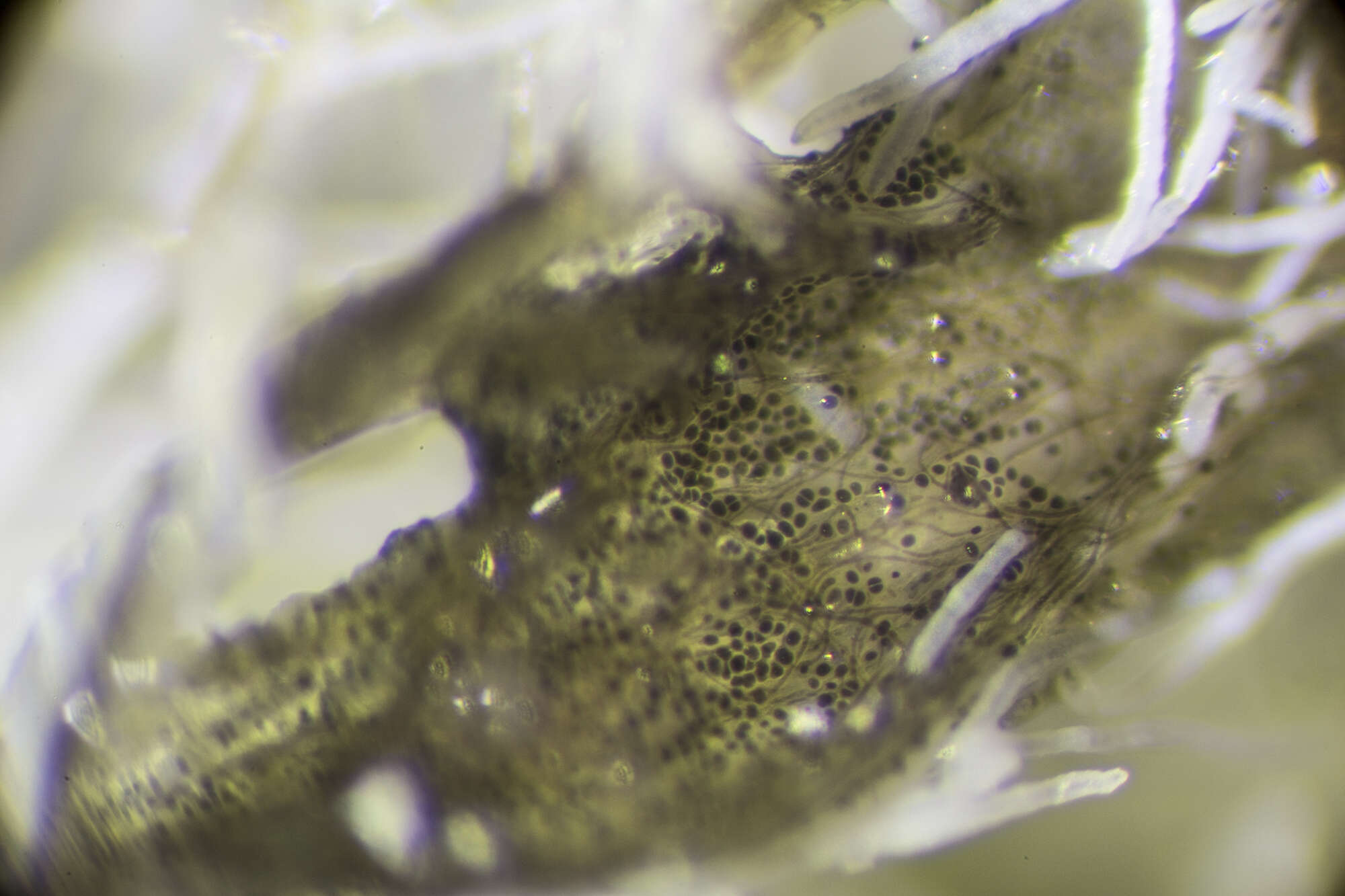
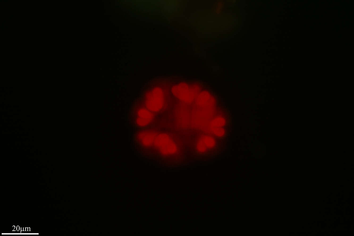
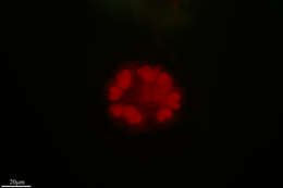
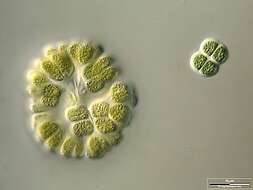
Microcystis flos-aquae together with Microcystis aeruginosa is common in Lake Kinneret throughout the year, and blooming periodically. The cells at the border and at the center of Microcystis colony looks different by chlorophyll and gas vesicles concentrations. The specimen was sampled from the shore of the lake in March 2006.
-
Arredondo, Cantabria, Espaa
-
Arredondo, Cantabria, Spain
-
Longuera-Toscal, Canary Islands, Spain
-
-
Arredondo, Cantabria, Spain
-
Longuera-Toscal, Canary Islands, Spain
-
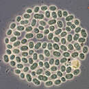
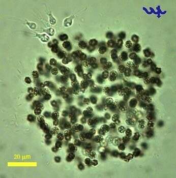
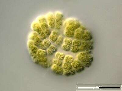
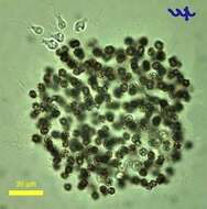
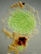
Microcystis flos-aquae (Cyanobacteria, Chroococcales) is a together with Microcystis aeruginosa are common in Lake Kinneret throughout the year, occasionally forming blooms with surface scums in winter or spring. The individual cells in a M. flos-aquae colony are 3-4 um in diameter, the colony is spherical or lens-shaped, with varying degree of spacing between cells within a colony. The dark spots clearly showing in the individual cells in this picture are due to the reflection of light from the gas vesicles. A bunch of choanoflagellates are attached at the upper left side of the colony.
-
Arredondo, Cantabria, Spain
-
Madrid, Madrid, Spain
-
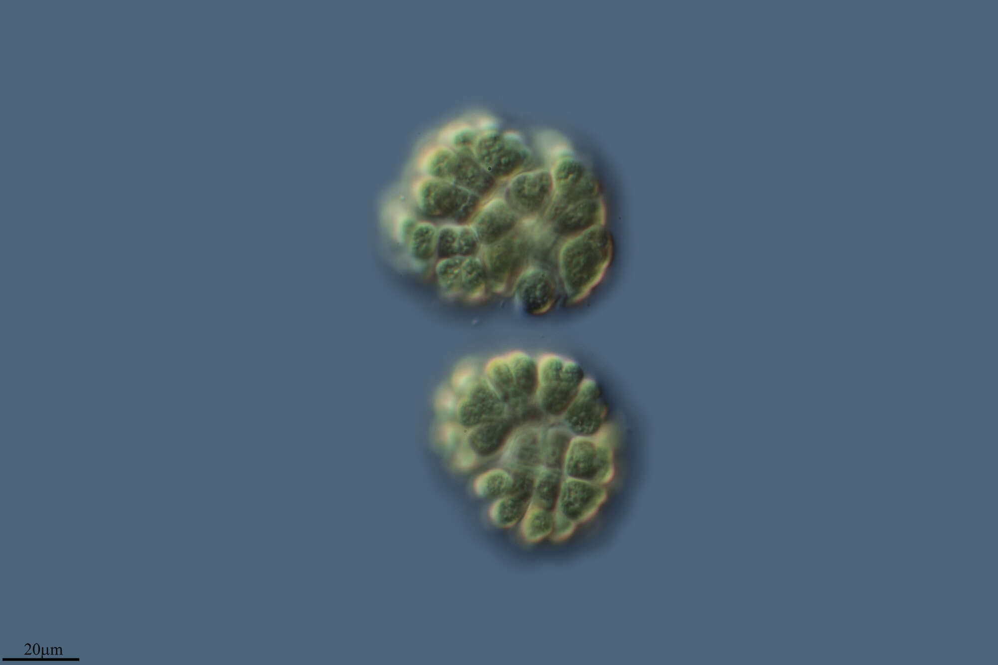
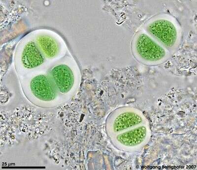
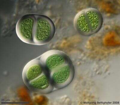
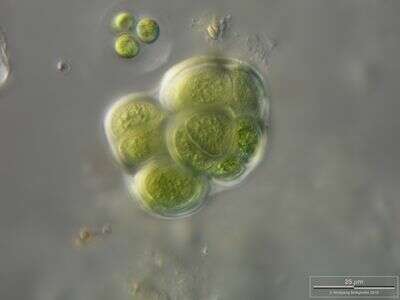
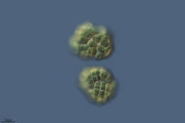
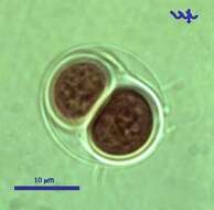
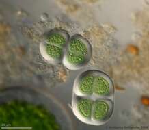
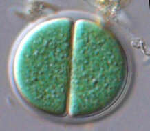
Chroococcus turgidus, (Cyanobacteria, Chroococales), from Lake Kinneret pelagic waters, April 2006, showing 2 daughter cells after division by simple binary fission â as characteristic for most Chroococcales species. This species is common in the plankton of Lake Kinneret throughout the year. Usually there are 2 â 8 cells in a colony. Clearly delimited colorless mucilaginous envelopes surround the individual cells following their contours, and the entire colony. Cell diameter: 8 â 11 µm.
-
Arredondo, Cantabria, Spain
-
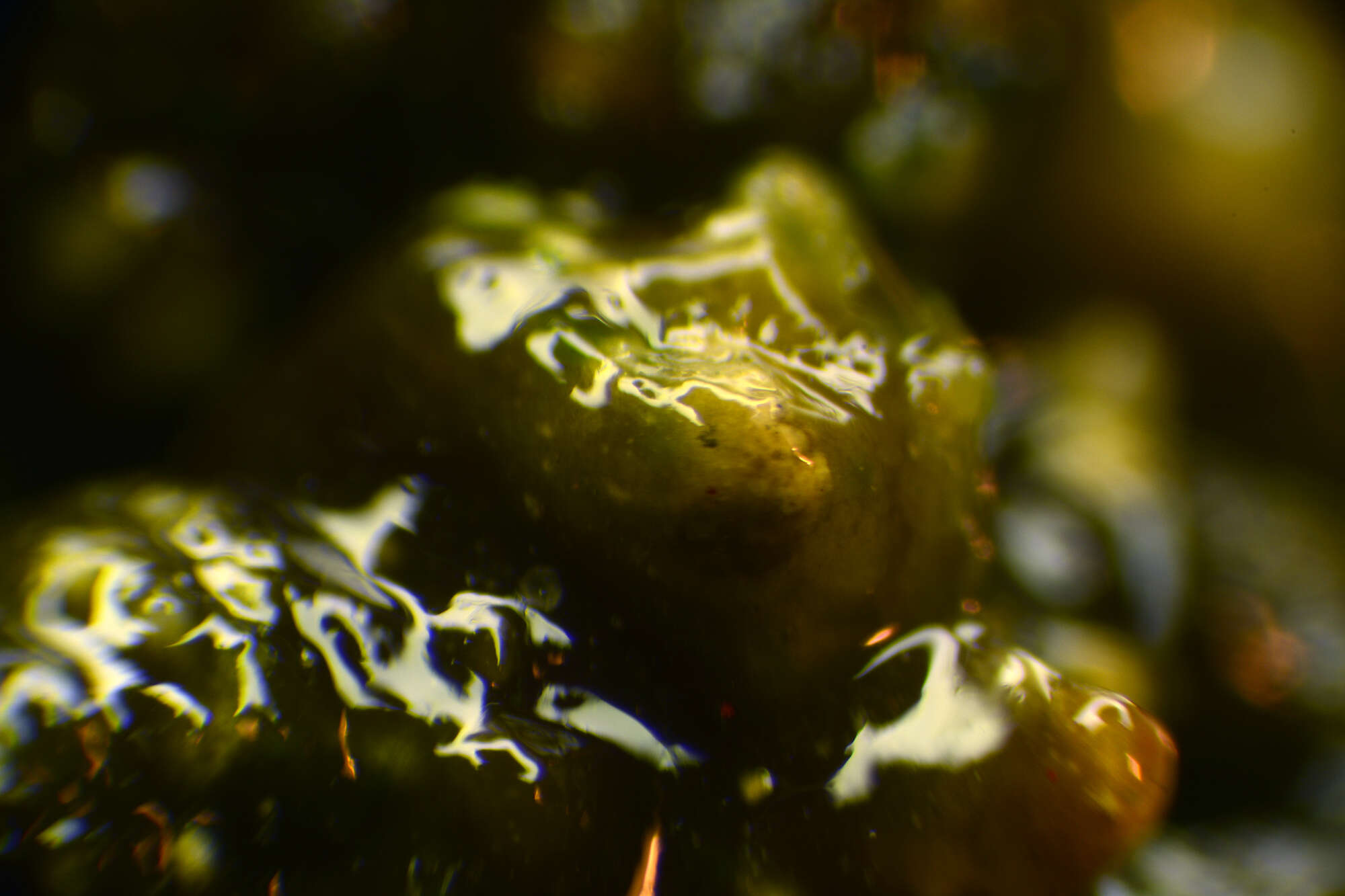
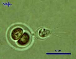
Chroococcus turgidus (Cyanobacteria, Chroococcales) is common in Lake Kinneret in recent years. In this unusual photograph a choanoflagellate is attached to its outer shell.
-
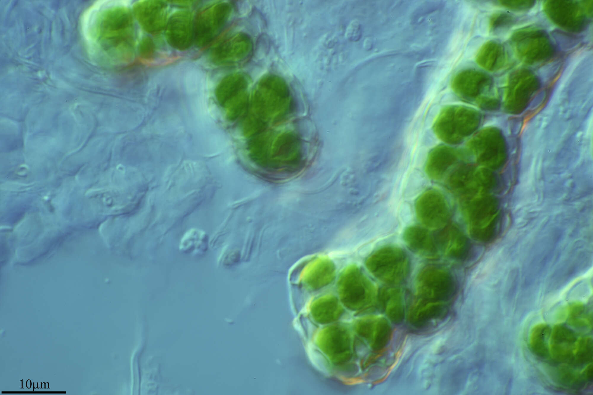
High resolution photo of Chroococcus turgidus using Planapo 63/1.4. Sample from sphagnum pond situated in the northern alpine region of Austria near Salzburg. Images were taken using Zeiss Universal with Olympus C7070 CCD camera.
-
High resolution photo of Chroococcus turgidus using Planapo 63/1.4 with DIC. Sample from sphagnum pond situated in the northern alpine region of Austria near Salzburg. Images were taken using Zeiss Universal with Olympus C7070 CCD camera.
-
High resolution photo of Chroococcus turgidus using Planapo 63/1.4 with DIC. Sample from sphagnum pond situated in the northern alpine region of Austria near Salzburg. Images were taken using Zeiss Universal with Olympus C7070 CCD camera.
-
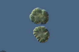
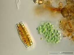
Colony accompanied by Epithemia adnata. Scale bar indicates 50 µm. Sample from a wetland at the Pillersee (Tyrol, Austria). The image was built up using several photomicrographic frames with manual stacking technique. Images were taken using Zeiss Universal with Olympus C7070 CCD camera.Image under Creative Commons License V 3.0 (CC BY-NC-SA).
-
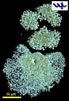
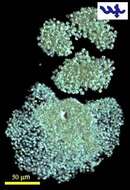
Scale bar indicates 100 µm. Sample from the pond Hegne Moor situated in the vicinity of Lake Constance. Images were taken using Zeiss Universal with Olympus C7070 CCD camera.Image under Creative Commons License V 3.0 (CC BY-NC-SA).
-
Scale bar indicates 50 µm. Sample from a wetland at the Pillersee (Tyrol, Austria). The image was built up using several photomicrographic frames with manual stacking technique. Images were taken using Zeiss Universal with Olympus C7070 CCD camera.Image under Creative Commons License V 3.0 (CC BY-NC-SA).
-
Colony accompanied by Chroococcus turgidus. Scale bar indicates 25 µm. Sample from a wetland at the Pillersee (Tyrol, Austria). The image was built up using several photomicrographic frames with manual stacking technique. Images were taken using Zeiss Universal with Olympus C7070 CCD camera.Image under Creative Commons License V 3.0 (CC BY-NC-SA).
-
Scale bar indicates 25 µm. Sample from sphagnum pond situated in the northern alpine region of Austria near Salzburg. Images were taken using Zeiss Universal with Olympus C7070 CCD camera.
-
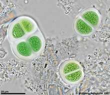
Chroococcus (crow-o-cock-us), large cyanobacterium, typically two (but sometimes one) cells enclosed within a mucus sheath. Photosynthetic pigment distributed through cytoplasm, which may have a granular texture, but does not have subcompartments (organelles). Differential interference contrast.