-
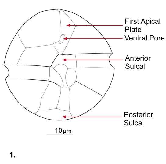
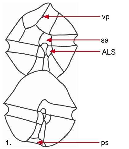
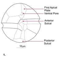
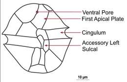
Fig 1: Alexandrium ostenfeldii Schematic drawing of a cell showing plate patterns on ventral side of cell
-
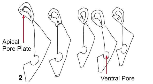
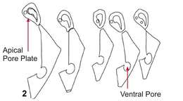
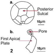
Fig 2: Alexandrium ostenfeldii Schematic drawing of a cell showing the morphological variation in APC and 1'
-
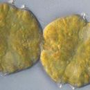
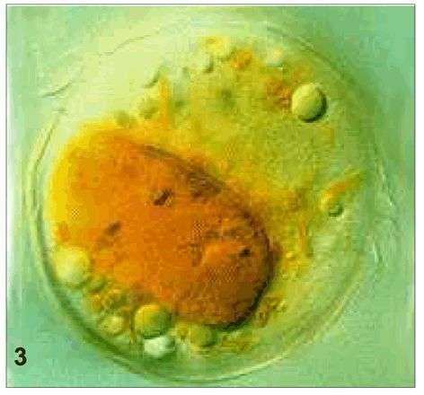
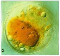
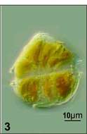
Fig 3: Alexandrium ostenfeldii whole cell with food vacuole
-
Fig 1: Schematic diagram of Alexandrium affine, showing ventral (top) and dorsal (bottom) views.
-
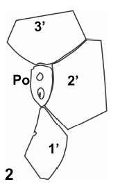
Fig 2: Schematic drawing of the Po and surrounding apical plates
-
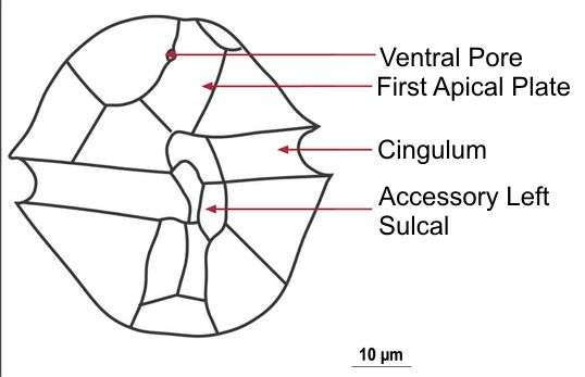
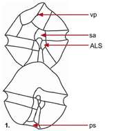
Fig 1: Alexandrium tamarense Schematic drawing of a cell showing plate patterns on ventral side of cell
-
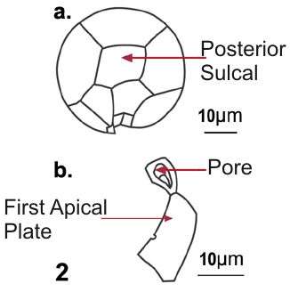
Fig 2: Alexandrium tamarense Schematic drawing of a cell in posterior view and b. apical pore coplex with 1'.
-
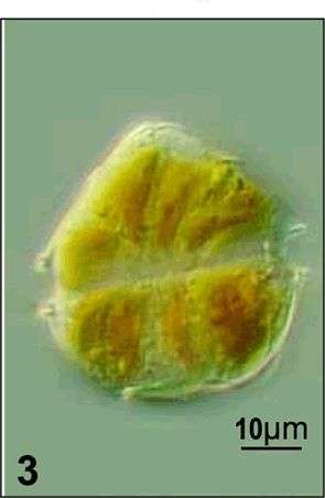
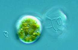
Fig 3: Alexandrium tamarense Live cell in dorsal view
-
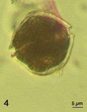
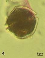
Fig 4: Alexandrium tamarense Lugol's preserved cell
-
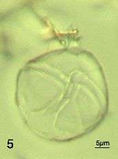
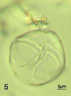
Fig 5: Alexandrium tamarense empty theca showing detail of sulal plates
-
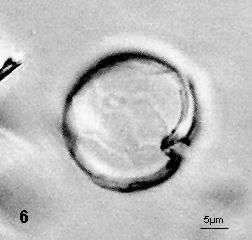
Fig 6: Alexandrium tamarense anterior part of theca showing outline of 1' plate
-
Fig 7: Image of Alexandrium tamarense