-
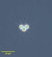
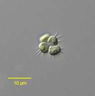
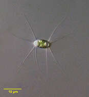
Micractinium pusillum (Fresenius,1858). Usually found in four-celled colonies (only three of the four cells of this colony are seen here). Each cell has two to seven long, delicate setae. Each cell has one cup-shaped plastid with a pyrenoid. 18S ribosomal RNA gene studies place this genus in the Trebouxiophyceae. One of the distinguishing features of trebouxiophytes is metacentric mitosis in which centrioles are located near the metaphase plate of the chromosomes rather than at the spindle poles. Other characteristics include formation of microtubules parallel to the dividing cell wall during cytokinesis (phycoplast). The similar genus, Errerella, has only one spine per cell.Collected from a freshwater pond near Boise, Idaho. July 2005. Phase contrast.
-
Dictyosphaerium spec. in mucilaginous envelope. Scale bar indicates 25 µm. Sample from sphagnum pond situated in the northern alpine region of Austria near Salzburg. Images were taken using Zeiss Universal with Olympus C7070 CCD camera.
-
Green algae live in a mucus ball. Phase contrast microscopy.
-
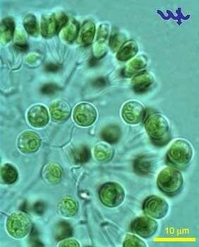
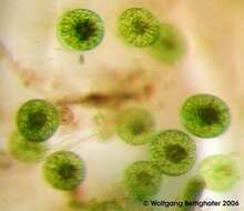
Portrait of Dictyosphaerium pulchellum (Nägeli,1849), a colonial green alga. The small cells are ovoid to reniform and are borne at the ends of colorless branching threads in groups of four. The threads are remnants of parental cell walls. Each cell has a cup-shaped chloroplast and solitary pyrenoid.The entire colony is embedded in a nearly invisible spherical gelatinous matrix. Collected from a freshwater pond near Boise, Idaho June 2004. DIC optics.
-
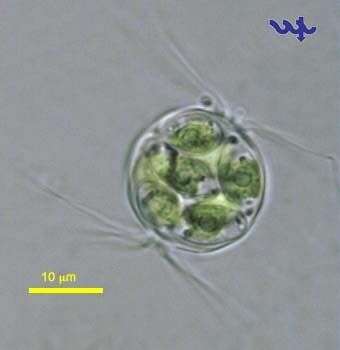
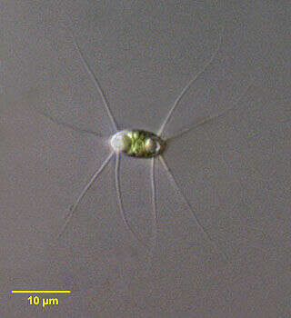
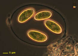
Chodatella citriformis containing 8 autospores, sampled from shore water of Lake Kinneret in March 2006. This Chlorophyte (Chlorococcales) can be found in the plankton of the lake at most times of the year, particularly in May-June, but is never dominant.
-
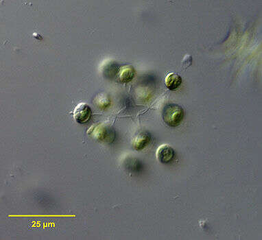
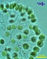
Portrait of the planktonic trebouxiophyte alga, Tetrastrum staurogeniaeforme (Schröder) Lemmermann.The colonies consist of four ellipsoid cells with four to six short spinous projections from the each cell surface. several four-celled colonies may join together to form compound colonie (compound coenobia). Each cell has one plastid and one pyrenoid.Collected from a freshwater pond near Boise,Idaho. July 2005.DIC.
-
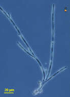
Microthamnion (mike-row-tham-knee-on), is a branching filamentous green alga. With cellulosic cell wall, plastids with chlorophylls a and b giving the plastids a green colour. Phase contrast.
-
Portrait of Dictyosphaerium pulchellum (Nägeli,1849), a colonial green alga. The small cells are ovoid to reniform and are borne at the ends of colorless branching threads in groups of four. The threads are remnants of parental cell walls. Each cell has a cup-shaped chloroplast and solitary pyrenoid.The entire colony is embedded in a nearly invisible spherical gelatinous matrix. Collected from a freshwater pond near Boise, Idaho June 2004. DIC optics.
-
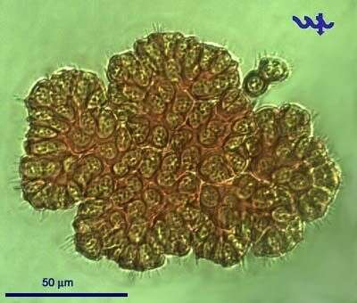
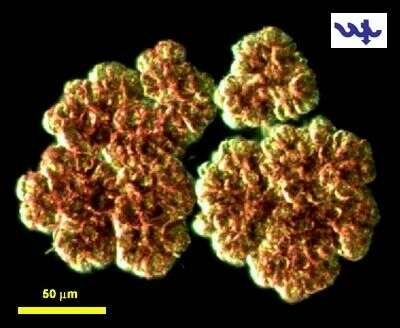
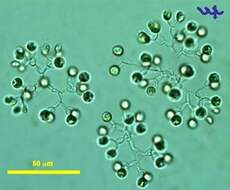
Botryococcus braunii (Chlorophyta, Chlorococcales) is a colonial chlorophyte, made of ovoid cells that are closely packed to form golden brown mucilaginous lumps. Older cells usually have large amounts of reserve food material (oil) such that the cell contents are obscured, and the colonies float. The species is abundant at times, forming surface scums at lee shores. On a single occasion in January 2000 a bloom of this species covered the entire lake for several days, giving it a golden color, then disappeared abruptly. This photo shows Botryococcus braunii colonies at x200 mag, with their typical mucilaginous strands connecting colonies and enveloping them.
-
Dictyosphaerium pulchellum is a common chlorophyte of Lake Kinneret, relatively more abundant in winter, but never dominant. This specimen was sampled at the shore of the lake in March 2006.
-
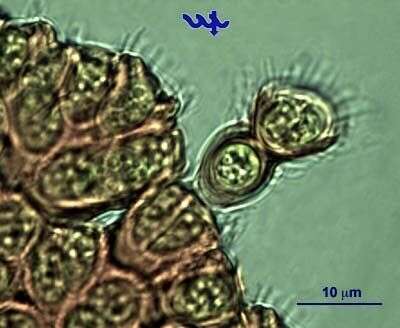
A young colony of Botryococcus braunii (Chlorophyta, chlorococcales) budding out of an old colony, photographed at x1000 mag. Note cup-shape of the mucilage in which each cell is embedded.
-
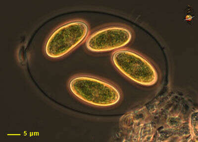
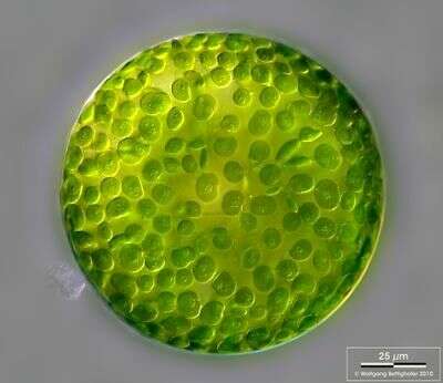
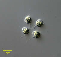
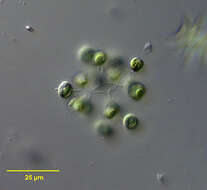
Dictyosphaerium pulchellum is a common chlorophyte (Chlorococcales) of Lake Kinneret, relatively more abundant in winter, never abundant or dominant. Note that the sphaerical cells are in clusters of 4, attached by parental cell wall fragments radiating from a common center. The chloroplasts are parietal and cup shaped with a single pyrenoid. This specimen was sampled at the shore of the lake in March 2006.
-
Botryococcus braunii (Chlorophyta, Chlorococcales) is a colonial chlorophyte, made of ovoid cells that are closely packed to form golden brown mucilagenous lumps. Older cells usually have large amounts of reserve food material (oil) such that the cell contents are aboscured, and the colonies float. The species is abundant at times, forming surface scums at lee shores. On a single occassion in January 2000 a bloom of this species covered the entire lake for several days, giving it a golden color, then disappeared abruptly. This photo shows Botryococcus braunii colonies at x200 mag, with their typical mucilagenous strands connecting colonies and enveloping them.
-
Portrait of the planktonic green alga Chodatella longiseta (Lemmerman,1898;Printz). This genus differs from the very similar Franceia in having long slender spinous processes limited to the poles of the ovoid cell. There is no gelatinous layer. There are two chloroplasts each with a prominent pyrenoid (seen in this image). Collected from a freshwater pond near Boise, Idaho June 2004. DIC optics.
-
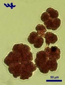
Colorful colonies of Botryococcus braunii (Chlorophyta, Chlorococcales) photographed at dark-field
-
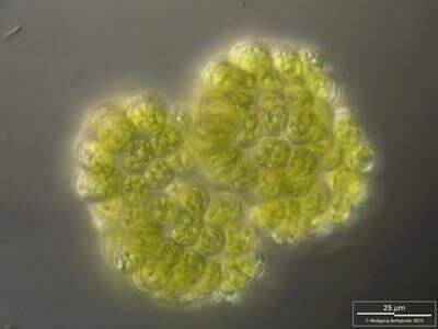
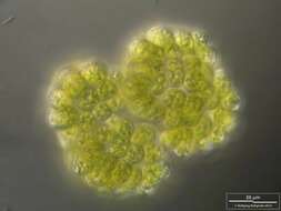
These green balls are often seen in sphagnum ponds. Looking through the stereomicroscope this group of specimen looked photogenic. The picture was generated from 5 shots using MicroPicS by Bernhard Wiedemann and Photoshop (depth of focus). Sample from sphagnum pond Dosenmoor near Neumuenster (Schleswig-Holstein, Germany). This image was taken using Zeiss Universal with Olympus C7070 CCD camera.
-
Eremosphaera viridis bears lots of chloroplasts, each with a central pyrenoid body. The brace like marks amidst the chloroplasts represent the amylum shells of the pyrenoids. High resolution multi layer image assembled of 15 Eremosphaera shots (manually stacked). See zip archive for details. Sample from sphagnum pond situated in the northern alpine region of Austria near Salzburg. Images were taken using Zeiss Universal with Olympus C7070 CCD camera.
-
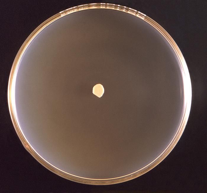
This 1971 image depicted a frontal view of a Petri dish culture in which a small colony of Prototheca wickerhamii algal organisms had been cultivated.Created: 1971
-
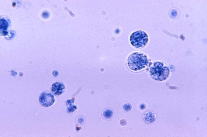
At a magnification of 1125X, this photomicrograph revealed the presence of a number of Prototheca wickerhamii algal organisms.Created: 1971
-
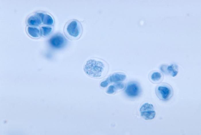
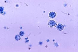
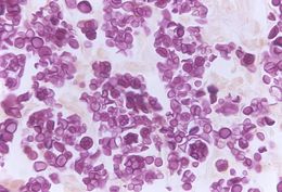
Under a magnification of 1125X, this photomicrograph revealed the presence of numbers of algal organisms, Prototheca wickerhamii, which were found within a tissue specimen. Though categorized taxonomically as an alga, it derives its sustenance as a saprophyte, consuming dead and decaying organic matter. This algal culture was prepared using a lactophenol cotton blue mount fixation technique.Under microscopic analysis, Prototheca spp. resemble a fungal organism, and can therefore, be mistaken when attempting to identify these algae.Similar to the members of the genus Chlorella, Prototheca spp. are heterotrophic, , which means these organisms require carbon in order to thrive, and obtains this nutrient through its consumption of organic substrates. This algal culture was prepared using a lactophenol cotton blue mount fixation technique.Created: 1972
-
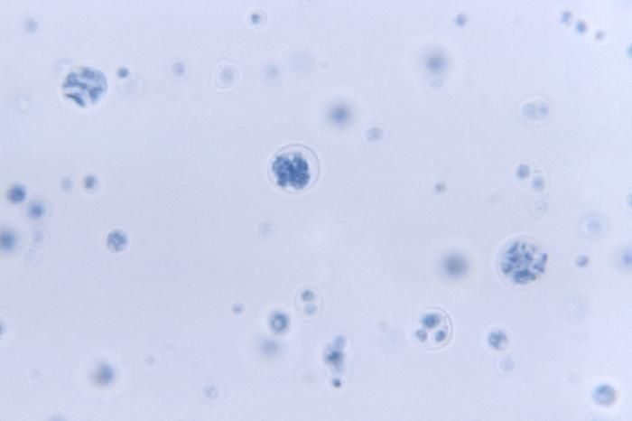
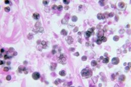
Under a magnification of 500X, this Gridley-stained photomicrograph revealed the presence of numbers of algal organisms, Prototheca wickerhamii, which were found within a specimen of deer tissue. Though categorized taxonomically as an alga, it derives its sustenance as a saprophyte, consuming dead and decaying organic matter.Under microscopic analysis, Prototheca spp. resemble a fungal organism, and can therefore, be mistaken when attempting to identify these algae.Similar to the members of the genus Chlorella, Prototheca spp. are heterotrophic, , which means these organisms require carbon in order to thrive, and obtains this nutrient through its consumption of organic substrates. This algal culture was prepared using a lactophenol cotton blue mount fixation technique.Created: 1972
-
This photomicrograph depicts the presence of Prototheca wickerhamii in a case of protothecosis.Created: 1971
-
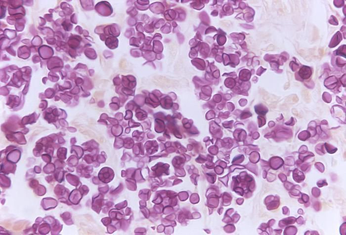
Note the histopathologic changes in protothecosis of the skin and mucous membrane of the nose due to P. wickerhamii.Created: 1973
-
This photomicrograph confirms the presence of Prototheca wickerhamii, an achlorophyllic algae.Created: 1972