-
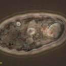
Samples from Sediment at Cedar swamps, Woods Hole, Massachusatts. Photographed by Hwan Su Yoon.
-
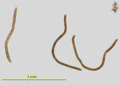
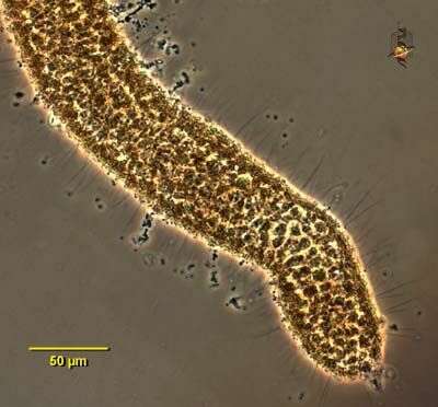
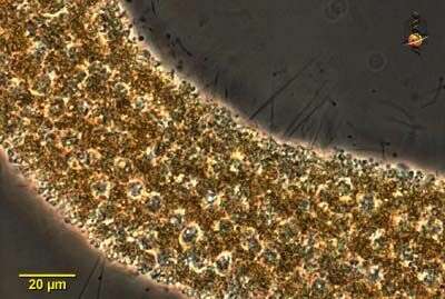
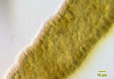
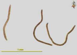
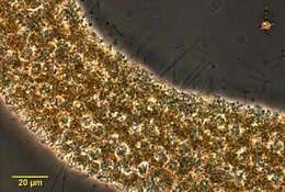
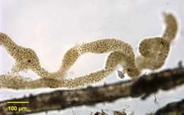
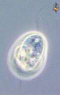
Spongomonas - a heterotrophic flagellate. The cells are spherical and give rise to two flagella that are slightly different in length. The cells form colonies, being embedded in a common mucous matrix that is made up of small globules of orange or brown mucus. This species forms sausage-shaped colonies that are up to a millimetre in length. Bright field illumination.
-
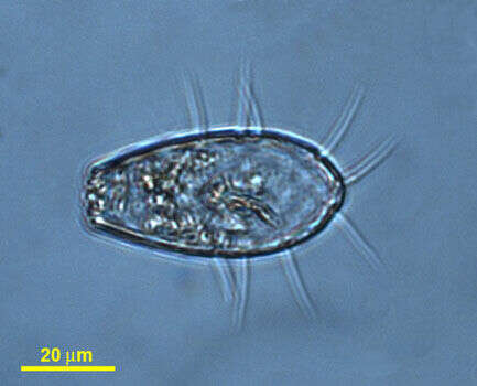
Galende, Castile and Len, Spain
-
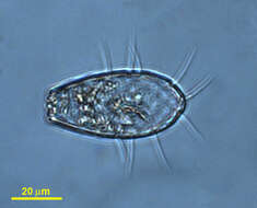
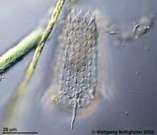
Members of testate amoebae group Euglyphidae form siliceous plates for constructing shell. Sample collection by Martin Kreutz from Simmelried near Konstanz(Baden-Wuerttemberg, Germany). This image was taken using Zeiss Universal with Olympus C7070 CCD camera.
-
Spongomonas - a heterotrophic flagellate. The cells are spherical and give rise to two flagella that are slightly different in length. The cells form colonies, being embedded in a common mucous matrix that is made up of small globules of orange or brown mucus. This species forms sausage-shaped colonies that are up to a millimetre in length. Phase contrast micrograph.
-
Galende, Castille and Leon, Spain
-
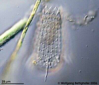
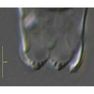
Apertural plates of Euglypha cristata Leidy, 1874. Found in a soil sample from Pyhä-Luosto National Park, Finland. DIC.
-
Spongomonas - a heterotrophic flagellate. The cells are spherical and give rise to two flagella that are slightly different in length. The cells form colonies, being embedded in a common mucous matrix that is made up of small globules of orange or brown mucus. This species forms sausage-shaped colonies that are up to a millimetre in length. Phase contrast micrograph.
-
Ribadelago, Castille and Leon, Spain
-
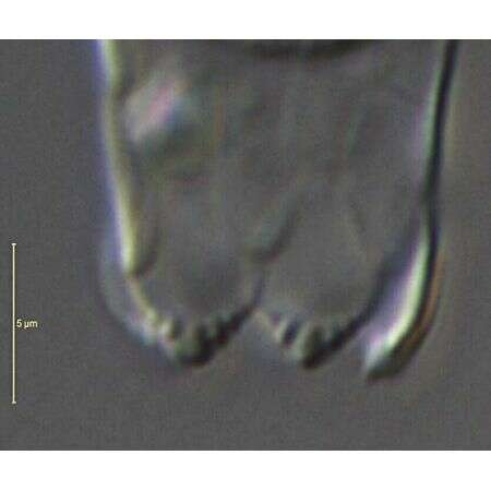
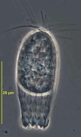
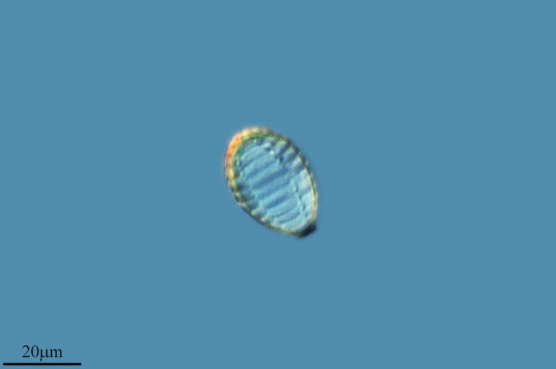
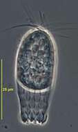
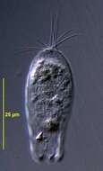
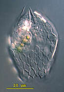
Euglypha (you-gligh-fah) cristata has an elongate shell that is composed of siliceous scales. The point of the shell shows a characteristic tuft of three to eight spines. The circular aperture is bordered by a single row of five to six denticulate scales. Pseudopodia are rarely extended. Differential interference contrast.
-
Spongomonas - a heterotrophic flagellate. The cells are spherical and give rise to two flagella that are slightly different in length. The cells form colonies, being embedded in a common mucous matrix that is made up of small globules of orange or brown mucus. This species forms sausage-shaped colonies that are up to a millimetre in length. Phase contrast micrograph.
-
Ribadelago de Franco, Castille and Leon, Spain
-
Euglypha cristata Leidy, 1874. Found in a soil sample from Pyhä-Luosto National Park, Finland. Phase contrast.
-
Portrait of Spongomonas intestinum colony. Colonies are composed of small spherical colorless individual flagellates each with two equal flagella embedded in a serpiginous gelatinous matrix. Flagella are about 3 times cell length. Some species form spherical colonies. Gelatinous matrix may be brownish-green as seen here or nearly colorless. From freshwater pond near Boise, Idaho. Brightfield.
-
Ribadelago de Franco, Castille and Leon, Spain
-
Euglypha cristata Leidy, 1874. Found in a soil sample from Pyhä-Luosto National Park, Finland. DIC.
-
Detail of Spongomonas colony showing individual cells embedded in gelatinous matrix. Each cell has two equal flagella about three times the cell length. From fresh water pond near Boise, Idaho. Oblique illumination.
-
Ribadelago, Castille and Leon, Spain
-
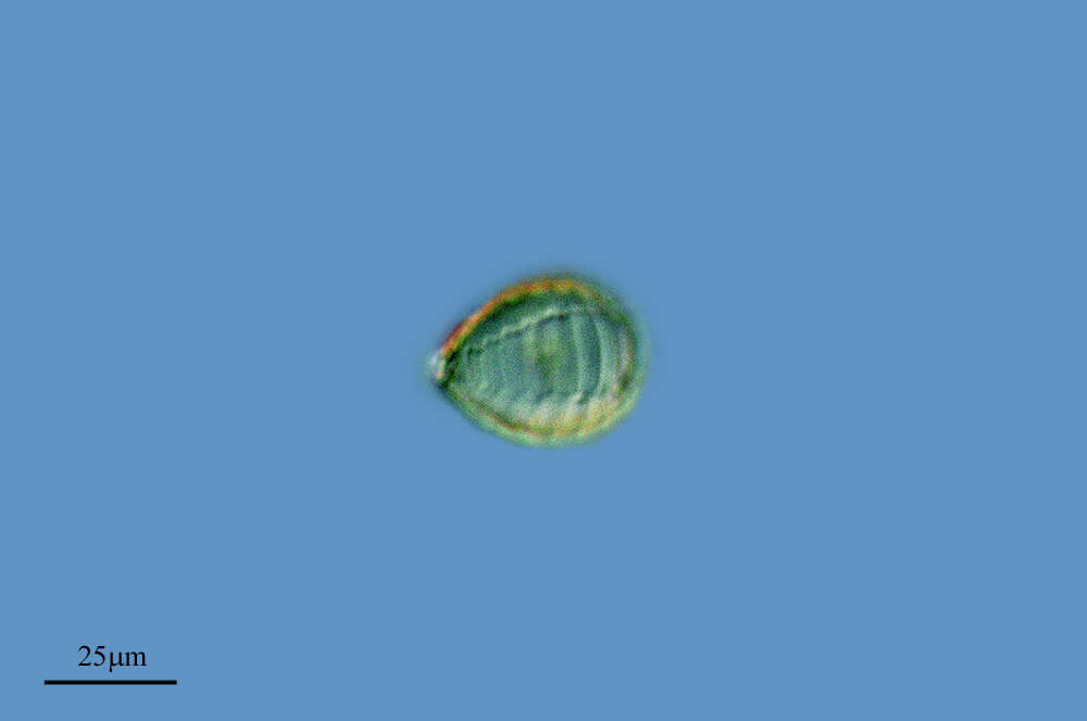
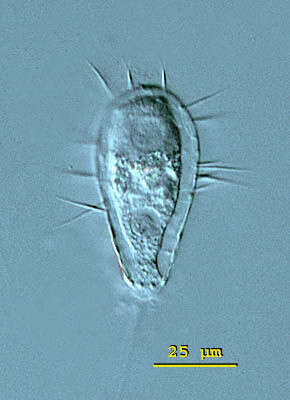
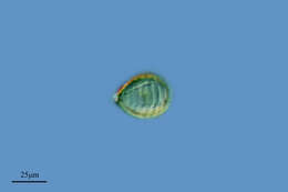
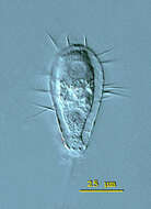
Euglypha (you-gligh-fah) filifera has an elongate shell that is composed of oval siliceous scales. The 15 micron long spines pointed out from the entire lateral edge of the shell. The nucleus is visible in the posterior third of the cell. Differential interference contrast.
-
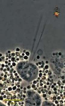
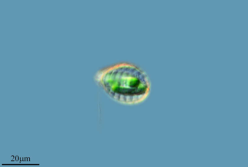
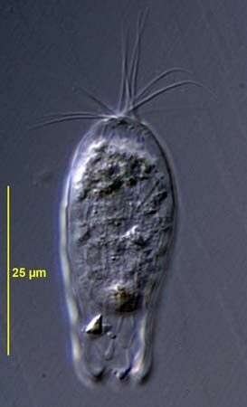
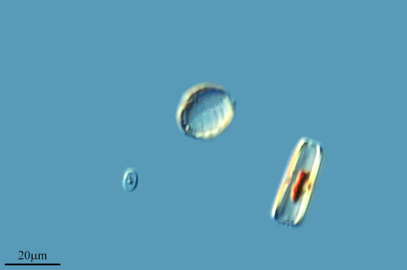
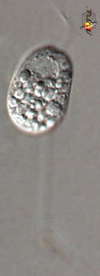
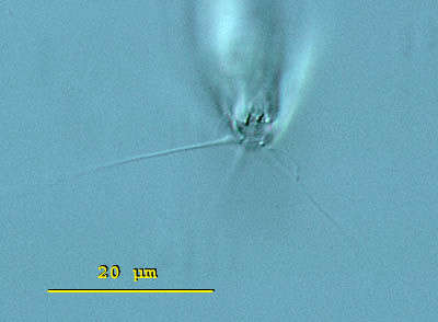
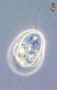
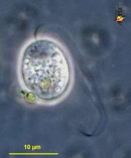
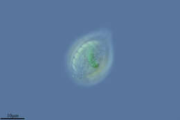
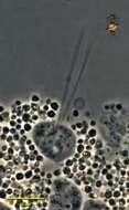
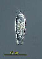
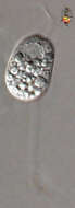
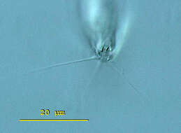
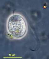
Protaspis (pro-tass-piss) A very common but little studied genus of gliding flagellates, two flagella inserted one in front of the other in a shallow ventral depression near the front of the cell. One flagellum trails behind the cell, one sweeps in front of the cell. There are caps (dictyosomes?) over the nucleus and these can be seen as two lines leading away from the site of flagellar insertion. Protaspis can produce pseudopodia and may eat diatoms. This individual has starchy inclusions. Phase contrast.
-
Euglypha (you-gligh-fah) filifera has an elongate shell that is composed of oval siliceous scales. The 15 micron long spines pointed out from the entire lateral edge of the shell. View of the aperture surrounded by denticulate scales. Long filopodia extended from the aperture. The aperture measures 6 microns in diameter. Differential interference contrast.
-
Protaspis (pro-tass-piss) A very common but little studied genus of gliding flagellates, two flagella inserted one in front of the other in a shallow ventral depression near the front of the cell. One flagellum trails behind the cell, one sweeps in front of the cell. There are caps (dictyosomes?) over the nucleus and these can be seen as two lines leading away from the site of flagellar insertion. Protaspis can produce pseudopodia and may eat diatoms. This individual has starchy inclusions. Phase contrast.
-
Euglypha (you-gligh-fah) filifera has an elongate shell that is composed of oval siliceous scales. The 15 micron long spines pointed out from the entire lateral edge of the shell. Image of Euglypha filifera lorica. The denticulate scales which border the circular aperture are visible. Each plate is measuring 6 X 8 microns on average. Differential interference contrast.
-
Protaspis (pro-tass-piss) A very common but little studied genus of gliding flagellates, two flagella inserted one in front of the other in a shallow ventral depression near the front of the cell. One flagellum trails behind the cell, one sweeps in front of the cell. Protaspis can produce pseudopodia and may eat diatoms. This individual has starchy inclusions. Phase contrast.