-
Ribadelago, Castille and Leon, Spain
-
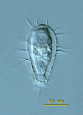
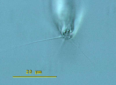
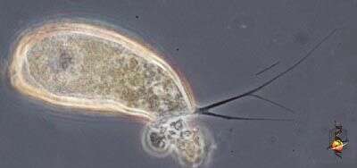
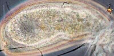
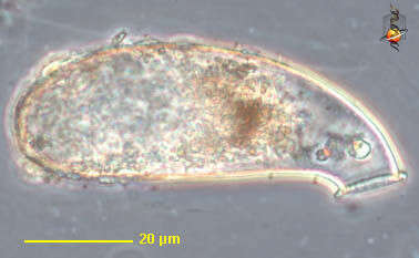
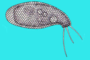
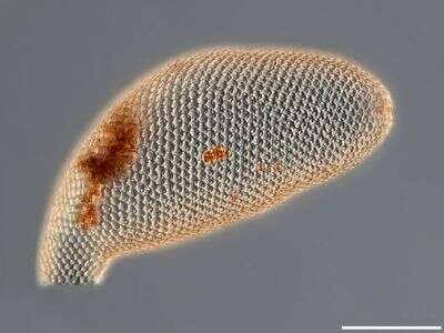
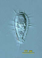
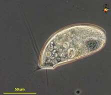
Euglypha (you-gligh-fah) filifera has an elongate shell that is composed of oval siliceous scales. The 15 micron long spines pointed out from the entire lateral edge of the shell. The nucleus is visible in the posterior third of the cell. Differential interference contrast.
-
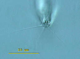
Euglypha (you-gligh-fah) filifera has an elongate shell that is composed of oval siliceous scales. The 15 micron long spines pointed out from the entire lateral edge of the shell. View of the aperture surrounded by denticulate scales. Long filopodia extended from the aperture. The aperture measures 6 microns in diameter. Differential interference contrast.
-
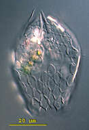
Euglypha (you-gligh-fah) filifera has an elongate shell that is composed of oval siliceous scales. The 15 micron long spines pointed out from the entire lateral edge of the shell. Image of Euglypha filifera lorica. The denticulate scales which border the circular aperture are visible. Each plate is measuring 6 X 8 microns on average. Differential interference contrast.
-
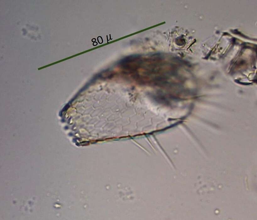
Euglypha (you-gligh-fah) filifera has an elongate shell that is composed of oval siliceous scales. The 15 micron long spines pointed out from the entire lateral edge of the shell. Apical view on the shell of Euglypha filifera. From this view it is evident that the shell is compressed and that the spines arise from the lateral edge of the shell. Differential interference contrast.
-
-
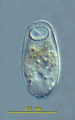
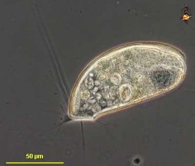
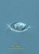
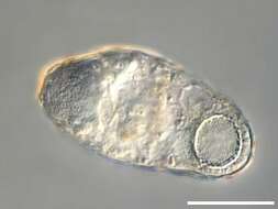
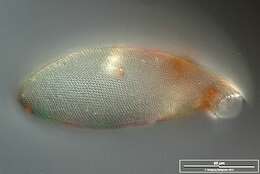
Trinema (try-knee-ma) enchelys, a testate amoeba. The focal plane on the circular aperture of the shell of Trinema enchelys. The aperture is located on the ventral surface and is surrounded by a number of rows of very minute scales. The central located contractile vacuole is visible and at the posterior end of the cell the macronucleus. This specimen was collected in a freshwater pond near Konstanz, Germany. Differential interference contrast.
-
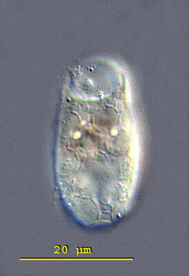
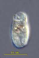
Trinema (try-knee-ma) enchelys, a testate amoeba. This specimen was collected in a freshwater pond near Konstanz, Germany. The focal plane is on the surface of the shell of Trinema enchelys. The shell is composed of transparent circular scales, each 5 - 6 mm in diameter. Differential interference contrast.
-
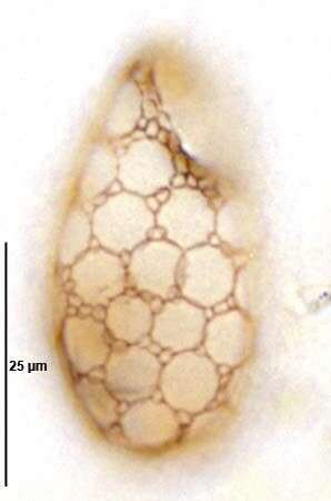
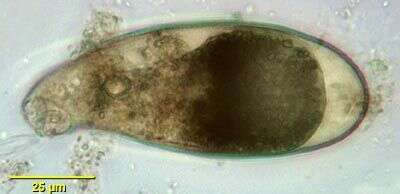
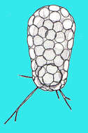
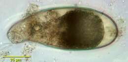
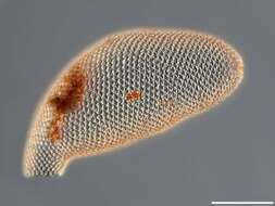
Trinema enchelys (EHRENBERG,1838) LEIDY,1878.Large incompletely overlapping circular shell plates with smaller peripherally placed oval shell plates. Collected from bottom sediments of an organically enriched freshwater pond near Boise, Idaho. December 2007. Protargol (see Foissner, W. Europ. J. Protistol., 27:313-330;1991).Brightfield.
-
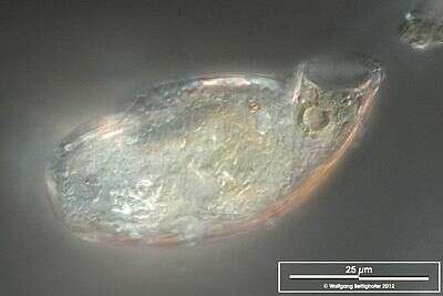
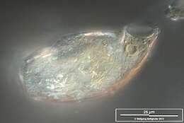
Scale bar indicates 25 µm. Sample from a wetland at the Pillersee (Tyrol, Austria). The image was built up using several photomicrographic frames with manual stacking technique. Images were taken using Zeiss Universal with Olympus C7070 CCD camera.Image under Creative Commons License V 3.0 (CC BY-NC-SA).
-
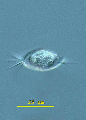
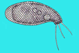
Cyphoderia (sigh-fo-dear-ee-a) is a shelled amoebae. This one has thin branching pseudopodia. The amoeba is protected by a loosely fitting lorica which has a single aperture through which the pseudopodia emerge. This form is thought to provide protection from the damaging effects of surface tension of water - something which many organisms living in the spaces between intertidal sand particles are likely to encounter as the tide goes out. Individual genera and species are mostly distinguished by the texture of the surface of the lorica. Not so common in marine habitats, more common in soils. Phase contrast.
-
Cyphoderia (sigh-fo-dear-ee-a) is a shelled amoebae. This one has thin branching pseudopodia. The amoeba is protected by a loosely fitting lorica which has a single aperture through which the pseudopodia emerge. The lorica of has thin scales attached to the external surface. These are evident towards the left of the picture. Phase contrast.
-
Cyphoderia (sigh-fo-dear-ee-a) is a shelled amoebae. This one has thin branching pseudopodia. The amoeba is protected by a loosely fitting lorica which has a single aperture through which the pseudopodia emerge. This is an empty shell. The shape of the lorica, and more importantly the texture of its surface and its composition are distinctive. Phase contrast.
-
-
Cyphoderia (sigh-fo-dear-ee-a) is a shelled amoebae. This one has thin branching pseudopodia. The amoeba is protected by a loosely fitting lorica which has a single aperture through which the pseudopodia emerge. This form is thought to provide protection from the damaging effects of surface tension of water - something which many organisms living in the spaces between intertidal sand particles are likely to encounter as the tide goes out. Individual genera and species are mostly distinguished by the texture of the surface of the lorica. Not so common in marine habitats, more common in soils. Phase contrast.
-
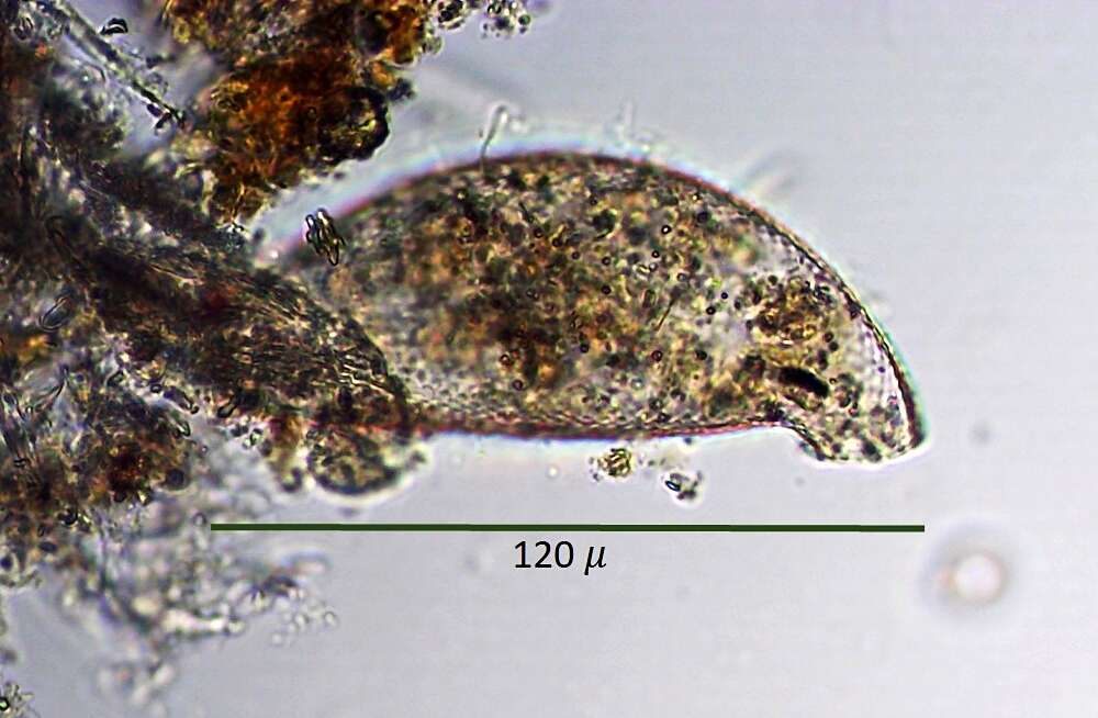
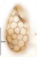
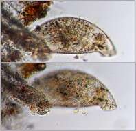
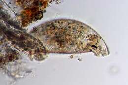
Flask shaped lorica with curved neck is composed of siliceous scales arranged on a chitinous membrane. Light brownish color. Cell body does not fill lorica completely. The circular pseudostome has a delicately scalloped edge. The s[pecies is probably ampulla. From freshwater pond sediment near Boise, Idaho. Composite brightfield image.
-
Scale bar indicates 25 µm. Sample from a pond situated in the region "High Fens" (Eifel highlands, Germany). The image was built up using several photomicrographic frames with manual stacking technique. Images were taken using Zeiss Universal with Olympus C7070 CCD camera.Image under Creative Commons License V 3.0 (CC BY-NC-SA).
-
Scale bar indicates 50 µm. Sample from a bog near Reith/Pillersee (Tyrol, Austria). The image was built up using several photomicrographic frames with manual stacking technique. Images were taken using Zeiss Universal with Olympus C7070 CCD camera.Image under Creative Commons License V 3.0 (CC BY-NC-SA).
-
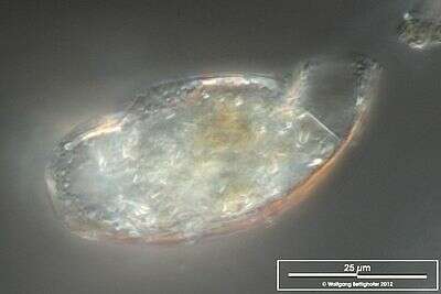
Scale bar indicates 25 µm. Sample from a bog near Reith/Pillersee (Tyrol, Austria). The image was built up using several photomicrographic frames with manual stacking technique. Images were taken using Zeiss Universal with Olympus C7070 CCD camera.Image under Creative Commons License V 3.0 (CC BY-NC-SA).
-
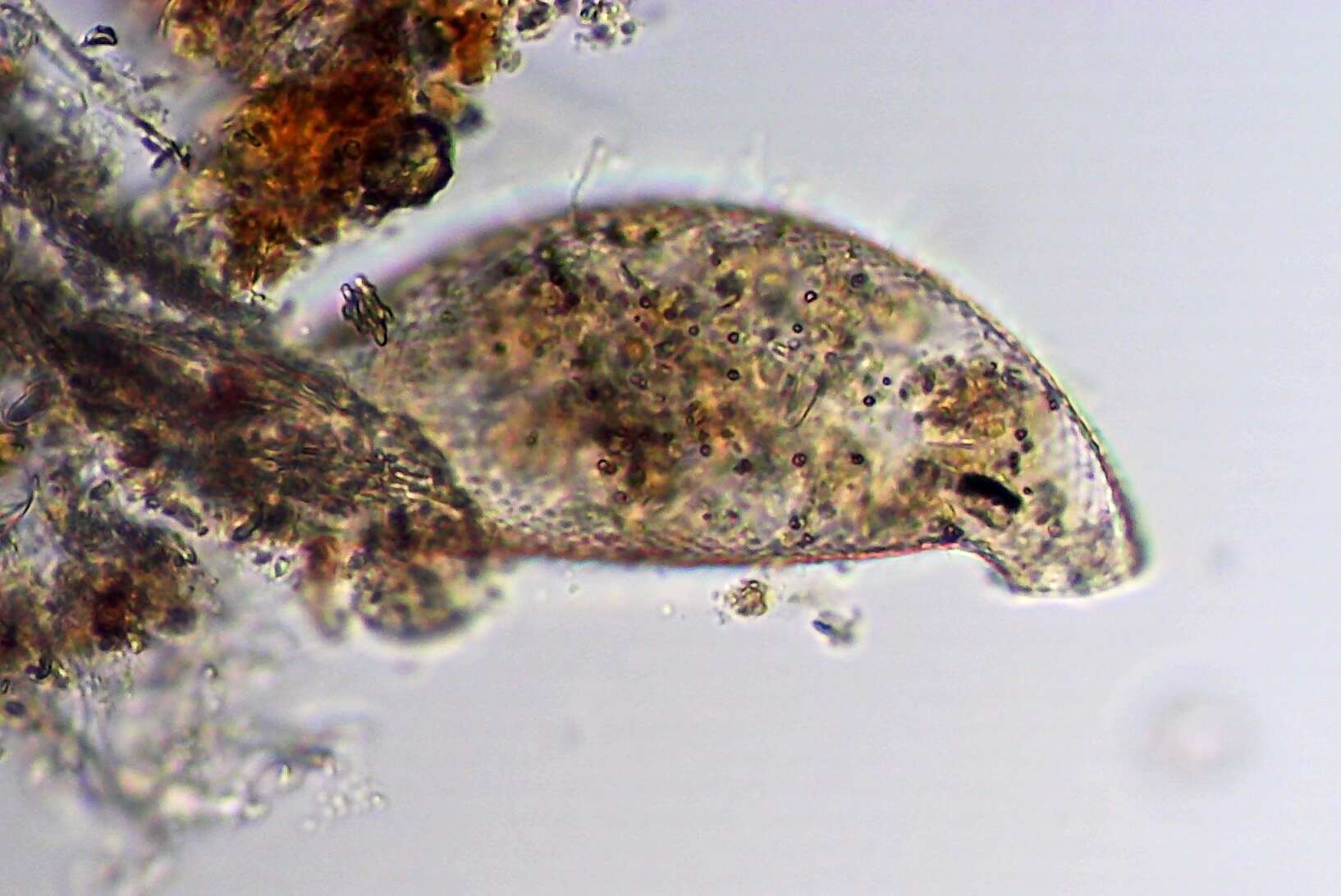
An optical cross-section. The edge at the end of the shell neck shows a temporary closure, possibly the beginning of an encystment. Scale bar indicates 25 µm. Sample from a bog near Reith/Pillersee (Tyrol, Austria). The image was built up using several photomicrographic frames with manual stacking technique. Images were taken using Zeiss Universal with Olympus C7070 CCD camera.Image under Creative Commons License V 3.0 (CC BY-NC-SA).
-
-
-
-