-
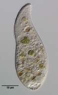
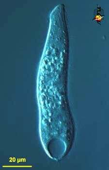
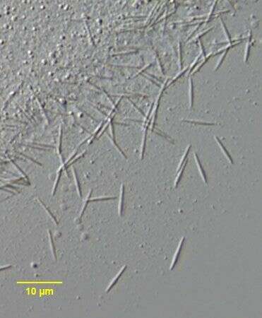
Collected from a non-flooded Petri dish culture of topsoil from a public park in Boise, Idaho. November 2006.DIC.
-
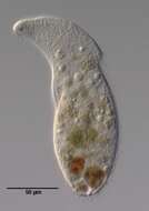
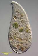
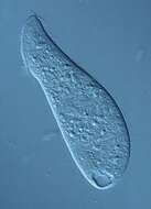
Spathidium (spa-thid-ee-um), a predatory ciliate. the mouth is the slightly expanded region at the front (top) of the cell and this is underlain with extrusomes which assist in the capture of food. The structure at the back end is the contractile vacuole. These guys usually eat other ciliates. Differential interference contrast. Material from Nymph Creek and Nymph Lake, thermal sites within Yellowstone National Park, photograph by Kathy Sheehan and David Patterson.
-
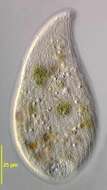
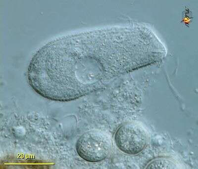
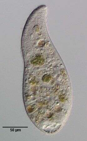
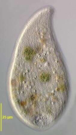
Spathidium (spa-thid-ee-um) is a predatory ciliate. The front end looks as if it is flattened, this is the mouth. Immediately inside the mouth are short extrusomes which are used to kill and capture food. May form cysts. Differential interference contrast.
-
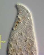
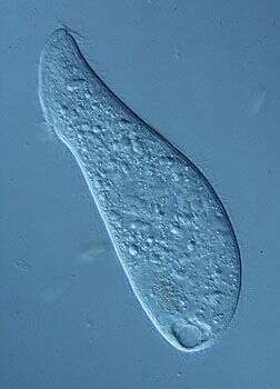
Differential interference contrast image, mouth to top.
-
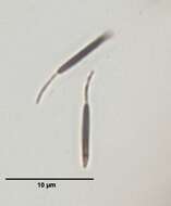
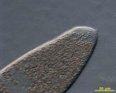
A litostome ciliate isolated from sandy sediments from Little Sippiwissett salt marsh. Micrograph taken by Jeffrey Cole.
-
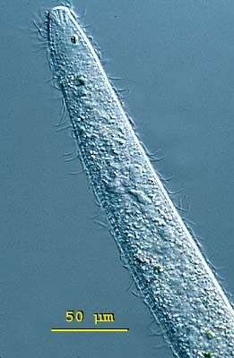
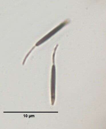
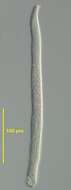
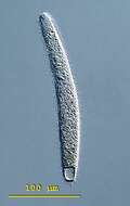
Spathidium (spa-thid-ee-um) moniliforme, the body is elongate, the posterior end is bluntly pointed or rounded but the anterior end is distinctively swollen - often fan-shaped and obliquely truncated. There is an ciliated apical ridge which is lined by toxicysts. The oral aperture is a slit that lies along the length of this ridge. The cilia are uniformly distributed in longitudinal parallel rows on both lateral surfaces. The macronucleus is highly variable, often elongate, ribbon-like or moniliform. The contractile vacuole is single and at the end of the cell. Spathidium feeds on other ciliates. It lives in fresh water ponds and lakes. This specimen was collected in a freshwater pond near Konstanz, Germany. This swimming cell is 250 microns long. Differential interference contrast.
-
Spathidium (spa-thid-ee-um) moniliforme, the body is elongate, the posterior end is bluntly pointed or rounded but the anterior end is distinctively swollen - often fan-shaped and obliquely truncated. There is an ciliated apical ridge which is lined by toxicysts. The oral aperture is a slit that lies along the length of this ridge. The cilia are uniformly distributed in longitudinal parallel rows on both lateral surfaces. The macronucleus is highly variable, often elongate, ribbon-like or moniliform. The contractile vacuole is single and at the end of the cell. Spathidium feeds on other ciliates. It lives in fresh water ponds and lakes. This cell is squashed allowing ribbon-like macronucleus and the fan-like arrangement of toxicysts at the front of the cell to be seen. Differential interference contrast.
-
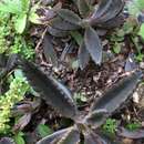
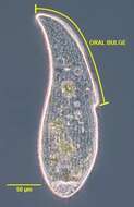
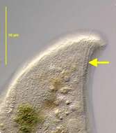
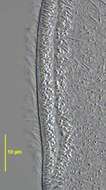
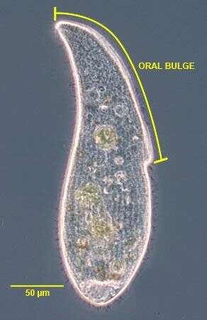
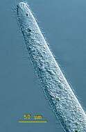
The long oral bulge (~50% of cell length) is one of the main distinguishing features of this subspecies of A. cultriforme. This specimen is somewhat stouter than the cells described by Foissner (Protistology 4 (1), 5-55 (2005) probably due to contraction after transfer from the culture dish to the slide. When observed undisturbed under the dissecting microscope the cells appear more slender.Phase contrast.
-
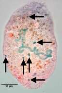
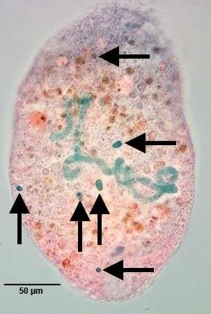
Six of the multiple (7-18) micronuclei are in the focal plane of this image. Stained by the methylgreen-pyroninY technique (see Foissner, W.Europ. J. Protistol.27:313-330;1991).Brightfield.
-
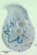
Band-form macronucleus of Arcuospathidum cultriforme scalpriforme (KAHL,1930) FOISSNER,2003.Stained by the methylgreen-pyroninY technique (see Foissner, W.Europ. J. Protistol.27:313-330;1991).Brightfield.
-
-
-
Stained by the silver carbonate technique (see Foissner, W.Europ. J. Protistol.27:313-330;1991).Brightfield.
-
-
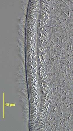
2 of the three dorsal brush rows are seen in this image.DIC.
-
-
-
-
-
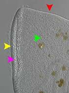
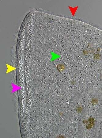
Complete circumoral kinety separate from somatic kineties (yellow). Extrusomes scattered randomly on each half of oral bulge (pink). extrusome (green). Clavate cilia of one of the dorsal brush rows (red).DIC.
-
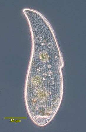
Arcuospathidium cultriforme scalpriforme (KAHL,1930) FOISSNER,2003. Phase contrast
-
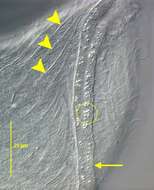
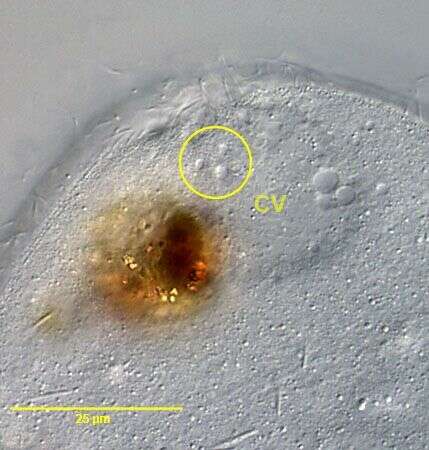
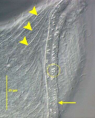
The posterior terminal contractile vacuole empties through multiple pores three of which are indicated within the yellow circle. DIC.
-
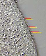
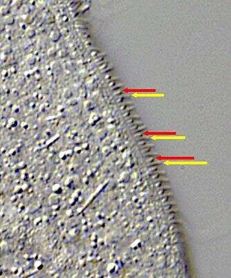
The dorsal brush rows have alternating longer and shorter cilia. In brush rows 1 and 2 the anterior cilium of each dikinetid is longer but in the 3rd dorsal brush row (seen here) the posterior cilium (yellow arrow) is longer than the anterior one (red arrow). DIC.
-
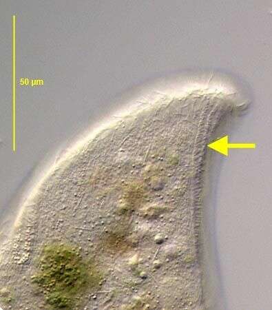
The oral bulge is indicated by the yellow arrow.The ends of the oral bulge extrusomes appear as bright spots here (yellow circle). Several right somatic kineties are indicated by the yellow arrowheads.The slit-like oral aperture is between the two halves of the oral bulge. DIC.