-
Ajamil, La Rioja, Spain
-
Ribadelago, Castille and Leon, Spain
-
Formentera, Balearic Islands, Spain
-
Illa de Arousa, A, Galicia, Spain
-
San Martn de Castaeda, Castilla y Len, Espaa
-
O Grove, Galicia, Spain
-
Tarragona, Catalunya, Espaa
-
Dehesa de Montejo, Castille and Leon, Spain
-
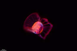
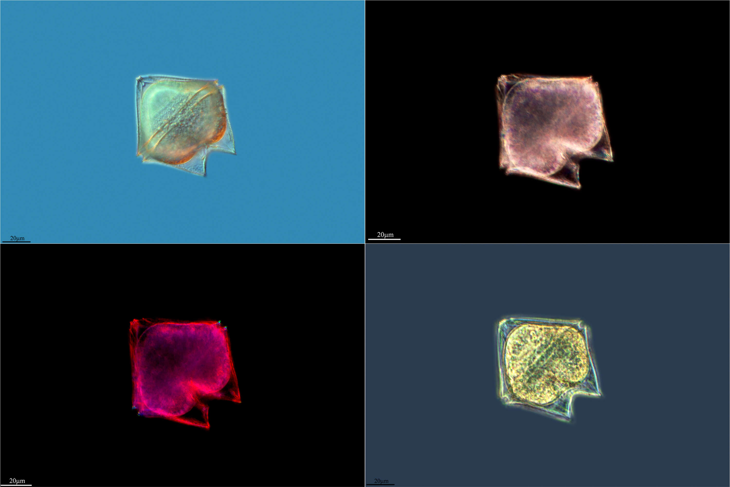
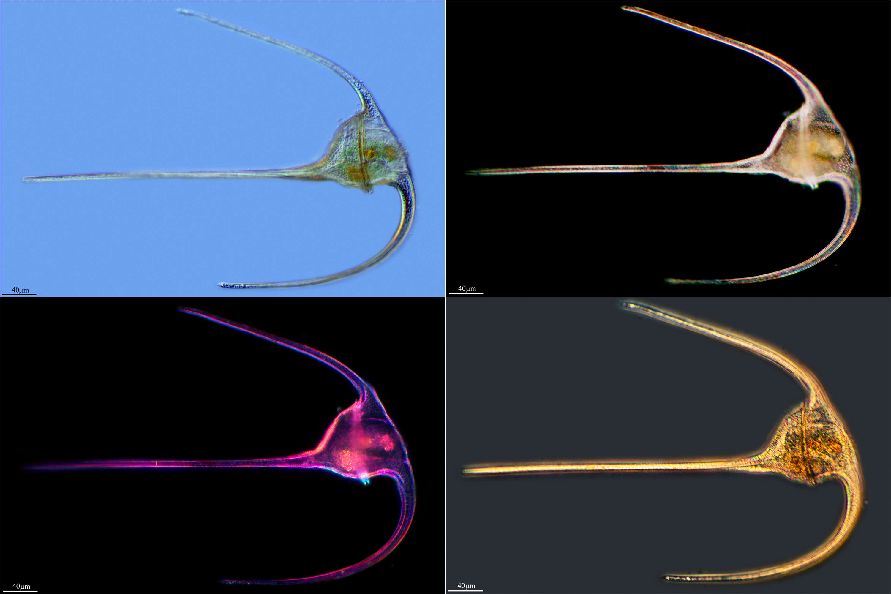
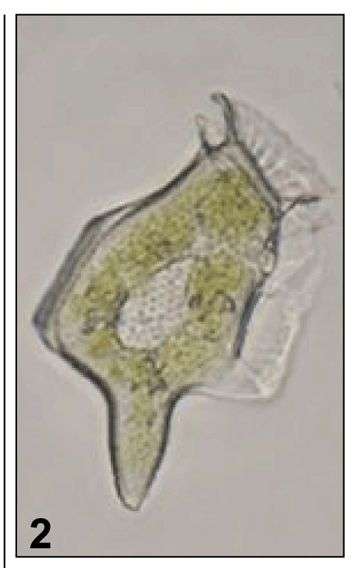
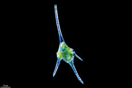
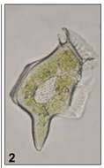
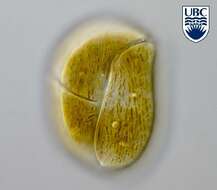
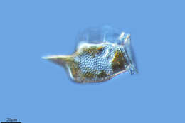
Fig 2: Dinophysis caudata Image of a live cell in lateral view
-
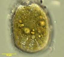
This is Amphidinium cf. glabrum in that it looks like but is not fully identical with the usual concept of this species.
-
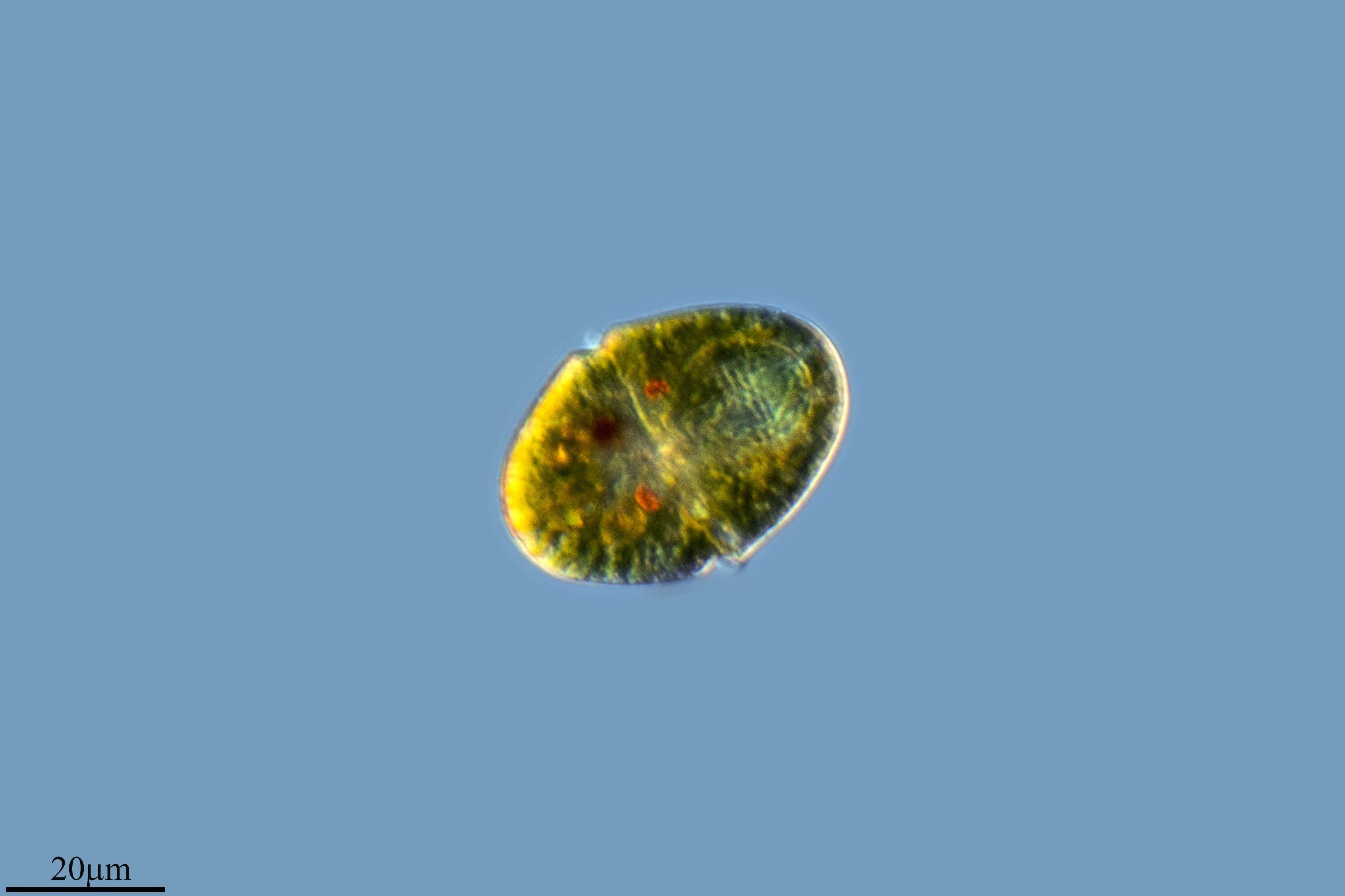
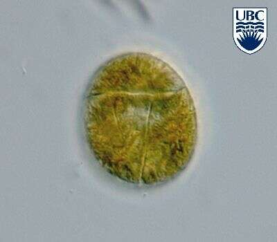
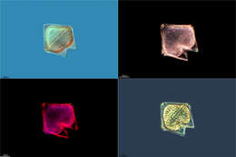
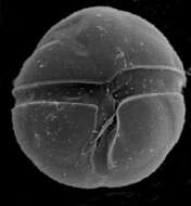
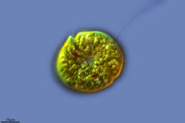
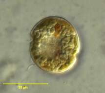
Durinskia baltica cells are round from the ventral side, slightly dorso-ventrally flattened, 16 - 28 microns long, 15 - 27 microns wide, and approximately 19 microns broad. Thecal plates present: an apical pore, a canal plate, 4 apical plates, 2 anterior intercalary plates, 7 precinguluar plates, 5 cinguluar plates, 4 sulcal plates, 5 postcingular plates, 2 posterior plates. Second apical intercalary plate much larger than the first and extending across the dorsal side. Cingulum 2-3 microns wide, consists of 5 plates, including a transitional plate. The large anterior sulcal plate forming a list over the left sulcal plate. Thecal plates smooth, with scattered pores (about 0.2 microns). Pores form a row along the edge of the cingulum. Nucleus round, in the centre or in the left side of the cell, approximately 7 microns. Reddish stigma, approximately 4 microns long, present in the sulcus. Yellow- brown plastids, 2 - 3 microns diameter, scattered throughout the cell.
-
Togula britannica (Herdman) Flo Jorgensen, Murray et Daugbjerg 2004
-
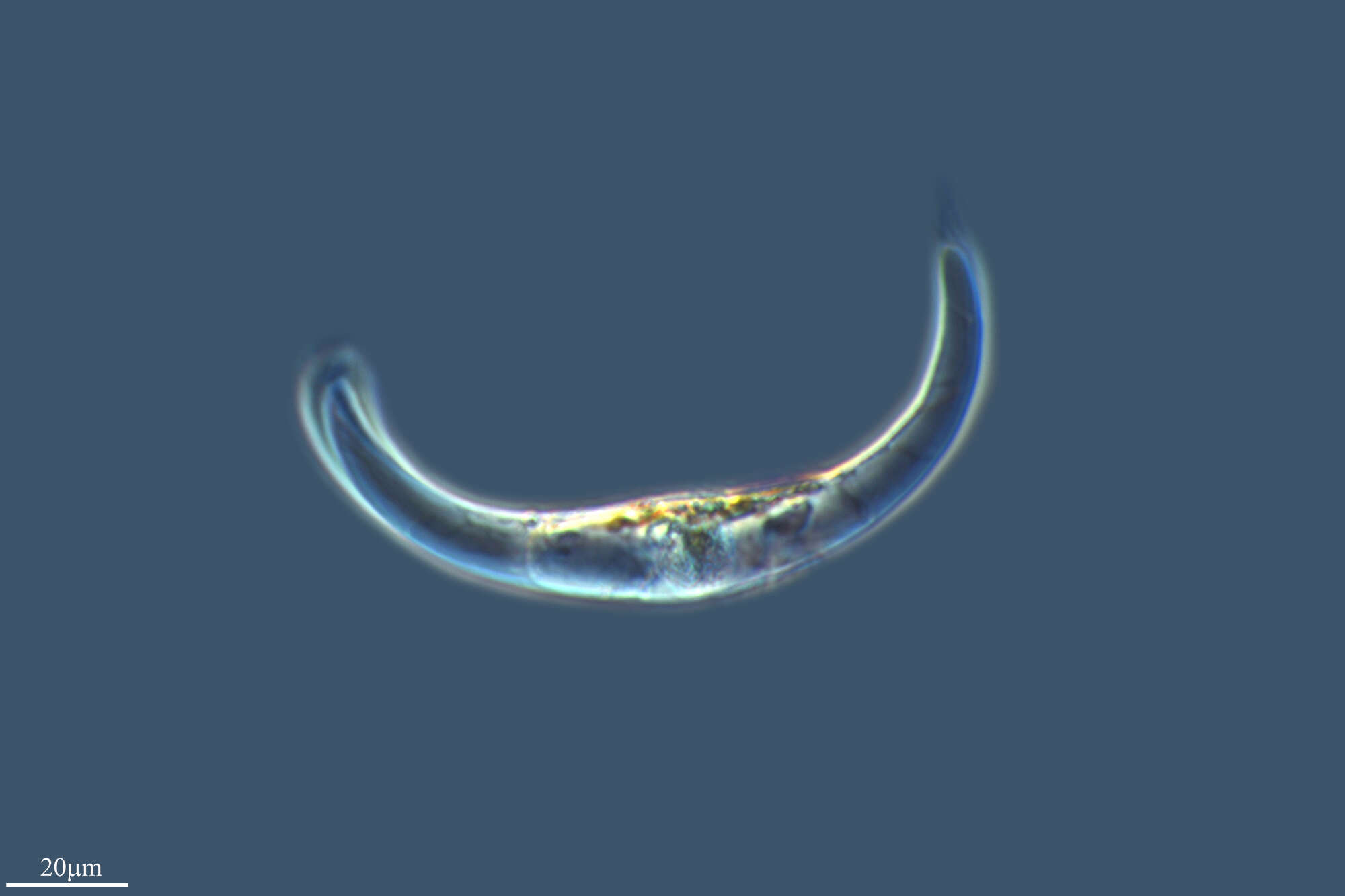
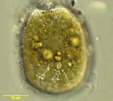
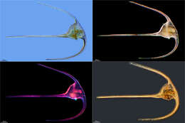
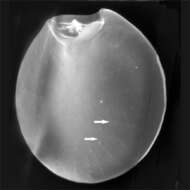
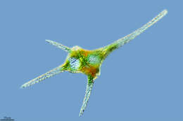
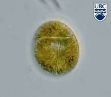
Prorocentrum clipeus cells are round, 37 - 44 long, 35 - 42 microns wide, length to width ratio 1.0-1.1. Apical area a wide rounded indentation, with a small list/collar present to the left side of the apical region. Small spine present, projecting from the apical region. Very small pores, approximately 0.1 microns diameter, present around the periphery of the valve and in short rows radiating towards the centre of the cell. Intercalary region with a horizontal banding pattern. Large yellow-brown plastid fills the cytoplasm. Large extrusomes (12 - 13 microns ) present in the anterior part of the valve, pointing towards the apical area. Nucleus large, approximately 20 microns by 10 microns , in the posterior part of the valve.
-
Galende, Castile and Len, Spain
-
Grove, O, Galicia, Spain
-
San Martn de Castaeda, Castilla y Len, Espaa
-
Formentera, Balearic Islands, Spain
-
Villar del Pedroso, Extremadura, Espaa
-
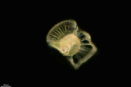
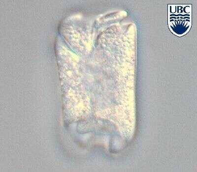
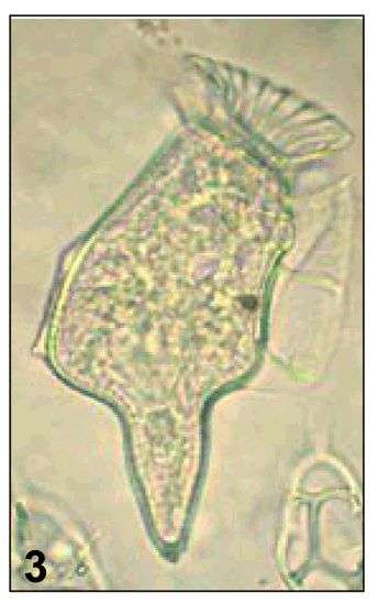
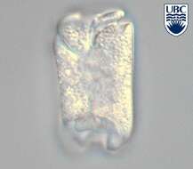
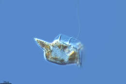
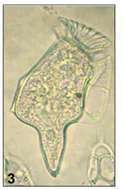
Fig 3: Dinophysis caudata Image of a Formalin preserved cell in lateral view
-
This is Amphidinium cf. glabrum in that it looks like but is not fully identical with the usual concept of this species.
-
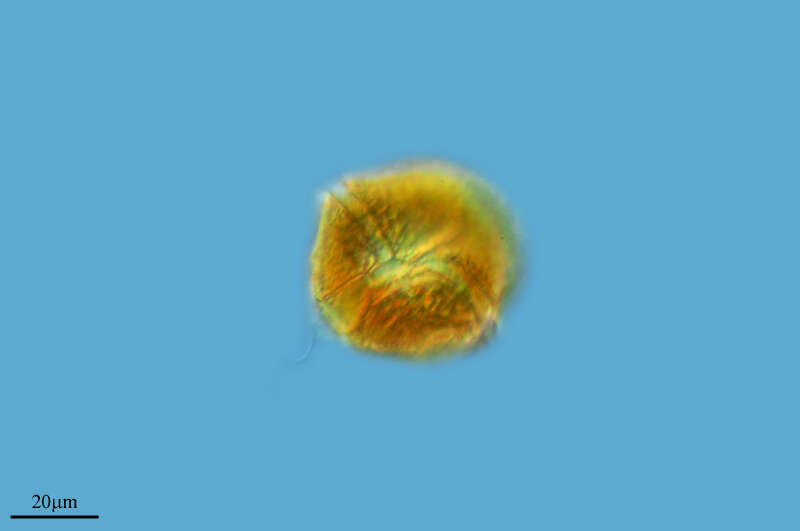
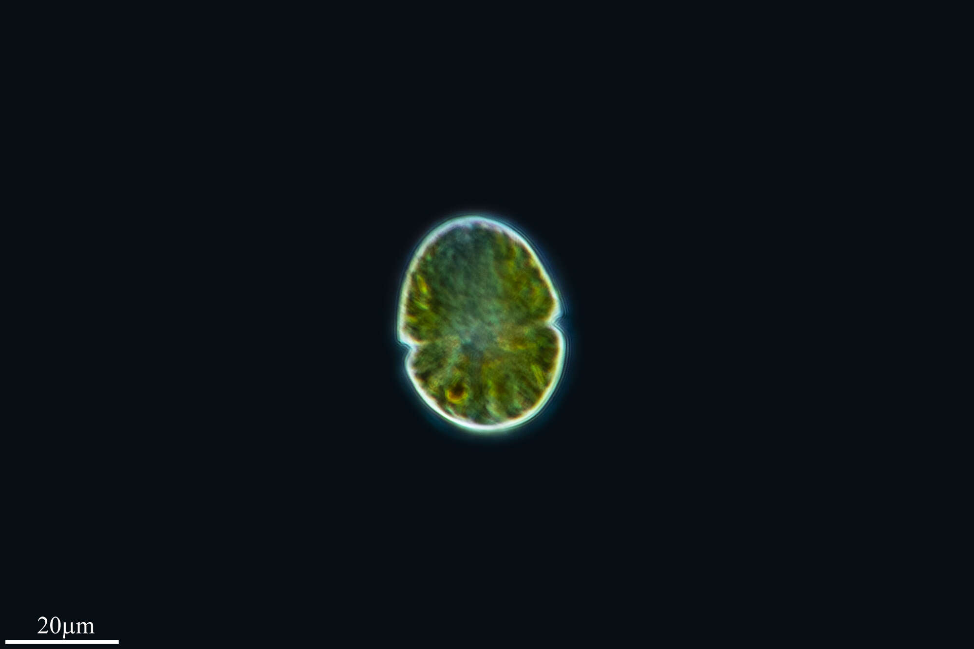
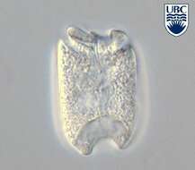
Durinskia baltica, observed in marine muds and sandy sediments in the vicinity of Broome, Western Australia in September 2003. This image was taken using differential interference contrast optics. This work was supported by the Australian Biological Resources Study.
-
Togula jolla Flo Jorgensen, Murray et Daugbjerg 2004
-
Prorocentrum emarginatum, showing plastids, observed in marine muds and sandy sediments in the vicinity of Broome, Western Australia in September 2003. This image was taken using differential interference contrast optics. This work was supported by the Australian Biological Resources Study.
-
Grove, O, Galicia, Spain