-
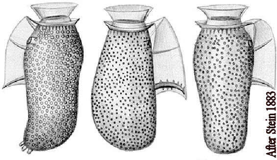
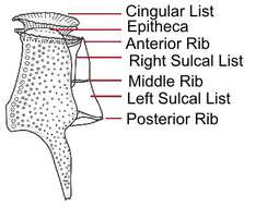
Fig 1: Dinophysis sacculus Schematic diagram (ventral view)from Stein 1883.
-
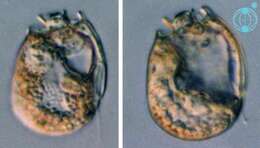
Sinophysis (sine-o-fi/fu-sis) ebriolum (Herdman) Balech 1956. The image on the left shows a cell in right lateral view. The cell is laterally compressed. The epicone is much smaller than the hypocone. The image on the right shows a cell in a mid-focal plane. The cells contain no plastids. The cells are thecate and have cingular lists.
-
Sinophysis (sine-o-fi/fu-sis) grandis Hoppenrath 2000. The image shows a rectangular cell in left lateral view. The cell is laterally compressed. The epicone is much smaller than the hypocone. The cell contains no plastids, but different coloured food particles are visible. The cell is thecate and has cingular lists.
-
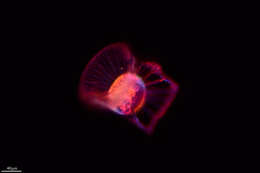
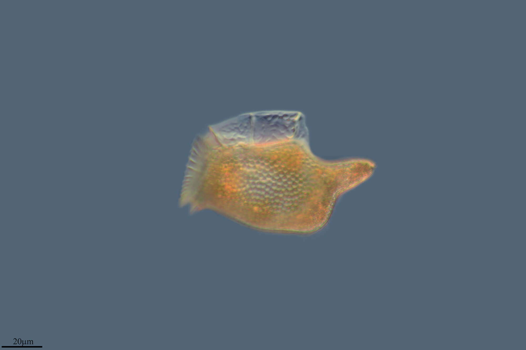
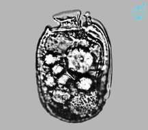
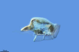
Dinophysis (die-know-fie-sis), common dinoflagellate in marine water column. This individual is clearly heterotrophic. Differential interference contrast.
-
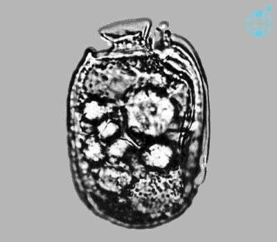
Sinophysis microcephalus observed in marine muds and sandy sediments in the vicinity of Broome, Western Australia in September 2003. This image was taken using phase contrast optics. This work was supported by the Australian Biological Resources Study.
-
Grove, O, Galicia, Spain
-
Formentera, Illes Balears, Espaa
-
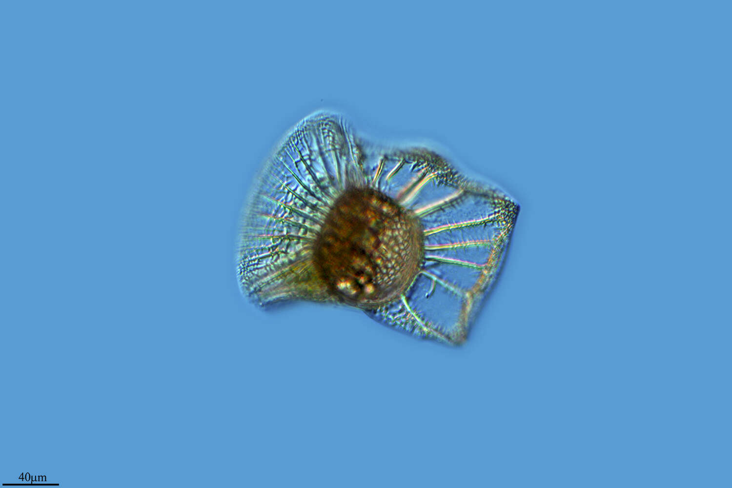
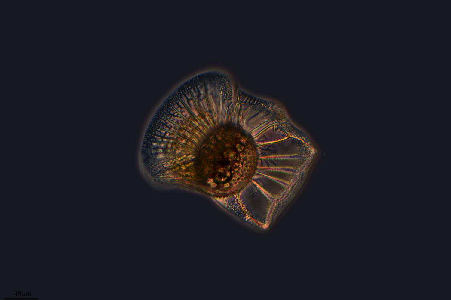
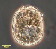
Histioneis elongata from the South Pacific, Tara Oceans Expedition station 111. Lugol's-fixed specimen, z-stack of images made using a 40x objective & DIC optics.
-
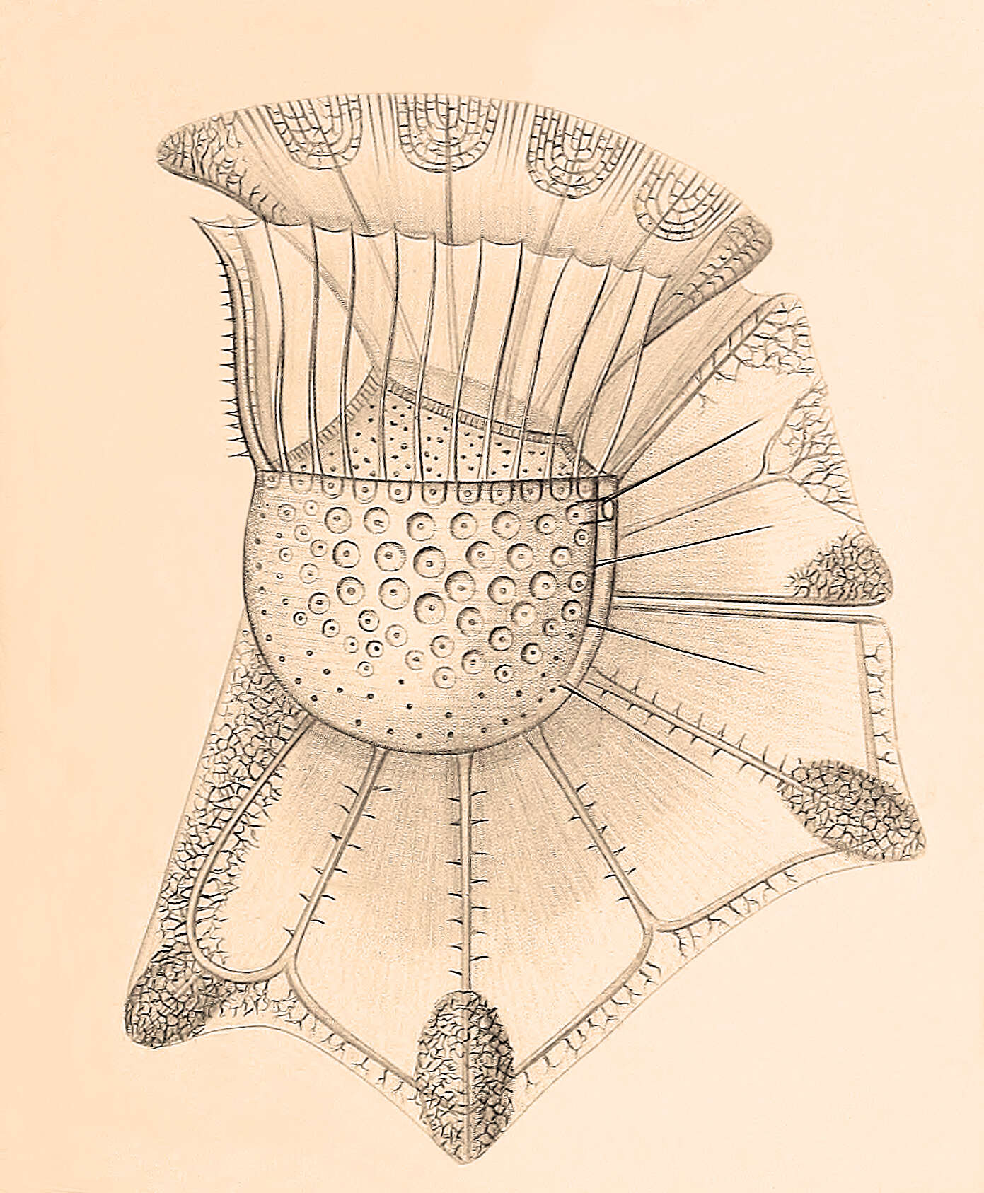
From the original species description: Figure 4 from plate 23 of Stein, F.R. (1883) Der Organismus der Arthrodelen Flagellaten. II. Hlfte. 30 pp & plates.
-
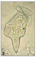
Dinoflagellate from the Ionian Sea in September 2010. Found in a sample from 74m depth.
-
Fig 1: Dinophysis caudata Schematic drawing of a cell in lateral view
-
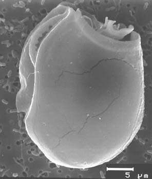
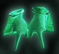
Sinophysis ebriolum, scanning electron microscope image. This image was taken by Mona Hoppenrath of a sample from Town Beach, Broome. This work was supported by the Australian Biological Resources Study.
-
Sinophysis (sine-o-fi/fu-sis) grandis Hoppenrath 2000. The image shows a cell in right lateral view. The cell is laterally compressed. The epicone is much smaller than the hypocone. The cell contains no plastids. The cell is thecate and has cingular lists.
-
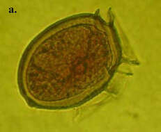
Two cells close to completing division.
-
Grove, O, Galicia, Spain
-
Formentera, Balearic Islands, Spain
-
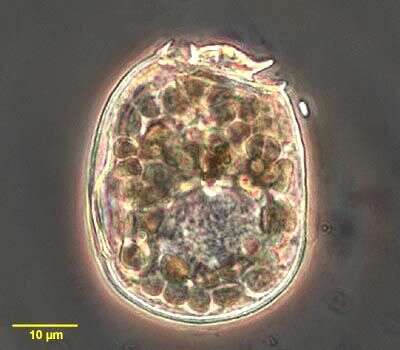
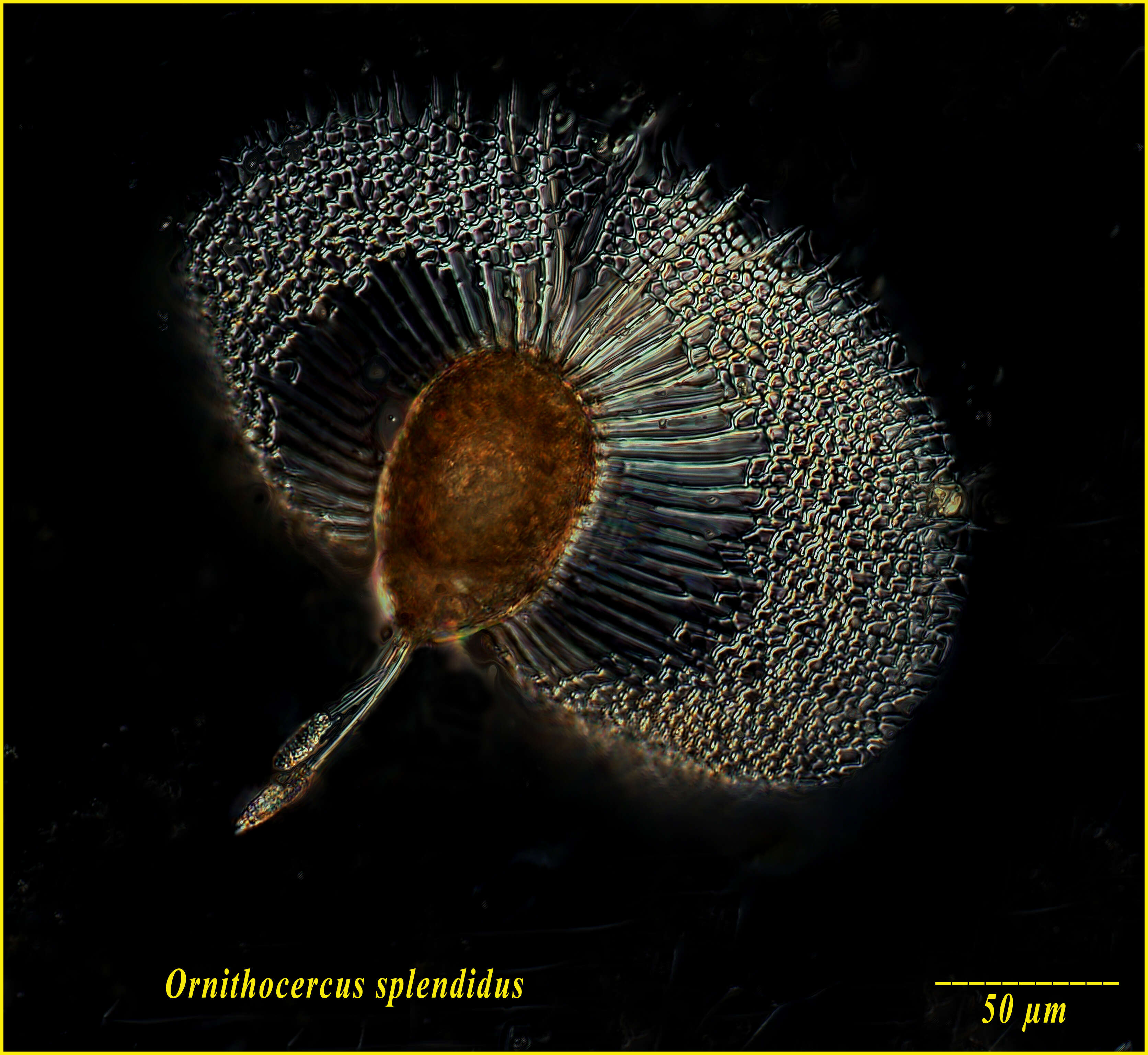
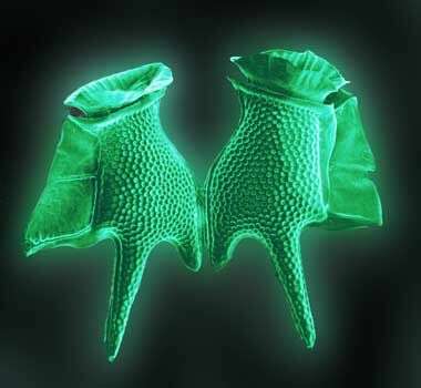
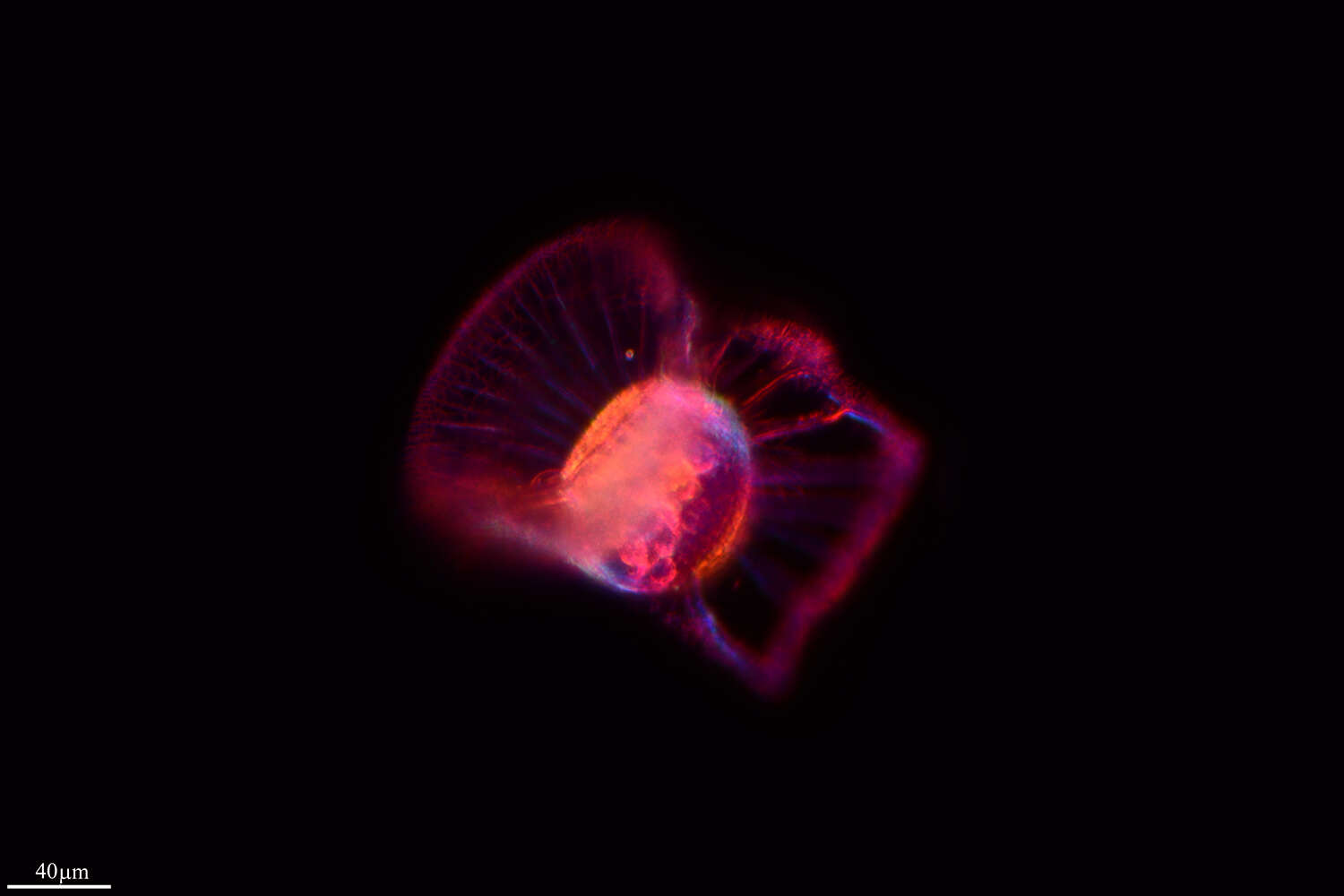
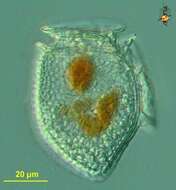
Ornithocercus magnificus from the Bay of Villefranche on Feb 18th 2014. The little orange balls are symbiotic cyanobacteria. Lugol's-fixed specimen, Z-stack of images made using a 40x objective and DIC optics.
-
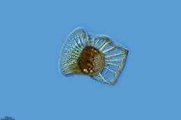
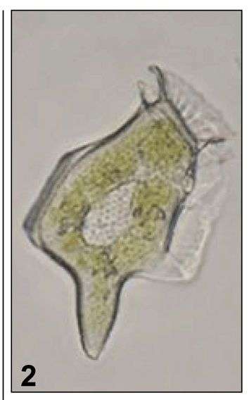
Fig 2: Dinophysis caudata Image of a live cell in lateral view
-
Sinophysis (sine-o-fi/fu-sis) stenosoma Hoppenrath 2000. The image shows a cell in right lateral view. The cell is laterally compressed. The epicone is much smaller than the hypocone. The cell contains no plastids. The cell is thecate and has cingular lists.
-
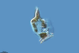
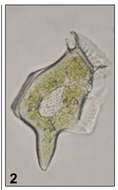
D. acuminata has an oval cell shape. The posterior end of the theca can bear small teeths. The left sulcal list is well developed and of a similar depth along its length. The pore structure is usually finer than in species such as D. norvegica, but is variable.
-
Grove, O, Galicia, Spain
-
Formentera, Balearic Islands, Spain
-
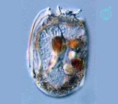
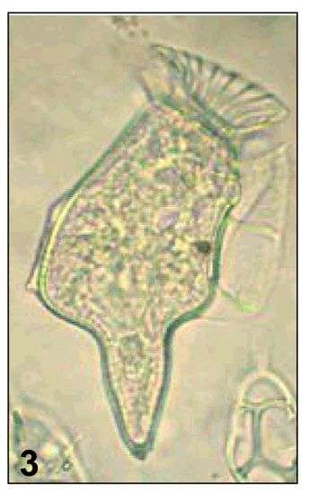
Fig 3: Dinophysis caudata Image of a Formalin preserved cell in lateral view
-
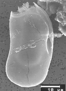
Sinophysis stenosoma, scanning electron microscope image. This image was taken by Mona Hoppenrath of a sample from Town Beach, Broome. This work was supported by the Australian Biological Resources Study.