-
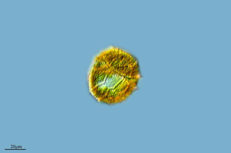
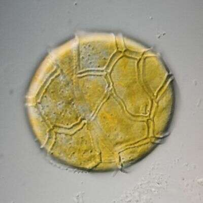
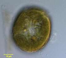
Bysmatrum, from the dorsal side, observed in marine muds and sandy sediments in the vicinity of Broome, Western Australia in September 2003. This image was taken using differential interference contrast optics. This work was supported by the Australian Biological Resources Study.
-
Sobrado, Galicia, Spain
-
Ribadelago de Franco, Castille and Leon, Spain
-
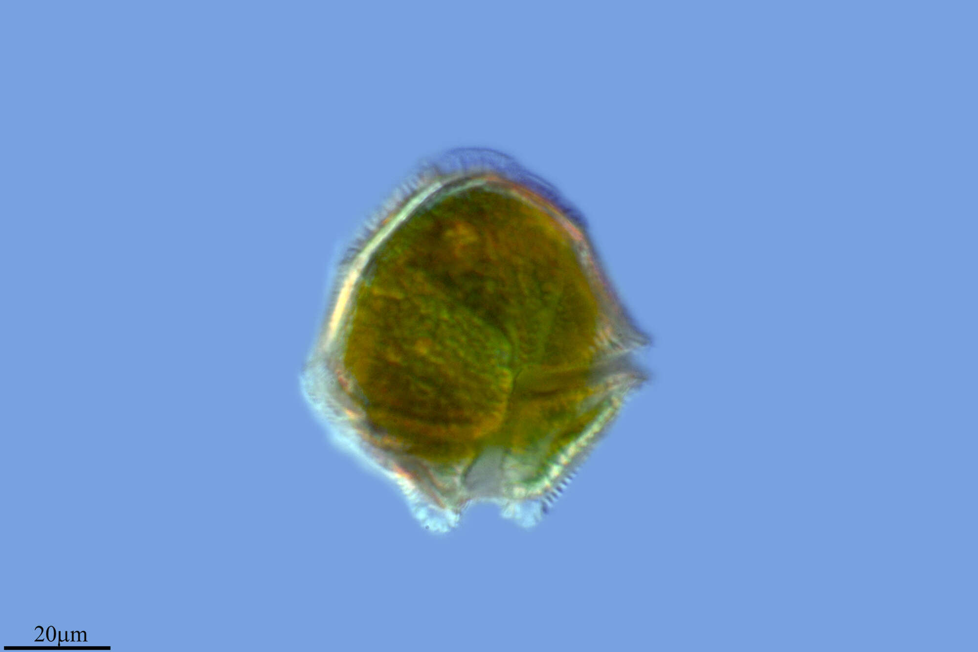
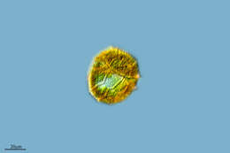
Bysmatrum, from the ventral side, observed in marine muds and sandy sediments in the vicinity of Broome, Western Australia in September 2003. This image was taken using differential interference contrast optics. This work was supported by the Australian Biological Resources Study.
-
Rabanera, La Rioja, Spain
-
Ribadelago de Franco, Castilla y Len, Espaa
-
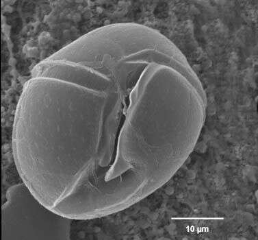
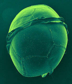
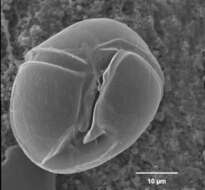
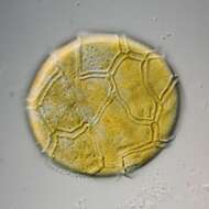
Bysmatrum observed in marine muds and sandy sediments in the vicinity of Broome, Western Australia in September 2003. This image was taken using scanning electron microscopy. This work was supported by the Australian Biological Resources Study.
-
Ajamil, La Rioja, Spain
-
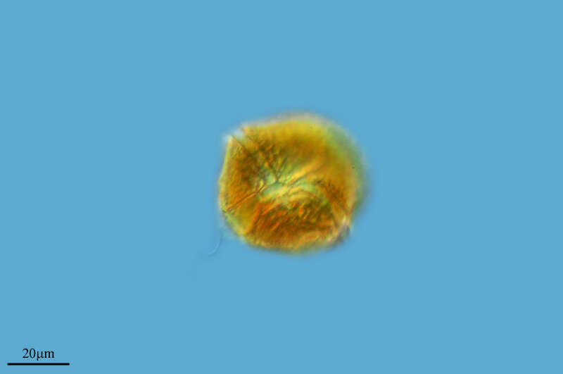
Peridinium (perry-din-ee-um) a dinoflagellate. This is one of the armoured dinoflagellate in which there are substantial skeletal elements in the cortical region of the cell. The groove which contains the circumferential flagellum has strongly developed margins. With chloroplasts. Differential interference contrast.
-
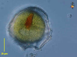
Peridinium (perry-din-ee-um) a dinoflagellate. This is one of the armoured dinoflagellate in which there are substantial skeletal elements in the cortical region of the cell. The groove which contains the circumferential flagellum has strongly developed margins. With chloroplasts. Encysted form - red inclusion resembles an eyespot but in this case we are advised this is more likely residues of food. Differential interference contrast.
-
Peridinium (perry-din-ee-um) a dinoflagellate. This is one of the armoured dinoflagellate in which there are substantial skeletal elements in the cortical region of the cell. The groove which contains the circumferential flagellum has strongly developed margins. With chloroplasts. Differential interference contrast.
-
-
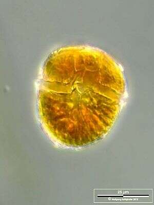
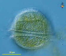
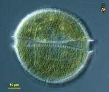
Ventral view of a dinoflagellate of the genus Peridinium (Ehrenberg,1832). Collected from a freshwater pond near Boise,Idaho. DIC
-
Dorsal view of the cell. The lists are the ridges on wirther side of the girdle.
-
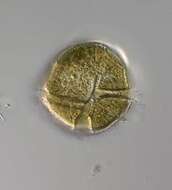
Peridinium penardii has skeletal elements (cellulose plates) in the cortical region of the cell. Species has chloroplasts. Collection from littoral region (stand of Phragmites) of a rain storage reservoir in Kiel (Schleswig-Holstein, Germany). This image was taken using Zeiss Universal with Olympus C7070 CCD camera.
-
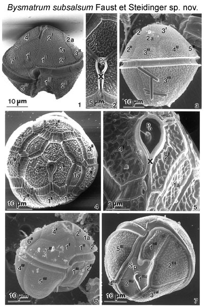
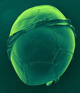
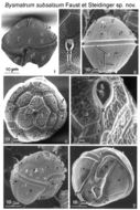
Figs 1-7. Lateral view of a cell with reticulated thecal surface, a conical epitheca, wide and deep, displaced cingulum, and a trapezoid hypotheca. The apical pore complex is situated ventrally. The apical plate 1' is asymmetric and pentagonal. The hypotheca is ventrally indented forming two lobes separating plates 2'" and 5'". The cingulum is displaced and finely striated with small pores aligned along the cingular lists. Fig. 2.The apical pore complex is a recessed chamber with a centrally located raised dome surrounded by a collar; it includes the apical pore plate (PO) and canal plate (X). Fig.3. Lateral view of a cell: a conical epitheca, wide and deep cingulum, and trapezoid hypotheca. Fig.4. Architecture of the epitheca including the position of the apical pore complex. Intercalary plates 2a and 3a are separated by plate 3'. The intercalary bands are striated.
EMu: Holotype SEM negative # 23040; SEM stub # ?; Field # 78-87; Accession # 407159; Catalog #1730; Figure # 1.
-
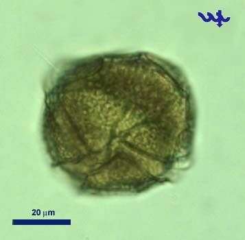
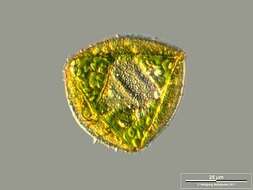
Scale bar indicates 25 µm. Sample from a wetland at the Pillersee (Tyrol, Austria). The image was built up using several photomicrographic frames with manual stacking technique. Images were taken using Zeiss Universal with Olympus C7070 CCD camera.Image under Creative Commons License V 3.0 (CC BY-NC-SA).
-
Cyst. Scale bar indicates 25 µm. Sample from a wetland at the Pillersee (Tyrol, Austria). The image was built up using several photomicrographic frames with manual stacking technique. Images were taken using Zeiss Universal with Olympus C7070 CCD camera.Image under Creative Commons License V 3.0 (CC BY-NC-SA).
-
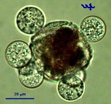
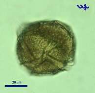
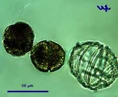
Peridinium gatunense is a large (ca. 50 um diameter) dinoflagellate, the most common bloom-forming species in Lake Kinneret, and the most studied species from this lake. Its winter-spring blooms give the water coffee-brown color. The blooms are patchy, the brown color patches can be observed from the distance. These blooms, reported to occur each spring since the 1950s, were characteristic of Lake Kinneret till the mid 1990s. In recent years Peridinium gatunense failed to bloom in some of the years, whereas in others its bloom was even more intense than recorded previously.
-
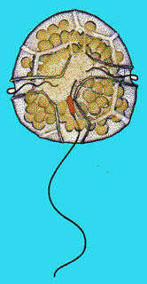
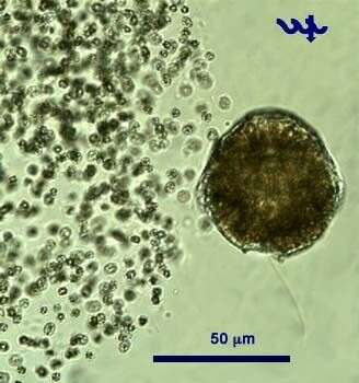
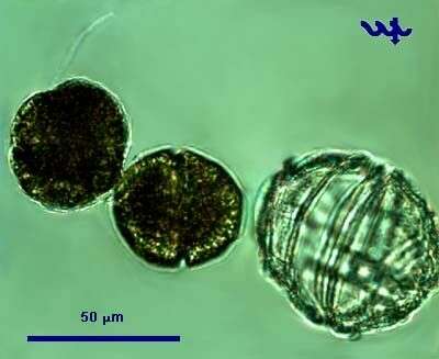
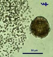
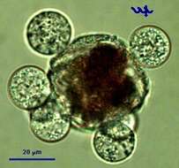
Peridinium gatunense, a large dinoflagellate (ca. 50 um diameter) is the main bloom-forming species in the plankton of Lake Kinneret. At the height of the bloom it forms patches in which cell densities are particularly high and the water gets coffee color. Peridinium has 2 unequal flagella, the longitudinal "whiplash" flagellum is seen in this picture, the second, transverse flagellum is hidden in the transverse groove or cingulum. A Microcystis colony is seen next to the Peridinium, note the large difference in cell size. This specimen was sampled in the littoral in June 2006.
-
A protoplast of Peridinium is leaving its thecae. Usually this happens during adverse conditions, e.g. lack of nutrients or when being subjected to very strong light - or as part of cell division. In this case, there is no cell division.
-
This cell of Peridinium gatunense is infected by several individuals of Phlyctochytrium sp, a chytrid fungus. Note that the Peridinium protoplasm is separated from the thecae and condensed, an indication that the cell is dead or dying. The fungal sporangia are at a developed stage with zoospores about to emerge out of in order to infect new Peridinium cells.
-
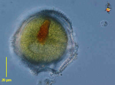
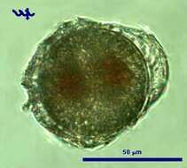
This cell of Peridinium gatunense is about to form a cyst in a process called encystation. Note the two red bodies within the protoplast, and the nearly perfect circular shape of the cyst emerging out of the thecae.
-
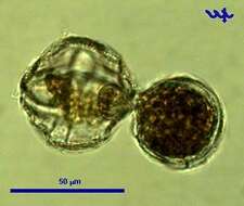
The two small daughter-cells have just emerged from the mother thecae. Note the considerably larger size of the empty, colorless thecae of the mother cell, note the flagellum of the cell on the left, note that both young cells are naked protoplasts, within a few hours they will be each covered by new thecae.