"The spionid Pygospio elegans reproduces both sexually and asexually. Using scanning electron and bright field microscopy, we examined morphogenesis following asexual reproduction to determine how "lost" body regions were generated after a worm spontaneously divided. Asexual reproduction occurred through transverse fission and divided the parent work into 2 to 6 fragments (architomy). All fragments retained their original anterior-posterior polarity. Regeneration in all fragments followed a specific series of events: wound healing (day 1); extension of blastema to generate lost body regions-specifically, the head and thorax for posterior fragments and the tail and pygidium for anterior fragments (day 2-3#; segmentation (days 3-6); and differentiation of segment- or region-specific structures (days 4-8). this pattern occurred regardless of where the original division took place. Subsequent growth occurred through addition of terminal setigers anterior to the pygidium followed by differentiation of tail setigers into abdominal setigers, leaving the tail region about 6 to 10 setigers in size. Fragments containing the original head (original mouth intact, generally much larger fragment) had a higher survivorship than fragments containing the original tail. Asexual reproduction has been widely reported in P. elegans, with most authors reporting its occurence or testing environmental factors that may influence rates of division. Pygospio elegans is a tubiculous polychaete that is common on mud and sand flats and has a cosmopolitan, temperate distribution. Adults grow to be 12mm long, and feed on detritus and phytoplankton. Pygospio elegans also reroduces sexually, and it exhibits considerable flexibility in reproduction, as both plantotrophic and adelphophagic (a farm of lecithotropy) larval development have been reported in worms from different populations (e.g., Thorson, 1946; Hannerz, 1956; Hobson and Green, 1968; Anger, 1984; Anger et al., 1986; Sholtzer-Schrerdhardst, 1991; Morgan et al., 1999)." (G. Gibson, J. Harvey, 2000).
"The overall body plan of Pygospio elegans is divided into four regions: the head, thorax, abdomen, and tail. The head is characterized by two ciliated palps, a prostomium with two or three pairs of eyes and paired nuchal organs. The thorax contains 10 to 12 abranchiate setigers, each with a single dorsal ciliary band, capillary notochaetae, and a lateral tuft of colia. Neurochaetae are simply capillary on setigers 1 to 8 and hooded hooks on setigers 9 to 12. The abdomen is 25 to 35 setigers in length. Each abdominal segment has paired branchiae and either a single (first few abdominal setigers) or double ciliary band, withh two closely apposed bands of tufted cilia. Abdominal setigers also have capillary notochaetae, a lateral tuft of cilia, and neurochaetae that are hooded hooks. The tail contains 6 to 12 a branchiate setigers. Tail setigers have capillary notochaetae, neurochaetae that are hooded hooks, and a lateral tuft of ciliar. There is a reduced ciliary band on the first few tail setigers only. The pygidium consists of four cirri, each with tufts of cilia on the inner surface. Male P. elegans have a pair of branchiae on nthe second setiger and dorsal organs on each setiger.
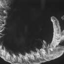
Morphogenesis following fission:
In all cases, fragments retained their original anterior-posterior polarity. Posterior fragments regenerated only the head and thorax, and anterior fragments regenerated only a new tail and pygidium. Subsequent growth involved elongation of the tail by the addition of terminal setigers. Segments are further developed in the posterior blastema as well, with 3 to 6 well-defined setigers, each with paired lateral pits in the region of the presumptive noto-and neurochaetae. Differentiation of the pygidium involves extension of the cirri and the appearance of small tufts of cilia on the inner surface. On the fifth day post-fission, the anterior blastema has regenerated the entire thoracic region and shows early differentiation of segment-specific structures. The number of thoracic setigers (10 to 12) that regenerated in the anterior blastema is similar in all specimens regardless of where fission occurred in the parent worm. The head has an elongate prostomium. The thoracic setigers develop neuropodial and notopodial buds, with a few small capially notochaetae and a small tuft of cilia between the neuropodium and the notopodium. The gut extends through the thorax, and the mouth is complete. On the same day, the 3 to 6 setigers of the posterior blastema also develop parapodial buds, a few notopodial capillary chaetae on setigers nearest the abdomen, and small lateral tufts of cilia. Regeneration on day 6 involves greater differentiation of segment-specific structures and addition of posterior setigers to restore the parental organization of the tail. The regenerated head has elongate, ciliated palps, a blunt prostomium, and 1 to 2 pairs of subdermal eyes. The thorax has dorsal bands of cilia on each setiger and well-developed notopodial chaetae throughout. Also in the thorax, the neuropodia exhibit short capillary chaetae on setigers 1-8 and a single hooded hook per setiger from setiger 8 poteriorly. The tail blastema has the 5 to 7 setigers characteristic of this reigion, with capillary notochaetae and notopodial hoooded hooks that decrease in number from three on the proximal, earliest-forming setiger, to one on the later-developing terminal setiger. Lateral tufts of cilia are present on all setigers. The pygidium has cirri that are mature in size and have well-developed tufts of cilia. By day 7, the anterior blastema has regenerated a head and thorax that are identical to those of parent worm except in setiger size and number of chaetae. Subsequent development in this region involves an increase in setiger size but not number. In the tail, setiger size and chaetae number also increases. By day 8, the tail regenerated thorax and tail have an increased number of chaetae, and are similar to the pre-fission organization except for setiger size. Also on day 8, the gut extends through the new tail to the pygidium. In all fragments, regeneration produces only specific body regions, regardless of where fission occurred in the parents. Anterior fragments regenerate only the pygidium and the 6 to 12 abranchiate setigers of the tail. Posterior fragments regenerate only the thorax (10 to 12 setigers) and head. Mid-worm fragments concurrently regenerate both anterior and posterior regions as described above, with the result that these gradments regenerate the head and thorax and tail and pygidium but not the abdoment, regardless of the original fragment. After regeneration, worms grow to their pre-fission size by increasing setiger size and setiger number. During the growth phase, new setigers will only form immediately anterior to the pygidium; new setigers do not form in thorax or abdomen once regeneration is complete. Newly formed terminal setigers develop chaetae and parapodial lobes typical of the tail region. As the tail region increases in setiger number, anterior tail setigers differentiate into abdominal setigers by forming dorsal ciliary bands and branchiae buds." (G. Gibson and M. Harvey, 2000).
Pygospio elegans is a species of marine polychaete worms in the family Spionidae. It is found in Western Europe (France, Belgium, and The Netherlands).
Pygospio elegans is a species of marine polychaete worms in the family Spionidae. It is found in Western Europe (France, Belgium, and The Netherlands).