-
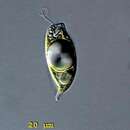
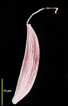
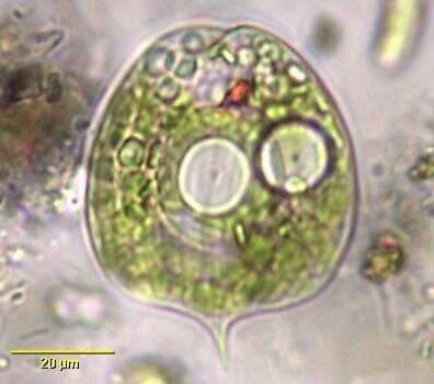
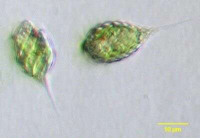
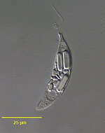
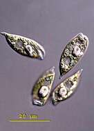
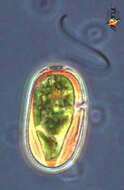
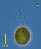
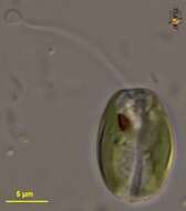
Phacus (fake-us) is a genus of autotrophic euglenids. It can be distinguished from Euglena by being flattened or twisted and rigid. The most species of the genus are flat and leaf-shaped often with ridges or fins running helically or longitudinally. The chloroplasts are small, discoid and pyrenoids are usually absent. There is a single red eyespot and one locomotive flagellum. Phacus contains conspicuous paramylon bodies, mostly with one ore two large bodies in the centre of the cell. The genus is common in freshwater ecosystems, in muds and associated with detritus. This specimen was collected in freshwater ponds near Konstanz, Germany. Phacus skujai is a small member of the genus. The cells are slender and ovoid. The large paramylon body (often accompanied by a second smaller body) in the mid-cell and the short spine point to the ventral side are said to be distinctive for the species. 28 µm.
-
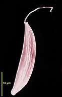
Portrait of the colorless euglenoid flagellate, Menoidium bibacillatum (Pringsheim, 1942). Strongly flattened. One side curved with the other more straight. One emergent flagellum. Stigma absent. Paramylon bodies are dimorphic with smaller round and larger elongate ring forms. Swims rotating on long axis. Highly refractile. From standing freshwater near Boise, Idaho. DIC.
-
-
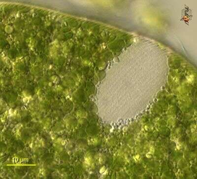
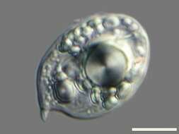
Scale bar indicates 10 µm. Sample from the pond Hegne Moor situated in the vicinity of Lake Constance. The image was built up using several photomicrographic frames with manual stacking technique. Images were taken using Zeiss Universal with Olympus C7070 CCD camera.Image under Creative Commons License V 3.0 (CC BY-NC-SA).
-
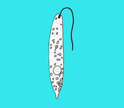
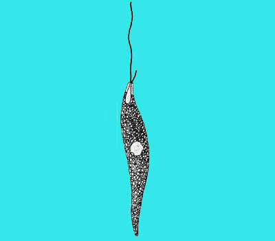
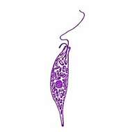
Distigma (die-stig-ma), heterotrophic euglenid flagellate. There are two flagella which are attached to the cell in a flagellar pocket which is an invagination that leads to the front of the cell by a tube called the flagellar canal. The light disc near the front is the contractile vacuole which is located alongside the flagellar pocket which cannot be seen in this image. One flagellum is long, the other short. The cytoplasm has large amounts of paramylon granules. The cell can squirm (is metabolic). Phase contrast.
-
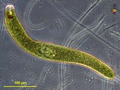
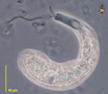
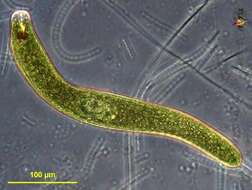
Euglena ehrenbergii. Cell observed in freshwater habitats in the vicinity of Broome, Western Australia in September 2003. This image was taken using phase contrast optics. This work was supported by the Australian Biological Resources Study.
-
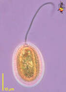
Phacus (fake-us) is a genus of autotrophic euglenids. It can be distinguished from Euglena by being flattened or twisted and rigid. The most species of the genus are flat and leaf-shaped often with ridges or fins running helically or longitudinally. The chloroplasts are small, discoid and pyrenoids are usually absent. There is a single red eyespot and one locomotive flagellum. Phacus contains conspicuous paramylon bodies, mostly with one ore two large bodies in the centre of the cell. The genus is common in freshwater ecosystems, in muds and associated with detritus. This specimen was collected in freshwater ponds near Konstanz, Germany. Phacus skujai is a small member of the genus. The cells are slender and ovoid. The large paramylon body (often accompanied by a second smaller body) in the mid-cell and the short spine point to the ventral side are said to be distinctive for the species. These cells are 'starved', the large paramylon body is considerably reduced and the nucleus is visible. 28 µm.
-
Portrait of the colorles euglenoid flagellate,Menoidium bibacillatum (Pringsheim, 1942). Strongly flattened. One side curved with the other more straight. One emergent flagellum. Stigma absent. Paramylon bodies are dimorphic with smaller round and larger elongate ring forms. Swims rotating on long axis. Highly refractile. From standing freshwater near Boise, Idaho. Phase contrast.
-
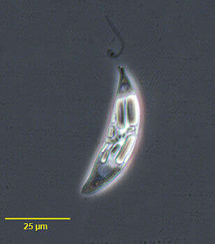
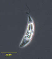
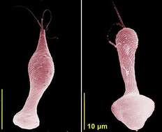
Distigma sennii Pringsheim, 1942. Metabolic swimming euglenid, cell typically club-shaped, broader anteriorly and truncated with an apical canal-opening, the posterior end of cell tapers, cells are 40 - 62 microns long. With two flagella, unequal in length. The longer flagellum is about 0.25-0.35 cell length and the recurrent flagellum is very short (5 - 6 microns), slightly curved and directed sideways or to the posterior of the cell. The pellicle seems smooth. The reservoir is in the middle of the cell with an associated contractile vacuole. A nucleus is located centrally or slightly behind the centre of the cell. The cell contains a large number of ellipsoidal or cylindical refractile grains and when swimming the cell moves quickly and rotates.
-
-
Euglena ehrenbergii. Cell observed in freshwater habitats in the vicinity of Broome, Western Australia in September 2003. This work was supported by the Australian Biological Resources Study.
-
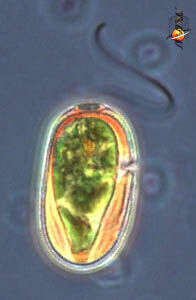
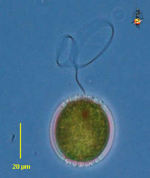
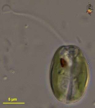
Trachelomonas (track-ell-owe-moan-ass) is an autotrophic euglenid flagellate. The genus is distinguished from others because the cells live within a loose fitting lorica with a small opening. The lorica of many species is elaborated with spikes or spines. The lorica accumulates metal salts with age, becoming brown and brittle and often obscuring the bright green colour of the chloroplasts within the cell. Red eyespot evident as dark region near the anterior of the cell. There is one emergent flagellum which emerges from the opening of the lorica. After division of the daughter cells will emerge from the opening of the lorica. Most freshwater but occasionally found in brackish habitats. Phase contrast.
-
Scanning electron micrograph showing the anterior flagellum and the cell shape.
-
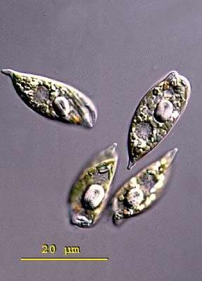
Phacus pleuronectes, euglenoid flagellate with a rigid, leaf-shaped pellicle with fine longitudinal striations and short curved spinous posterior. Many discoid chloroplasts. Usually with one large circular central paramylon body (although two are seen in this individual). Red stigma. From freshwater pond near Boise, Idaho. Brightfield
-
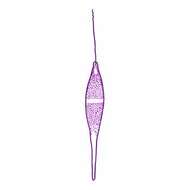
Distigma proteus Ehrenberg, 1838. Body highly metabolic, scarcely ever presenting the same contour, usually more or less elongate, with irregular constrictions and distensions, longer flagellum nearly equaling the body in length, the shorter one scarcely one quarter that length, endoplasm transparent, enclosing numerous dark coloured refringent corpuscles whose positions are constantly shifting from one extremely to the other in accordance with the peristaltic motions of the body, two minute, blackish, eye-like pigment-spots usually developed at the anterior extremity, tubular pharynx slender, greatly prolonged, contractile vacuole conspicuous, located close to the termination of the pharynx, nucleus ovate, subcentral. Length 44-106 microns
-
Euglena ehrenbergii. Cell observed in freshwater habitats in the vicinity of Broome, Western Australia in September 2003. This image was taken using differential interference contrast optics. This work was supported by
The Australian Biological Resources Study
-
Trachelomonas (track-ell-owe-moan-ass) is an autotrophic euglenid flagellate. The genus is distinguished from others because the cells live within a loose fitting lorica with a small opening. The lorica of many species is elaborated with spikes or spines. The lorica accumulates metal salts with age, becoming brown and brittle and often obscuring the bright green colour of the chloroplasts within the cell. Red eyespot evident as dark region near the anterior of the cell. There is one emergent flagellum which emerges from the opening of the lorica. After division of the daughter cells will emerge from the opening of the lorica. Most freshwater but occasionally found in brackish habitats. Phase contrast.
-
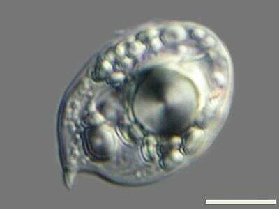
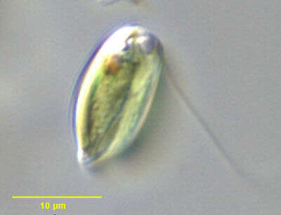
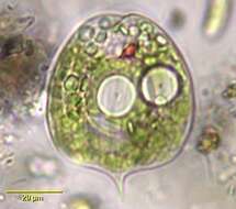
Cryptoglena pigra. Although Cryptoglena has been considered a euglenid flagellate the genus should probably be considered of uncertain affinity. The cells are solitary and have a single emergent flagellum slightly longer than the cell body. The cell body is rigid, dorsoventrally flattened and ovoid in outline with rounded anterior and bluntly pointed posterior. There is an obvious longitudinal furrow seen well in this image. Described as having one or two laminate parietal chloroplasts without pyrenoids. The posterior nucleus is not visible in this image. The prominent anterior stigma is seen well here. The genus has been described as monospecific by some authors and as containing five or six species by others. From a slow flowing freshwater stream near Boise, Idaho. Differential interference contrast illumination.
-
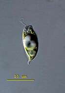
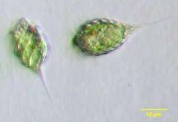
Phacus pyrum or P. rudicula, a small euglenoid flagellate having a pyriform rigid pellicle thrown into folds (P. pyrum is described as round in cross section while P. rudicula is said to be more flattened as is the cell shown here, but it is likely the two are different forms of the same species). With oblique ridges and tapering, pointed posterior. Red stigma. These individuals have shed their flagella which are usually about 1½ body length. From freshwater pond near Boise, Idaho. Oblique illumination.
-
Distigma proteus clavatum Playfair, 1921. Cells are 18-44 microns long and 8-12 microns wide. see Menoidium pellucidum var clavatum.
-
Phacus agilis. Cell observed in freshwater habitats in the vicinity of Broome, Western Australia in September 2003. This image was taken using differential interference contrast optics. This work was supported by the Australian Biological Resources Study.
-
Trachelomonas (track-ell-owe-moan-ass) is an autotrophic euglenid flagellate. The genus is distinguished from others because the cells live within a loose fitting lorica with a small opening. The lorica of many species is elaborated with spikes or spines. The lorica accumulates metal salts with age, becoming brown and brittle and often obscuring the bright green colour of the chloroplasts within the cell. Red eyespot evident as dark region near the anterior of the cell. There is one emergent flagellum which emerges from the opening of the lorica. After division of the daughter cells will emerge from the opening of the lorica. Most freshwater but occasionally found in brackish habitats. Phase contrast.
-
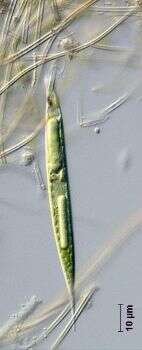
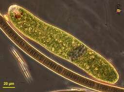
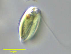
Portrait of euglena acus (Ehrenberg,1830). Cell body is an elongate cylinder with a sharply pointed posterior end. The flagellum is about one-fourth the body length; Delicate pellicular striations are difficult to see without DI. The small discoid chloroplasts are numerous. Paramylon bodies are rod-shaped. The nucleus iscentral. The stigma is prominen. Moves sluggishly among debris. Collected from the slow-moving organically enriched outflow from a pond near Boise, Idaho. DIC.
-
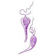
Scanning electron micrographs showing two stages of contraction (metaboly).