The species-rich phylum Microsporidia includes obligate intracellular parasites that infect a wide range of vertebrate and invertebrate hosts. Microsporidians have evolved an elaborate mechanism for invading animal host cells, but have otherwise greatly reduced biological complexity. The smallest known autonomous nuclear genome is that of a microsporidian, Encephalitozoon intestinalis (Keeling and Corradi 2011). Although the taxonomic affiliation of the Microsporidia has long been controversial, they are now known to either fall within the Fungi or to be extremely closely related to the Fungi.
Microsporidians produce resistant spores and possess a unique organelle, the polar tubule, or polar filament, which is coiled inside the spore (see below). The spores of microsporidians known to infect humans range from 1 to 4 microns in diameter. At least 14 microsporidian species have been identified as human pathogens (see Table from U.S. CDC): Brachiola algerae, B. connori, B. vesicularum, Encephalitozoon cuniculi, Enc. hellem, Enc. intestinalis (formerly known as Septata intestinalis), Enterocytozoon bieneusi, Microsporidium ceylonensis, M. africanum, Nosema ocularum, Pleistophora sp., Trachipleistophora hominis, T. anthropophthera, and Vittaforma corneae. Some domestic and wild animals may be naturally infected with Enc. cuniculi, Enc. intestinalis, and Ent. bieneusi (e.g., Santin and Fayer 2011). Birds, especially parrots (parakeets, lovebirds, budgies) are naturally infected with Enc. hellem. Enterocytozoon bieneusi and V. corneae have been identified in surface waters and spores of "Nosema" (probably B. algerae) have been identified in ditch water.
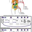
The infective form of microsporidians is the resistant spore, which can survive for long periods in the environment. The spore extrudes its polar tubule and infects the host cell. The spore injects the infective sporoplasm into the eukaryotic host cell through the polar tubule. Inside the cell, the sporoplasm undergoes extensive multiplication either by merogony (binary fission) or schizogony (multiple fission). This development can occur either in direct contact with the host cell cytoplasm (e.g., Enterocytozoon bieneusi) or inside a vacuole known as a parasitophorous vacuole (e.g., Encephalitozoon intestinalis). Either free in the cytoplasm or inside a parasitophorous vacuole, microsporidia develop by sporogony to mature spores. During sporogony, a thick wall is formed around the spore, which provides resistance to adverse environmental conditions. When the spores increase in number and completely fill the host cell cytoplasm, the cell membrane is disrupted and releases the spores to the surroundings. These free mature spores can infect new cells, continuing the cycle. (Centers for Disease Control Parasites and Health Website)
Microsporidia are being increasingly recognized as opportunistic infectious agents worldwide (Didier and Weiss 2011). Cases of microsporidiosis have been reported from developed as well as developing countries, including Argentina, Australia, Botswana, Brazil, Canada, Czech Republic, France, Germany, India, Italy, Japan, The Netherlands, New Zealand, Spain, Sri Lanka, Sweden, Switzerland, Thailand, Uganda, United Kingdom, United States of America, and Zambia. (Centers for Disease Control Parasites and Health Website)
Nosema microsporidians have been suspected of playing an important role in the dramatic population declines of certain bumblebee species (e.g., Cameron et al. 2011 and references therein, but see Kissinger et al. 2011) and some researchers believe Nosema has played a role in honeybee colony collapse disorder in at least some regions (Paxton 2010).
Vertical transmission (i.e., from parent host to offspring) is unusually common among microsporidians. Because this mode of transmission depends on a host surviving long enough to reproduce, it is often associated with low pathogenesis. Smith (2009) reviewed the ecology and evolution of microsporidians, emphasizing both what is known and what is not known.