The adult Diphyllobothrium latum may reach up to 12 meters, increasing throughout its life. These whitish/yellow worms are dorso-ventrally flattened, and much more narrow than they are long. They have no oral opening, thus feeding through absorption. The finger-shaped scolex has no hooks, and narrow weakly-muscular grooves (bothria) run longitudinally along the body (scolex) (Roberts and Janovy, 2000).
Diphyllobothrium latum is hermaphroditic, carrying both sets of sex organs (Vergeer, 1932). In each proglottid, testes and vitelline follicles are found (Roberts and Janovy, 2000). The uterus is a bilobed structure that loops, extending from the ovary to the uterine pore on the midventral surface of the segment, through which the mature proglottids (the term for a complete set of sex organs) release the eggs. The eggs are continually produced (Swiderski, 2000). This species is anapolytic, meaning that they shed their proglottids after usage. The eggs are unembryonated and have a lid-like operculum (USFDA, 2001).
Range length: 12 (high) m.
Other Physical Features: ectothermic ; bilateral symmetry
Diphyllobothrium latum is found in and around freshwater lakes and streams. Each stage inhabits a different habitat. The eggs inhabit fecal matter from the definitive host, the larvae live first in a copepod and then in the flesh of fish, and the adults inhabit mammalian intestines.
Habitat Regions: temperate ; freshwater
Terrestrial Biomes: forest
Aquatic Biomes: lakes and ponds; rivers and streams
Other Habitat Features: agricultural ; riparian
This tapeworm is found in Palearctic and Nearctic areas: in the freshwater lakes and streams of North America and the Great Lakes, as well as the Mediterranean and Baltic Seas.
Biogeographic Regions: nearctic (Introduced ); palearctic
This parasitic organism feeds on the contents of the host mammals' intestines through absorption. It has no gut or mouth, thus does not contain a complete digestive system. This tapeworm especially depletes the host of vitamin B-12, cleaving and sequestering almost all of the host's B-12. The worm may also interfere with the host's ability to take up the vitamin, thus supplements are needed to combat the deficiency (Roberts and Janovy, 2000; USFDA, 2001).
Animal Foods: body fluids
Primary Diet: carnivore (Eats body fluids)
Ecosystem Impact: parasite
Species Used as Host:
Diphyllobothrium latum has no positive effects on humans, but can be very harmful. Infestation (diphyllobothriasis) in humans can lead to anemia, due to depletion of vitamin B-12. Treatment for the anemia may be as simple as taking vitamin supplements. The worm must be irradicated medically, however, by the use of a drug called praziquantel (USFDA, 2001).
Negative Impacts: injures humans (causes disease in humans )
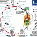
The adults are hermaphroditic and capable of self- fertilization. Some are known to develop a second set of reproductive organs. Adults living in mammalian hosts produce eggs, which exit the host in fecal matter. Eggs may survive up to three years until conditions are right for development. The eggs are typically washed into freshwater lakes and streams where they are eaten by a copepod (Pappas, 1999). The coracidium larva, which hatches from the egg, is ciliated and bears six terminal hooks. The coracidium sheds its epithelium and furthur develops into the procercoid inside a copepod, the first intermediate host. Transformation into the fully infective procercoid takes several weeks to be completed (Roberts and Janovy, 2000). From there, the procercoid transfers hosts to a fish (such as trout) via ingestion of the copepod intermediate host. There it migrates to the flesh of the fish and further develops into the plerocercoid. The plerocercoid is characterized by a ribbon-like body with an undivided scolex. The plerocercoid may pass through other paratenic hosts until finally consumed by a mammalian definitive host (Roberts & Janovy, 2000). Mammals such as bears, dogs, and humans eat those fish and aquire the worms, which grow and reproduce in the host's intestines.
Diphylobothrium latum was introduced to North America by immigrants from Scandinavia and has been spread greatly by domestic dogs that are fed raw fish. A severe broad tapeworm infection in humans is known to cause anemia due to the lack of vitamin B-12, which the tapeworm absorbs through the lining of the gut. The parasite can be avoided by thoroughly cooking fish and taking care when working with fish flesh.
Cestodes in general have sensory organs in the scolex, which are attached to longitudinal nerves extending down the body. The nerves are attached to organs and the cestodes can detect tactile stimulation.
Communication Channels: tactile
Perception Channels: tactile
Because of their potential harm to humans, efforts are being taken to prevent the spread of these worms.
CITES: no special status
The adults are hermaphroditic and capable of self- fertilization. Some are known to develop a second set of reproductive organs. Adults living in mammalian hosts grow and reproduce in the host's intestinges. The eggs exit the host in fecal matter.
Key Reproductive Features: simultaneous hermaphrodite; sexual
Parental Investment: no parental involvement
The cestode (tapeworm) Diphyllobothrium latum (Broad Tapeworm or Fish Tapeworm) is the largest human tapeworm, sometimes exceeding 10 m in length. In the human intestine, it may live for over two decades. Transmission to humans occurs via the consumption of infected fish. Several other Diphyllobothrium tapeworms have been reported to infect humans, but less frequently (these include D. pacificum, D. cordatum, D. ursi, D. dendriticum, D. lanceolatum, D. dallliae, and D. yonagoensis). The familiar pork and beef tapeworms are in a different genus, Taenia (Taenia solium and Taenia saginata, respectively).
Immature eggs of D. latum are passed in feces from the definitive host (i.e., the host harboring adults, or "final" host). Under appropriate conditions, the eggs mature (after approximately 18 to 20 days) and yield oncospheres (first stage larvae), which develop into coracidia (swimming larvae that attract potential first intermediate hosts). After ingestion by a suitable freshwater crustacean (the copepod first intermediate host), the coracidium develop into procercoid larvae. Following ingestion of the copepod by a suitable second intermediate host, typically a minnow or other small freshwater (or, possibly, anadromous or marine fish), the procercoid larvae are released from the crustacean and migrate into the fish flesh, where they develop into plerocercoid larvae (spargana). The plerocercoid larvae are the infective stage for humans. Because humans do not generally eat undercooked minnows or similar freshwater fish, these do not represent an important source of infection. However, these small second intermediate hosts can be eaten by larger predator species such as trout, perch, and walleyed pike. In this case, the plerocercoid can migrate to the musculature of the larger predator fish and humans can acquire the disease by eating these later intermediate infected host fish raw or undercooked. After ingestion of the infected fish, the plerocercoids develop into immature adults and then into mature adult tapeworms, which will reside in the human host's small intestine. The adults of D. latum attach to the intestinal mucosa by means of the two bilateral grooves (bothria) of the scolex (the anterior part of the tapeworm that is specialized for attachment to the gut wall of the host). The adult tapeworms can exceed 10 m in length, with more than 3,000 proglottids (the bisexual reproductive units strung along the length of the body, which in most cestodes each represent a single worm segment). Immature eggs are discharged from the proglottids (up to 1,000,000 eggs per day per worm) and are passed in the feces. Eggs appear in the feces 5 to 6 weeks after infection. In addition to humans, many other mammals can also serve as definitive hosts for D. latum. Most Diphyllobothrium species exhibit fairly low host specificity as adults, suggesting that the natural hosts of this parasite could be carnivorous mammals or even fish-eating birds.
Infection by D. latum is known from the Northern Hemisphere (Europe, states of the former Soviet Union, North America, Asia) and Uganda and Chile. Freshwater fish infected with Diphyllobothrium larvae may be transported to and consumed in geographic areas where active transmission does not occur, resulting in human diphyllobothriasis. For example, cases of D. latum infection associated with consumption of imported fish have been reported in Brazil.
(Centers for Disease Control Parasites and Health Website; Scholz et al. 2009 and references therein)
In the early 1970s, diphyllobothriosis was estimated to affect 9 million people globally in Europe, Asia, and North America. A more recent estimate indicated that 20 million people are infected worldwide, although infection appears to have declined in some regions and increased in others. Scholz et al. (2009) provide a recent broad review of the biology and epidemiology of D. latum. (Scholz et al. 2009 and references therein)
Der Fischbandwurm oder Grubenkopf (Diphyllobothrium latum) gehört zu den Bandwürmern. Er parasitiert vor allem im Haushund, sehr selten auch in der Hauskatze und im Menschen. Die Erkrankung wird als Diphyllobothriasis bezeichnet.
Als erster Zwischenwirt dienen Ruderfußkrebse, hauptsächlich der Gattungen Cyclops und Diaptomus. Den zweiten Zwischenwirt stellen vor allem Karpfenfische oder andere sich von Plankton ernährende Fische. Oft sind auch Hechte als paratenischer Wirt beteiligt.
Die Verbreitungsgebiete erstrecken sich hauptsächlich über Nord-[1] und Südamerika,[2] Europa und Teile Asiens.[3] Er tritt jedoch auch vereinzelt weltweit in Binnengewässern auf. Besondere Befallsgebiete sind überall dort vorhanden, wo Fisch roh als Nahrung Verwendung findet. Das trifft besonders auf Skandinavien, insbesondere Finnland, das Baltikum und die Kurische Nehrung zu.[4] Heute jedoch ist aufgrund der veränderten Ernährungsgewohnheiten beim Menschen ein Rückgang des Befalls zu beobachten.
Diphyllobothriasis kann in Europa und Nordamerika von einer der drei vorkommenden Arten ausgelöst werden, die meist undifferenziert überwiegend als Diphyllobothrium latum bezeichnet werden:[5]
In Skandinavien sind es überwiegend die beiden letzteren,[10][11] in Polen überwiegend Diphyllobothrium ditremum.[9]
In anderen Gebieten können auch andere Arten fälschlich als Diphyllobothrium latum als solche bezeichnet werden, auch in wissenschaftlichen Veröffentlichungen.[6]
Der Fischbandwurm ist der größte Vertreter der Bandwürmer, der Kopf (Scolex) besitzt im Gegensatz zu anderen Cestoda keine Haken (Rostellum), sondern nur zwei Sauggruben, um sich an der Darmwand des Wirts festzusetzen. Sein Körper ist in bis zu 4000 Proglottiden (Segmente) unterteilt. Er wird bis zu 20 Meter lang und bis zu 20 Millimeter breit. Im letzten Segment werden die Eier produziert, dort sind männliche und weibliche Geschlechtsorgane vorhanden. Die Eier werden über eine Öffnung am letzten Segment, dem Tocotrem, ausgeschieden.
Die Eier gelangen mit dem Kot des Endwirts (Mensch, Hund, Katze) in geeignete stehende Gewässer, wo sie sich zu einer Hexacanthenlarve, beim Fischbandwurm als Coracidium bezeichnet, entwickeln. Das Coracidium wird von Ruderfußkrebsen der Gattungen Diaptomus und Cyclops aufgenommen. In den Ruderfußkrebsen reift das Procercoid heran. Die Krebse werden von Karpfenfischen als Nahrung aufgenommen, die Parasiten durchdringen dann die Darmwand der Fische und entwickeln sich so weiter zum Plerocercoid. Dabei kann es vorkommen, dass der Fisch von einem anderen Raubfisch aufgenommen wird, wobei dieser dann als paratenischer Wirt dient. Als Beispiel sei hier der Hecht genannt. Der Endwirt (Mensch, Hund, Katze) infiziert sich dann durch die Aufnahme des Zwischenwirts (Karpfenfische) oder des paratenischen Wirts Hecht.
Beim Verzehr von rohem Fischfleisch kommt es zur Aufnahme der Plerozerkoiden.[1] Die Plerozerkoiden entwickeln sich im Darm beispielsweise der Katze oder des Menschen zum adulten Wurm. Das tägliche Wachstum des Wurmes im Darm beträgt 9 bis 15 cm. Nach 3 bis 5 Wochen werden die Würmer geschlechtsreif und beginnen mit der Eiproduktion. Der Parasit kann bis zu 25 Jahre im Darm persistieren. Da fast immer nur ein Wurm in einem Endwirt vorkommt, wird der Befall häufig nicht bemerkt.[12] Die Diphyllobothriasis kann selten (bei etwa 2 % der Befallenen) eine makrozytäre Anämie durch Mangel an Vitamin B12,[13] verursachen, der durch die Aufnahme in großen Mengen[14] durch den Fischbandwurm im Darm hervorgerufen wird. Nur in Einzelfällen wurde von schweren Auswirkungen berichtet.[15]
Die wirksamste Maßnahme ist die Vermeidung des Verzehrs von rohem Süßwasserfisch.[1] Bei einer behandlungsbedürftigen Infektion durch den Fischbandwurm kommen als Antiparasitika Praziquantel oder Niclosamid zum Einsatz.[16]
Der Fischbandwurm oder Grubenkopf (Diphyllobothrium latum) gehört zu den Bandwürmern. Er parasitiert vor allem im Haushund, sehr selten auch in der Hauskatze und im Menschen. Die Erkrankung wird als Diphyllobothriasis bezeichnet.
Als erster Zwischenwirt dienen Ruderfußkrebse, hauptsächlich der Gattungen Cyclops und Diaptomus. Den zweiten Zwischenwirt stellen vor allem Karpfenfische oder andere sich von Plankton ernährende Fische. Oft sind auch Hechte als paratenischer Wirt beteiligt.
El gusano ancho de los peces (Diphyllobothrium latum) es una especie de platelminto parásito de la clase de los cestodos, que provoca en humanos la enfermedad llamada difilobotriasis, botriocefalosis o botriocefaliasis. Tiene dos huéspedes intermediarios, el primero son crustáceos copépodos y el segundo son los peces de agua dulce, después de ingerir copépodos infectados.[1] La transmisión a los humanos (y a otros mamíferos) ocurre al consumir pescado fresco crudo. Se ha cambiado el nombre de género recientemente como Dibothriocephalus por lo que ahora debería llamarse Dibothriocephalus latus.
D. latum tiene un escólex alargado y, a diferencia de otros cestodos, tiene discos succionadores en lugar de ventosas. Los huevos son ovales y tienen un opérculo en forma de tapa.
Los huevos no embrionados son liberados a través de las heces; en el agua se vuelven embrionados y son consumidos por crustáceos o peces en forma de coracidios. Después de ser ingeridos por los peces predadores, las larvas jóvenes o procercoides migran a la musculatura y se desarrollan las larvas maduras o plerocercoides. Si las plerocercoides son ingeridos por los humanos se desarrolla el gusano adulto, el cual que pueden producir numerosos huevecillos. En el intestino humano pueden alcanzar una longitud que alcanza 13 metros y llegar a vivir durante 20 años. En el intestino las proglótides liberan los huevos inmaduros, siendo expulsados con las heces, iniciándose un nuevo ciclo.
El hallazgo patológico principal es la presencia de gusanos en el intestino adulto con datos de inflamación local.
Cursan generalmente asintomáticas. Malestar abdominal, pérdida de peso, ataque al estado general, diarrea. Infrecuente pero muy característico: absorción de grandes cantidades de vitamina B12 por parte de los céstodos, provocando anemia megaloblástica por déficit, datos clínicos de anemia y síntomas neurológicos.
También la presencia del parásito puede ocasionar una reacción tóxico-alérgica.
Demostración de huevos con opérculo en las evacuaciones o proglótides que son más anchas que alargadas (con patrón en roseta de las ramas uterinas) en heces o vómito.
El gusano ancho de los peces (Diphyllobothrium latum) es una especie de platelminto parásito de la clase de los cestodos, que provoca en humanos la enfermedad llamada difilobotriasis, botriocefalosis o botriocefaliasis. Tiene dos huéspedes intermediarios, el primero son crustáceos copépodos y el segundo son los peces de agua dulce, después de ingerir copépodos infectados. La transmisión a los humanos (y a otros mamíferos) ocurre al consumir pescado fresco crudo. Se ha cambiado el nombre de género recientemente como Dibothriocephalus por lo que ahora debería llamarse Dibothriocephalus latus.
Harilik laiuss ehk laiuss (Diphyllobothrium latum) on laiussiliste sugukonna laiuslaste perekonda kuuluv parasiit.
Laiussi pikkus on tavaliselt üle 10 meetri, ulatudes isegi 20 meetrini.[1] Ussi laius 1–2 cm. Laiuss areneb täiskasvanuks umbes ühe kuu jooksul ja võib peremeesorganismis elada isegi kuni 25 aastat.[2]
Laiuss põhjustab inimesel haigust, mida nimetatakse laiusstõveks ehk difüllobotrioosiks.
Harilik laiuss ehk laiuss (Diphyllobothrium latum) on laiussiliste sugukonna laiuslaste perekonda kuuluv parasiit.
Laiussi pikkus on tavaliselt üle 10 meetri, ulatudes isegi 20 meetrini. Ussi laius 1–2 cm. Laiuss areneb täiskasvanuks umbes ühe kuu jooksul ja võib peremeesorganismis elada isegi kuni 25 aastat.
Laiuss põhjustab inimesel haigust, mida nimetatakse laiusstõveks ehk difüllobotrioosiks.
Le ténia du poisson (Dibothriocephalus latus[1], anciennement Diphyllobothrium latum), est une espèce de cestodes causant la dibothriocéphalose (anciennement diphyllobothriose, ou même bothriocéphalose). Ce parasite a pour hôte définitif différents mammifères (dont l'homme), qui s'infestent par la consommation de poisson cru ou mal cuit, eux-mêmes parasités par la consommation de crustacés (tels les copépodes) ayant ingéré les œufs présents dans les fèces d'un hôte définitif.
Des œufs de D. latus ont été identifiés dans des coprolithes humains du néolithique de l'ancien et du nouveau monde[2],[3]. Ils sont aussi retrouvés sur des sites de l'âge du Bronze et d'Europe médiévale, ce qui montrerait la permanence de pratiques culinaires à base de poissons crus ou peu cuits[4].
Les vers plats de grande taille sont connus dès l'Antiquité et mentionnés dans les principaux traités médicaux (papyrus Ebers, Corpus hippocratique, Celse, Avicenne...). Ils sont toutefois confondus entre eux et seulement différenciés des vers ronds.
Le médecin suisse Félix Plater (1536-1614) aurait été le premier auteur à distinguer D. latus des autres vers plats ou ténias, au début du XVIIe siècle. Il donne aussi une première description de la maladie chez l'homme[5].
Une première illustration (mais sans le scolex) est celle de Nicolas Andry en 1718[6]. La morphologie précise de ces vers en anneaux détachables ou proglottis est le fait d'un autre suisse, le naturaliste Charles Bonnet en 1750, quoique l'illustration de cette publication indique plutôt un scolex de ténia, erreur qu'il corrigea lui-même en 1777[5].
Vers le milieu du XVIIIe siècle, par les observations du médecin finlandais Herman Spöring le vieux (de) (1701-1747), il est acquis que la présence de D. latus chez l'homme est en rapport avec un régime alimentaire à base de poissons[6]. Mais le cycle parasitaire reste méconnu du fait de sa complexité (trois hôtes successifs : copépode, poisson, humain). Ce cycle ne sera complètement élucidé qu'au début du XXe siècle, en trois étapes marquantes[5] :
Dibothriocephalus latus est le plus grand des cestodes humains, avec une taille moyenne de 2 à 8 mètres mais qui peut dépasser les 15 mètres, et exceptionnellement aller jusqu'à 20 mètres[7],[8].
Sa « tête » ou scolex mesure de 1 à 5 mm. Il a une forme de massue portant deux fentes allongées faisant office de ventouse, appelées bothridies. Le cou est court et grêle[7].
Le corps ou strobile est composé de plusieurs centaines de segments ou proglottis, de forme trapézoïde plus larges que hauts. Ces segments, qui peuvent atteindre le nombre de 4 000[9], sont hermaphrodites avec un orifice génital médio-ventral. Les segments mûrs portent en leur centre une tache noirâtre qui est un utérus rempli d'œufs[7].
L'adulte demeure dans la partie moyenne de l'intestin grêle, où il peut vivre plus de dix ans. Ce n'est pas un « ver solitaire », des sujets pouvant être polyparasités par plusieurs dizaines d'individus[7].
L'œuf est brun, ovoïde, long de 60-70 μm et large de 40-45 μm[8], présentant un opercule à un pôle. L'œuf est non-embryonné à la ponte, il contient le zygote proprement dit et des cellules vitellines[7].
Les œufs sont pondus dans la lumière intestinale de l'hôte définitif, et éliminés dans les selles au nombre de plusieurs millions par jour. Les anneaux vidés de leurs œufs se désagrègent et sont évacués sous la forme de débris non identifiables. Cependant, il est possible de retrouver des fragments de chaînes d'anneaux[7].
Pour que l'œuf évolue et se segmente, il faut qu'il tombe dans de l'eau douce à une température favorable. Il devient alors embryon en 10 à 14 jours. À la température de + 1° C, l'œuf peut rester vivant plusieurs mois, mais sans évoluer[7].
Lorsque le développement de l'embryon est terminé, l'opercule de l'œuf se soulève, et il en sort un embryon hexacanthe ou coracidium, portant des cils vibratiles pour se déplacer. Ces coracidiums sont la proie de crustacés minuscules, les copépodes (comme les cyclops). Dans un cyclops, le coracidium évolue en deux semaines environ pour devenir un procercoïde, d'environ 0,5 mm de long avec un appendice globuleux[7].
Lorsque le crustacé est avalé par un petit poisson (gardon par exemple), le procercoïde migre dans les viscères ou les muscles de son nouvel hôte où il se transforme en plérocercoïde, qui est la forme larvaire infestante, de 1 à 2 cm de long, présentant des bothridies et un début de segmentation[8]. Les plérocercoïdes peuvent se retrouver chez les poissons prédateurs des précédents, carnassiers d'eau douce comme le brochet, la perche ou la lotte d'eau douce[7].
Lorsque ces poissons sont consommés par un hôte favorable, humain ou autre mammifère (chien, chat, renard, porc, ours, phoque...), la larve infestante se fixe à la muqueuse du grêle pour devenir adulte en 5 à 6 semaines. La taille de l'adulte s'adapte à celle de son hôte (plusieurs mètres chez l'homme, moins d'un mètre chez le chat)[7],[8].
L'humain s'infeste en consommant du poisson cru ou mal cuit (les larves sont détruites à plus de 50° C). Les femmes préparant des plats traditionnels tels que le Gefilte fish seraient les plus infestées car elles goûtent le poisson émincé avant de le faire cuire, de même pour les pêcheurs qui consomment des abats crus de divers poissons[7].
Classiquement, la répartition géographique s'explique par les modalités du cycle. L'infestation par D. latum se retrouve dans les régions de lacs en zone tempérée ou froide de l'hémisphère nord. Elle se voit surtout dans les pays riverains de la mer du Nord et de la mer Baltique ; en Europe, autour des lacs alpins de Suisse et d'Italie, elle est rare en France sauf près de la frontière suisse. À l'est, l'aire de répartition englobe le delta du Danube, l'ex-URSS, le Japon, et l'Amérique du nord[7].
Des cas sont aussi rapportés dans des pays chauds (en Israël autour du Lac de Tibériade) et dans l'hémisphère sud (Afrique, Amérique du Sud, Australie...)[7].
Bien que peu connue du grand public et des médecins, cette parasitose est en expansion, probablement liée à des modifications des habitudes alimentaires par la mondialisation et aux nouvelles techniques de surgélation et refroidissement rapide[2].
Le ténia du poisson (Dibothriocephalus latus, anciennement Diphyllobothrium latum), est une espèce de cestodes causant la dibothriocéphalose (anciennement diphyllobothriose, ou même bothriocéphalose). Ce parasite a pour hôte définitif différents mammifères (dont l'homme), qui s'infestent par la consommation de poisson cru ou mal cuit, eux-mêmes parasités par la consommation de crustacés (tels les copépodes) ayant ingéré les œufs présents dans les fèces d'un hôte définitif.
Diphyllobothrium latum est species vermium parasiticorum.
Diphyllobothrium latum est species vermium parasiticorum.
Platusis (žuvinis) kaspinuotis (lot. Diphyllobothrium latum) – parazitinė plokščioji kirmėlė. Galutinis šeimininkas – mėsėdžiai bei žmogus, tarpiniai šeimininkai – irklakojai vėžiagyviai (pvz., ciklopai) ir žuvys.
Ilgis 7-16 m. Nareliai trumpi ir platūs. Galvutė pailga, šonuose yra 2 prisisiurbimo vagelės – botrijos. Galvutė su kūnu susijungusi kakleliu. Nesubrendusio narelio sandara panaši į kitų kaspinuočių, bet turi skirtumų. Lytinės kloakos gumburėlis ne narelio šone, o narelio pilvinėje pusėje. Gimda ne šakota, o vingiuota, gimdos vingiai rozetės formos. Subrendusiame narelyje sunykę visi lytiniai organai, išskyrus gimdą. Ji yra pripildyta bręstančių kiaušinėlių.
Kaspinuotis vystosi galutiniame šeimininke ir bent 2 tarpiniuose. Iš galutinio šeimininko kiaušinėliai išeina į aplinką ir turi patekti į vandenį. Vandenyje iš kiaušinėlio išeina koracidija (apvali, judri, plaukioja vandenyje). Joje yra onkosfera. Vandenyje korocidijas praryja vėžiagyviai. Jų žarnyne koracidijos numeta blakstienėles ir skverbiasi į organizmo ertmes. Vėžiagyvio organizme susiformuoja procerkoidas. Kai vėžiagyvį suėda žuvis, procerkoidas prasiskverbia į žuvies raumenis ar kitus organus ir virsta plerocerkoidu. Žinduoliai užsikrečia suėsdami užsikrėtusią žuvį. Jų žarnyne vystosi suaugęs kaspinuotis.
Platusis (žuvinis) kaspinuotis (lot. Diphyllobothrium latum) – parazitinė plokščioji kirmėlė. Galutinis šeimininkas – mėsėdžiai bei žmogus, tarpiniai šeimininkai – irklakojai vėžiagyviai (pvz., ciklopai) ir žuvys.
Diphyllobothrium latum é a "tênia do peixe", causadora da difilobotríase ou esparganose.[1]
Os ovos, na água limpa, liberam coracídios que são ingeridos por pequenos artrópodes; os artrópodes são ingeridos por peixes, e as larvas procercóides infestam o organismo do peixe e, se esse for ingerido por um peixe maior, o maior infestar-se-á. A infecção humana se dá pelo consumo de peixe cru contendo esparganos. No homem, o verme adulto se localiza no jejuno, e mede entre 3 a 15 metros de comprimento (é o maior cestódeo que pode parasitar o homem).[2][3]
Entre as manifestações clínicas, pode ocorrer dor epigástrica, dor de fome, anorexia, náuseas, vômitos, astenia, perda de peso, eosinofilia. Uma complicação peculiar dessa helmintose é a anemia megaloblástica (anemia hipocrômica macrocítica), pois o parasito adulto tem a capacidade de absorver intensamente a vitamina B12 no intestino do hospedeiro. Contudo, esta infecção pode ser assintomática. O diagnóstico consiste em observação dos ovos nas fezes do paciente.[2]
A prevenção se faz pelo cozimento adequado dos peixes, dar destino higiênico aos excretos humanos, inspeção do pescado e congelamento adequado dos peixes nos frigoríficos.[2]
Diphyllobothrium latum é a "tênia do peixe", causadora da difilobotríase ou esparganose.
Os ovos, na água limpa, liberam coracídios que são ingeridos por pequenos artrópodes; os artrópodes são ingeridos por peixes, e as larvas procercóides infestam o organismo do peixe e, se esse for ingerido por um peixe maior, o maior infestar-se-á. A infecção humana se dá pelo consumo de peixe cru contendo esparganos. No homem, o verme adulto se localiza no jejuno, e mede entre 3 a 15 metros de comprimento (é o maior cestódeo que pode parasitar o homem).
Entre as manifestações clínicas, pode ocorrer dor epigástrica, dor de fome, anorexia, náuseas, vômitos, astenia, perda de peso, eosinofilia. Uma complicação peculiar dessa helmintose é a anemia megaloblástica (anemia hipocrômica macrocítica), pois o parasito adulto tem a capacidade de absorver intensamente a vitamina B12 no intestino do hospedeiro. Contudo, esta infecção pode ser assintomática. O diagnóstico consiste em observação dos ovos nas fezes do paciente.
A prevenção se faz pelo cozimento adequado dos peixes, dar destino higiênico aos excretos humanos, inspeção do pescado e congelamento adequado dos peixes nos frigoríficos.
광절열두조충(Diphyllobothrium latum, broad tapeworm, 廣節裂頭條虫)은 촌충 중에서 열두조충의 한 종류이며 또는 광절열두조충과, 대형 촌충, 긴촌충이라고도 한다.
북미, 시베리아, 일본 북부 지역 및 대한민국에 분포하며 이 중에서 대한민국에서는 이 광절열두조충과 왜소열두조충(D. parvum), 요나고열두조충(D. yonagoense)의 감염례가 알려져 있다.
이 조충의 몸 색깔은 대체적으로 회백색인 경우가 많으며 성충 기준으로 다 성장한 길이는 약 2~10m, 너비는 약 1.5~2cm 정도가 된다. 몸은 약 3천 ~ 4천 개 정도의 편절이 서로 연결된 형태이며, 머리에 해당하는 두절은 몸에 비해 작고 방추형이며 흡구가 한쌍으로 이루어져 있다.
본래 조충에 감염되더라도 초창기에는 별다른 증상이 없는 경우가 많으나, 이 조충이 움직이거나 할 경우 장이 자극을 받아 설사, 복통, 무력증, 입맛 소실, 체중 감소, 영양결핍이 나타난다.[1] 또한 이 조충이 생성하는 대사산물에 의해서 혈색소증가증이나 악성 빈혈이 유발되는 경우도 있는데, 이러한 원인으로 발생한 빈혈을 열두조충성 빈혈(bothriocephalus anemia)라고 한다. 원인은 조충이 비타민 B12를 과량 섭취하여 인체에서 필요한 비타민을 흡수하지 못해 발생하는 것으로 알려져 있다.
광절열두조충은 사람 말고도 개, 고양이, 곰, 여우 등도 감염될 수 있다.[2] 감염된 숙주로부터 배출된 충란이 약 15~25℃ 수온의 물에서 부화하면 섬모유충 단계까지 성장한다. 이 후 섬모를 이용해 물 속을 헤엄치다가 물벼룩에 의해 잡아먹히면 물벼룩 안에서 기생하게 된다. 여기서 물벼룩은 제1중간숙주역할을 하게 된다. 물벼룩 속에서 섬모유충 단계의 조충 유충은 성장하면 섬모가 없어지고 아메바 모양의 소체가 되어 약 2주 정도가 되면 원미충 단계까지 성장한다. 이 후 감염된 이 물벼룩을 송어, 연어 등이 잡아먹으면 이 어류가 제2중간숙주 역할을 한다. 제2중간숙주 몸 안으로 들어온 원미충은 어류의 장벽을 뚫고 장 안으로 이행해 다시 근육 안으로 침범하여 충미충 단계로 성장한다. 충미충 단계의 조충 유충은 아직 두절, 편절 등이 없고 흡구가 있어야 할 두절은 그냥 함몰된 형식으로만 남아 있다. 이 후 감염된 어류를 사람이 생식하거나 불완전하게 조리해서 섭취할 경우 감염되며, 인체 내로 들어온 충미충 단계의 유충은 소장 장벽에 달라붙어 성장한다. 인체에서 성장한지 약 3주 정도가 되면 1m 정도로 성장하게 되며, 이 때부터 산란할 수 있어 충란을 배출하기 시작한다. 조충의 성장 속도는 빠른 편에 속하여 하루에 약 30편절 정도를 생성하며, 수명은 약 6~14년 정도까지 살 수 있는 것으로 알려져 있다.
광절열두조충(Diphyllobothrium latum, broad tapeworm, 廣節裂頭條虫)은 촌충 중에서 열두조충의 한 종류이며 또는 광절열두조충과, 대형 촌충, 긴촌충이라고도 한다.
북미, 시베리아, 일본 북부 지역 및 대한민국에 분포하며 이 중에서 대한민국에서는 이 광절열두조충과 왜소열두조충(D. parvum), 요나고열두조충(D. yonagoense)의 감염례가 알려져 있다.


