Comprehensive Description
provided by Smithsonian Contributions to Zoology
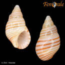
Angiola lineata (da Costa, 1778)
Buccinum lineatum da Costa, 1778:130.
Planaxis lineatus (da Costa).—Abbott, 1974:102: fig. 974.
This small, colorful, gregarious species is common throughout the Caribbean and southeastern Florida. Large discrete populations live in the rubble of sheltered littoral habitats such as bays, lagoons, and other low energy environments. Angiola lineata is an active crawler while submerged, but when exposed by falling tides, it gathers into large aggregations under stones and in the interstices of gravel, where it can remain moist until the next high tide. In the laboratory, it is strongly photonegative and lives well in petri dishes with daily changes of water for as long as three months.
SHELL MORPHOLOGY (Figure 14).—The small, thick shell is up to 10 mm in length and comprises 8 or 9 moderately inflated whorls. It is glossy and smooth, with a creamy white base color broken by numerous brown to black spiral bands (Figure 14A,C,D). The shell matrix is more dense than that of S. nucleus and does not fracture into fine lamellae when broken. The tan protoconch (Figure 14F,H) comprises protoconch I (sensu Robertson, 1974:227) of 1.5 smooth whorls, and protoconch II of 2 whorls. It is sculptured with two strong median spiral cords, with prominent axial plications above and below the spiral elements. There is a strong sinusigeral notch. The early whorls have strong spiral cords that gradually become broader on succeeding whorls and begin to merge (Figure 14F). The last two whorls are smooth except for the shell base which has spiral incised lines (Figure 14B). The suture is deeply impressed and there is a narrow abapical ramp. The aperture is large, spanning a little less than one-half the shell length. The outer lip is thick, beveled on the inside and denticulate within. Shells of females are significantly larger than those of males (T = 2.19; p = 0.02, n = 20). A very thin hispid periostracum is sometimes seen in specimens from protected areas but is abraided from the shell in most cases. The thin, tan operculum is lenticular with a subterminal nucleus (Figure 14E).
EXTERNAL ANATOMY.—The animal has an overall light tan color. The dark brown head-foot is overlain by a network of iridescent blue with the large red buccal mass showing through the snout. The sole of the foot is white. The tentacles are tapering and very long in relation to the animal, spanning about one-third the shell length.
The mantle is large, covering much of the head-foot, thin, somewhat transparent, and its edge is smooth. The middorsal surface of the mantle edge contains a luminous organ, comprised of tissue located just below the epithelium, that emits blue-green light when stimulated. Haneda (1958:154–155) stated that the luminous organ is localized in a limited area of the mantle, is comprised of many folds of tissue that run parallel to each other, and that this structure is identical in all five species examined. The luminescence is intracellular. Histological sections showed that it is confined to small, closely packed cells beneath the thin epithelium (Haneda, 1958:155–156). The area of the mantle which Haneda's (1958, fig. 3) illustration indicates as containing the luminescent cells appears to be the hypobranchial gland. His figure of the longitudinal section through the mantle containing this luminescent organ (Haneda, 1958, fig. 5) also looks like typical hypobranchial gland tissue. The cells he identified as being bioluminescent appear to lie between the outer mantle epithelium and the hypobranchial gland. Although I observed bioluminescence in these snails, I was unable to detect any specific area of the mantle that could be considered the light organ. My histological studies of Angiola lineata do not show any specific tissue that I can identify as distinctive tissue supporting bioluminescent cells. Emission of the blue-green light is caused by strong stimulation, such as shaking or mechanical irritation of the body, which I observed in Angiola fasciata in Guam and A. lineata in Florida. The natural function of the bioluminescence was not ascertained, but it may serve as a recognition mechanism or have an antipredation function.
There is no ciliated groove leading to the brood pore, which is a simple raised area on the right side of the neck, conspicuous because of the unpigmented epithelium that surrounds it. A tiny central hole enters the brood pouch (Figure 16G,H, bp). In ripe snails, female gonads occupy about 3 coils and male gonads about 2.5 coils.
MANTLE CAVITY ORGANS.—The osphradium is typically planaxoid with a raised broad band of cilia on each side (Figure 16F, OS). The distal osphradium deviates from the ctenidium and becomes narrow as it curves into the inhalant siphon. The long, narrow ctenidium is whitish in color, somewhat transparent, and comprised of about 30 long triangular filaments. The hypobranchial gland begins about 7 mm from the mantle edge and consists of thick, transverse ridges of papillate, glandular tissue that partially covers the rectum.
ALIMENTARY TRACT.—The mouth is a small slit at the edge of the flattened, ovoid snout tip. There is a moderately sized buccal mass (Figure 16H, bm).
Radula (Figure 16A–E). The radula is short, about one-fourth the shell length, and has about 14 rows of teeth. The rachidian tooth (Figure 16B,D) is wider than tall, having a basal plate with tapering lateral extensions that curve downward on each side. There is a central, triangular basal extension. The tip of the rachidian is concave and has a cutting edge with a wide, central, triangular cusp flanked by 3 or 4 tiny serrations on each side. The rachidian formula is . The lateral tooth (Figure 16C) is subtriangular and has a long distal lateral extension and a cutting edge of one large, pointed cusp with two inner denticles and two outer denticles. The marginal teeth (Figure 16C, E) are long and tapering with curved, wide tips and have cutting edges of numerous fine, nearly fused, denticles. A description of the radula of A. lineata from Santa Marta, Colombia, was given by Bandel (1984:35–36, fig. 57).
The paired salivary glands originate behind the nerve ring and lie closely adpressed to it as coiled tubes. They pass through the nerve ring as single thin tubes and run over the dorsal surface of the central buccal mass where they enter the buccal cavity.
The esophagus widens immediately behind the nerve ring to form the esophageal gland, which is similar to that described in Planaxis sulcatus. In section, the epithelium of these folds stains deeply with hematoxylin (Figure 16F, eg).
The stomach has a moderately sized, narrow, bilobed pad that becomes single lobed as it approaches the short style sac.
REPRODUCTIVE SYSTEM.—The male pallial gonoduct is folded transversely interiorly and divided by a transverse internal ridge into a proximal and distal portion. The proximal portion is a whitish, glandular area that stains dark purple with methylene blue and probably serves as the prostate-spermatophore gland; the distal portion is less glandular and stains light blue. Euspermatozoa and paraspermatozoa are present in about equal numbers in the vas deferens. Euspermatozoa are about 12 μm long and have a short, well-defined acrosome and a relatively long midpiece. Paraspermatozoa are a little larger and have a broad triangular head and five flagellae.
The female pallial oviduct (Figure 15) is conspicuously swollen in ripe females. The oviducal groove (Figure 15, og) lies at the base of the two lamina that comprise the open duct and is demarcated by a transversely folded, thickened glandular area along its length. The proximal end of the pallial oviduct comprises the albumin gland (Figure 15, ag) and, in section, appears as a dark staining bulge along the base of the lateral lamina (Figure 15, ll). The median part of the pallial oviduct is much thicker and glandular, and is probably the capsule gland. The wall of the lateral lamina is thin and highly ciliated. The medial lamina has a short, wide, sperm collecting gutter (Figure 15, sg) along its distal edge that becomes a duct (Figure 15, osb) leading into a large median spermatophore bursa (Figure 15, sb). Adjacent to this duct is another highly ciliated opening (Figure 15, osr) in the outer median part of the medial lamina that leads into a median distal seminal receptacle (Figure 15, sr) located in the inner wall of the medial lamina, adjacent to the spermatophore bursa.
The small dumbbell-shaped brood pouch is located in the right side of the head-foot (Figure 16G, bp) and is lined with ciliated tissue. It is small in comparison to those of Planaxis, Fissilabia, and Supplanaxis species. Histological sections through the chamber show a deeply stained ciliated epithelium throughout. The brood pouch is subdivided into interconnected locula or chambers by thin invaginations of the internal wall (Figure 16H, bp), which greatly increase its surface area. Larvae were rarely observed in the brood pouch, but the few examined contained about 30 larvae that are brooded until the veliger stage. The brood pouch is adjacent to and innervated by large, swollen extensions of the pedal ganglia (Figure 16G, exp). This species was examined at various times throughout the year and no trace of a brood pouch on the left side of the foot was seen. It thus seems certain that it is limited to the right side.
NERVOUS SYSTEM.—Angiola species have a typical planaxoid nervous system and an RPG ratio of 0.50 (n = 5).
TYPE-SPECIES.—Planaxis sulcatus (Bruguière, 1789), by original designation.
DIAGNOSIS.—Shell conical, obese, black and smooth except for early whorls, part of body whorl and area beneath suture, where there are incised spiral grooves. Aperture wide with thick outer lip, grooved within, and thick columella with broad siphonal notch. Rachidian tooth broad, wider than long, with pair of small basal cusps close to outer edges. Cutting edge with large, rounded, central denticle flanked by smaller denticles. Seminal receptacle in proximal oviduct. Large dumbbell-shaped brood pouch in both sides of head-foot beneath buccal cavity and esophagus. Many small embryos incubated to veliger stage in brood pouch, followed by planktotrophic development. Large pair of coiled salivary glands pass through nerve ring. Midesophagus forms wide esophageal gland. Habitat low rocky, high energy, intertidal zone.
- bibliographic citation
- Houbrick, Richard S. 1987. "Anatomy, reproductive biology, and phylogeny of the Planaxidae (Cerithiacea: Prosobranchia)." Smithsonian Contributions to Zoology. 1-57. https://doi.org/10.5479/si.00810282.445
Comprehensive Description
provided by Smithsonian Contributions to Zoology
Planaxis lineatus (Da Costa, 1778)
Buccinum lineatum Da Costa, 1778:130, pl. 8: fig. 5 [Cornwall and the West Indies].
Planaxis lineatus Da Costa.—Smith, 1890b:319.—Stearns, 1893:333.
PREVIOUS ASCENSION RECORDS.—Smith (1890).
PRESENT MATERIAL.—3 specimens (4.6 mm) Ascension, K. M. Hutchfield; 75+ specimens (7.2 mm) ASC 5; 50+ (4.8 mm) ASC 8; 10 (5.9 mm) ASC 12; 17 (4.2 mm) ASC 13; 4 (5 mm) ASC 15; 2 (4.4 mm) ASC 16; 9 (4.6 mm) ASC 18; 44 (4.6 mm) ASC 20; 100 (5.5 mm) ASC 21; 1 (4.4 mm) ASC 23, R. B. Manning, May 1971.
DISTRIBUTION.—Bermuda, Florida, West Indies, Caribbean coast of Central America, Cape Verde Islands, St. Helena, Ascension.
DISTRIBUTION.—Tropical east Atlantic islands.
- bibliographic citation
- Rosewater, Joseph. 1975. "An annotated list of the marine mollusks of Ascension Island, South Atlantic Ocean." Smithsonian Contributions to Zoology. 1-41. https://doi.org/10.5479/si.00810282.189