-
Valvata ouscubakus Nykk,.
-
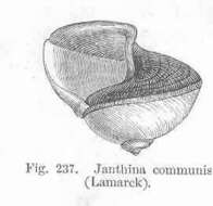
Janthina communis (Lamarck).
-
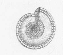
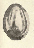
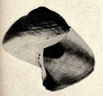
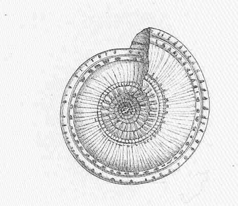
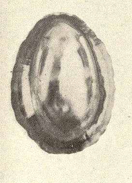
Solarium perspectivum Lam., from the under side.
-
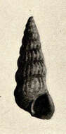
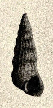
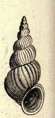
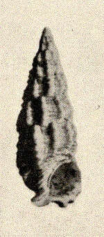
Scala lineata.
-
Manuel Caballer, Jesus Ortea, Samuel Narciso
Zookeys
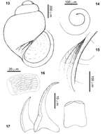
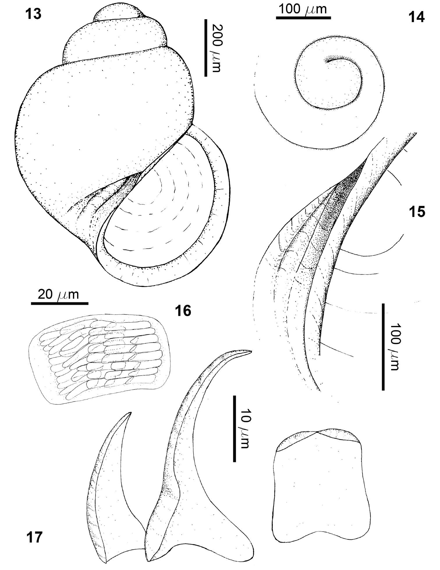
Figures 13–17.Rissoella morrocoyensis sp. n.: shell (13), protoconch (14), detail of the umbilicus (15), odontophoral cartilages (16), radular teeth (17).
-
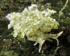
Plate 15.Cyerce bourbonica sp. n., 12 mm paratype, La Réunion, photo: H Flodrops.
-
Cerithidea scalariformis.
-
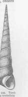
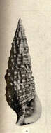
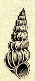
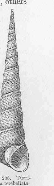
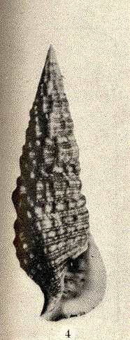
Turritella terebellata (Lamarck).
-
Lottia gigantea, inside view.
-
Janthina fragilis.
-
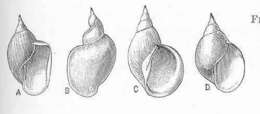
Four examples of Limnaea stagnalis L., from marshes in the Aral district which are salt for several months in the year, illustrating variation produced by changes in the environment.
-
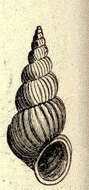
Scala multistriata.
-
Manuel Caballer, Jesus Ortea, Samuel Narciso
Zookeys
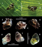
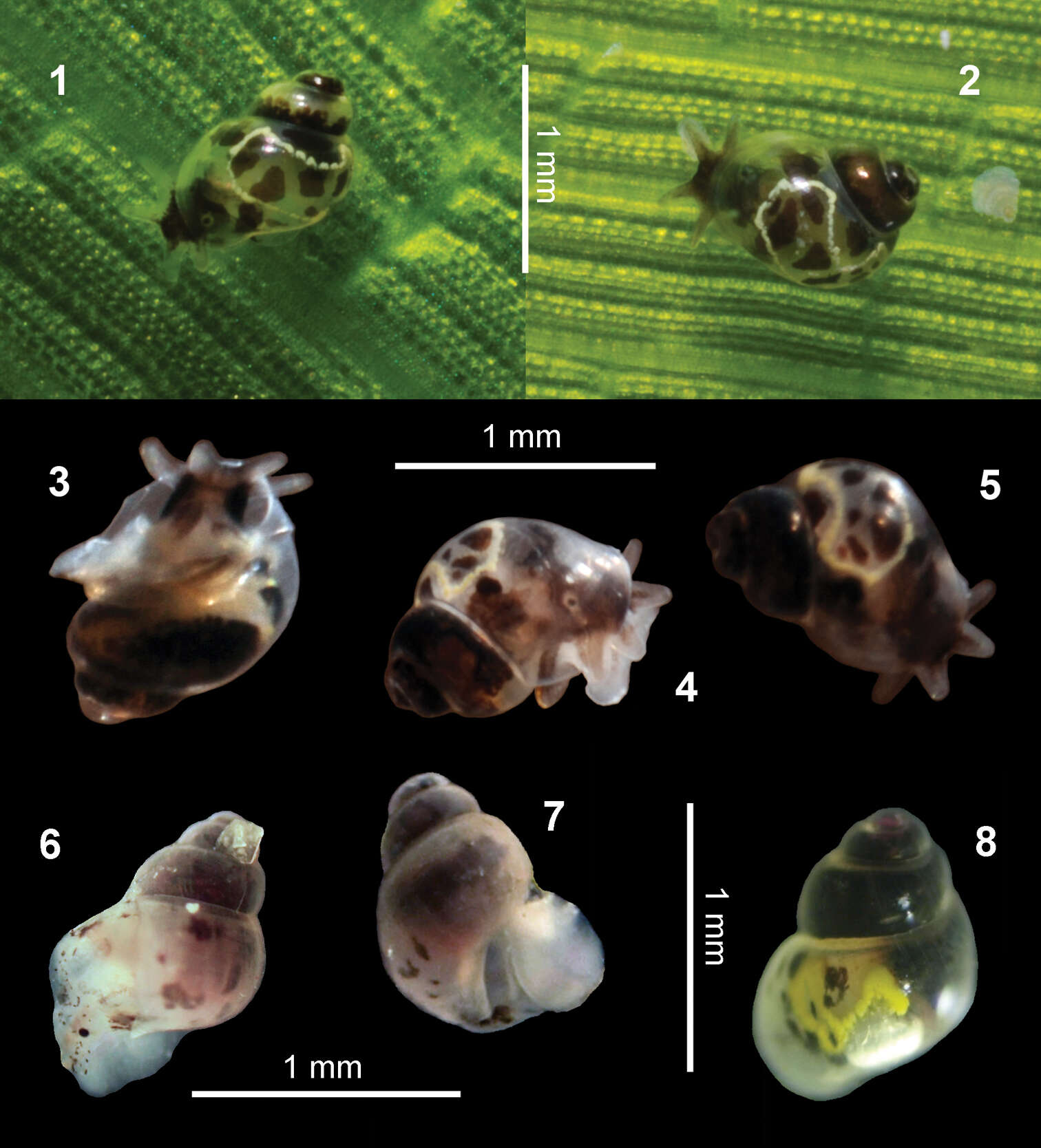
Figures 1–8.Rissoella morrocoyensis sp. n. 1–5: on a leaf of Thalassia testudinum (1–2), ventral view (3), lateral view (4), dorsal view (5). Rissoella venezolanicola sp. n. 6–8: lateral view (6), ventral view (7), dorsal view (8).
-
Peter Glöer, Vladimir Pešić
Zookeys
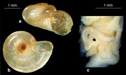
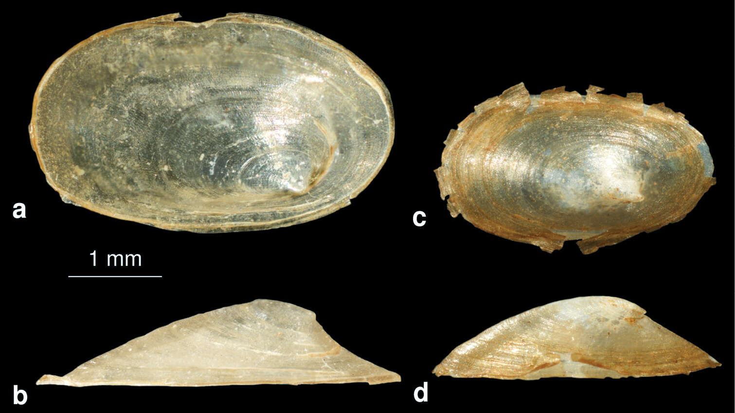
Figure 15.a–b Acroloxus pseudolacustris sp. n.: shell c–d Acroloxus lacustris (from Hamburg, Germany): shell.
-
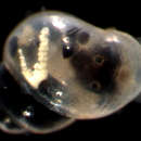
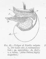
Veliger of Patelia vulgata L, 130 hours old; f, rudimentary foot; op, operculum; sh, shell; v v, velum.
-
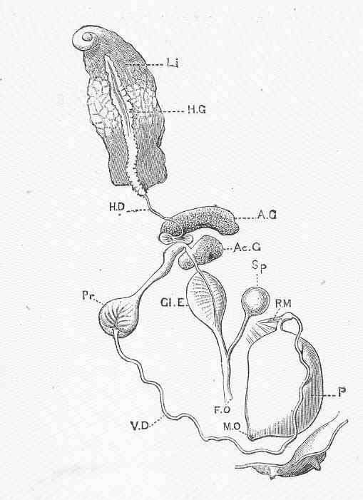
Genitalia of Limnaea stagnalis L. (from a dissection by F. B. Stead).
-
Manuel Caballer, Jesus Ortea, Samuel Narciso
Zookeys
Figures 9–12.Rissoella morrocoyensis sp. n. 9–10: view of the aperture and the umbilicus (9) detail of the umbilicus (10). Rissoella venezolanicola sp. n. 11–12: view of the aperture and the umbilicus (11), detail of the umbilicus 12.
-
Peter Glöer, Vladimir Pešić
Zookeys
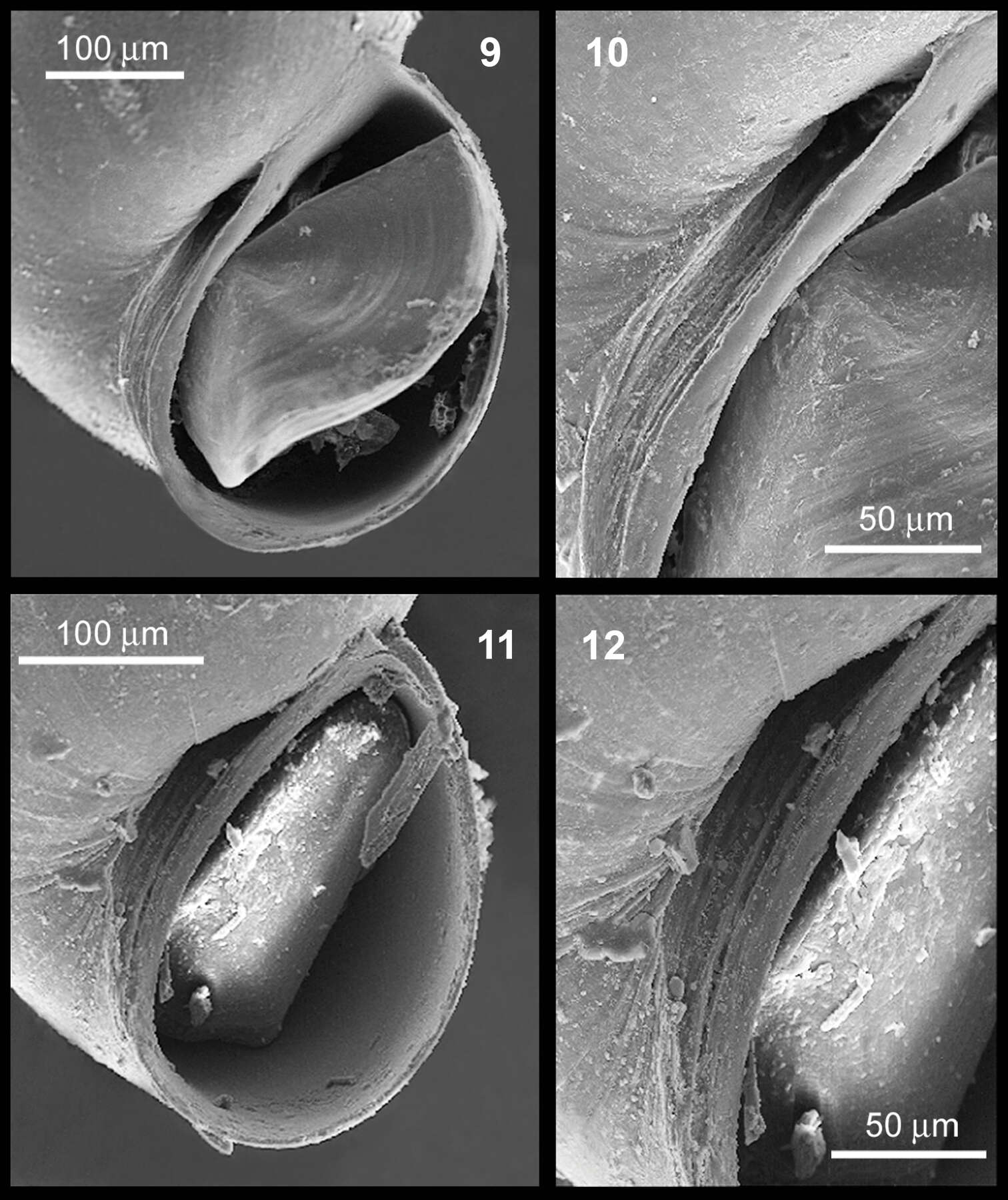
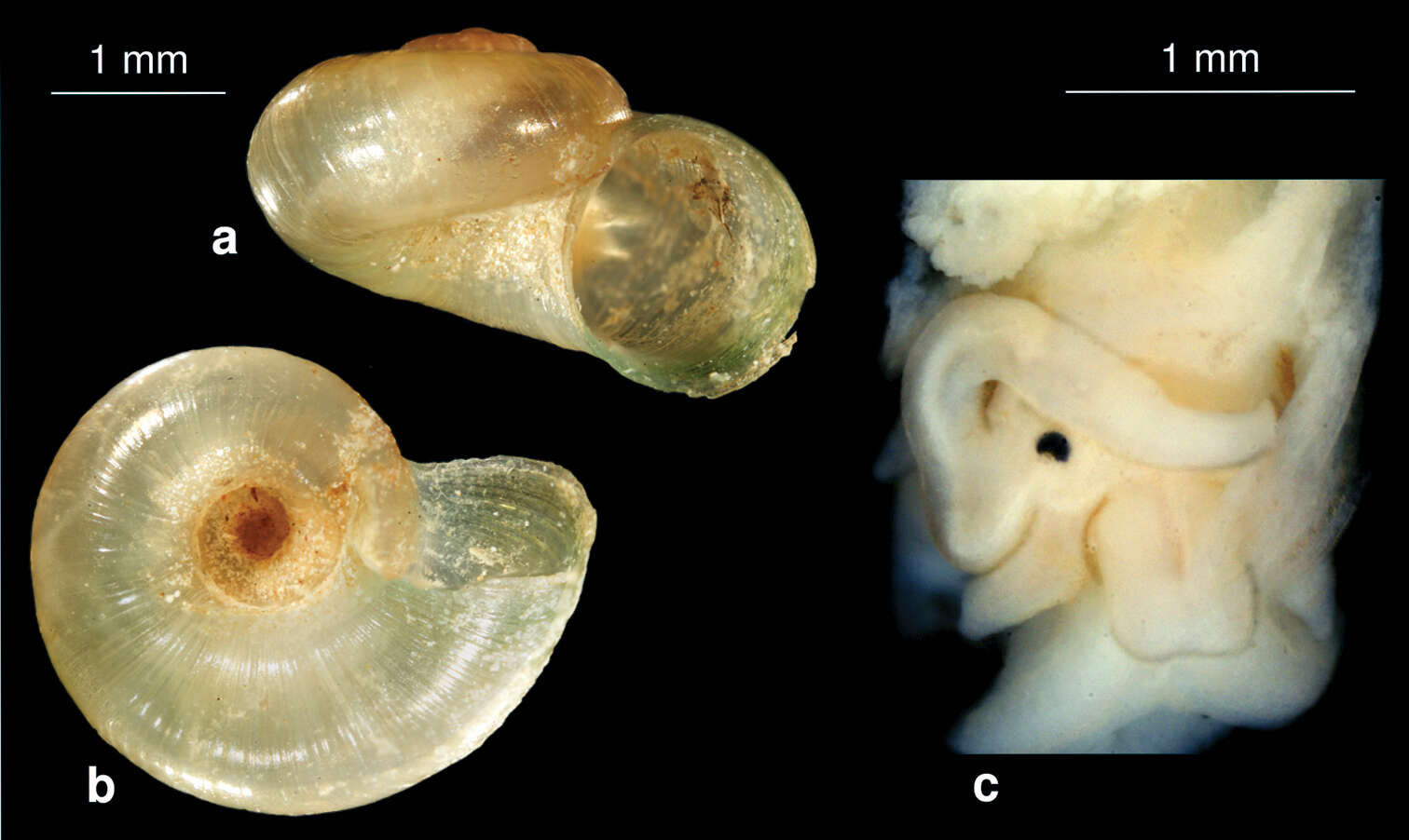
Figure 14.Valvata nowshahrensis sp. n. a shell b ventral view on the umbilicus c head with penis in situ.
-
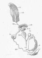
Patella vulgata L., seen from the ventral side.
-
Manuel Caballer, Jesus Ortea, Samuel Narciso
Zookeys
Figures 13–17.Rissoella morrocoyensis sp. n.: shell (13), protoconch (14), detail of the umbilicus (15), odontophoral cartilages (16), radular teeth (17).
-
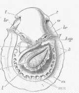
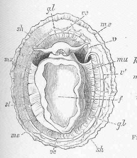
Patella vulgata L., seen from the dorsal side after the removal of the shell and the black pigment covering the integument; the anterior portion of the mantle is cut away or turned back.
-
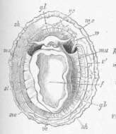
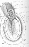
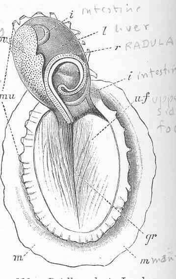
Patella vulgata L., showing the normal position of the radula, which is doubled back in a bow; the shell has been removed, and the whole visceral mass is turned forward, exposing the dorsal surface of the muscular foot.
-
Cerithium muscarum.
-
Cerithium floridanum.