-
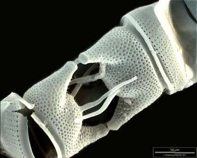
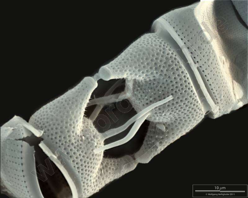
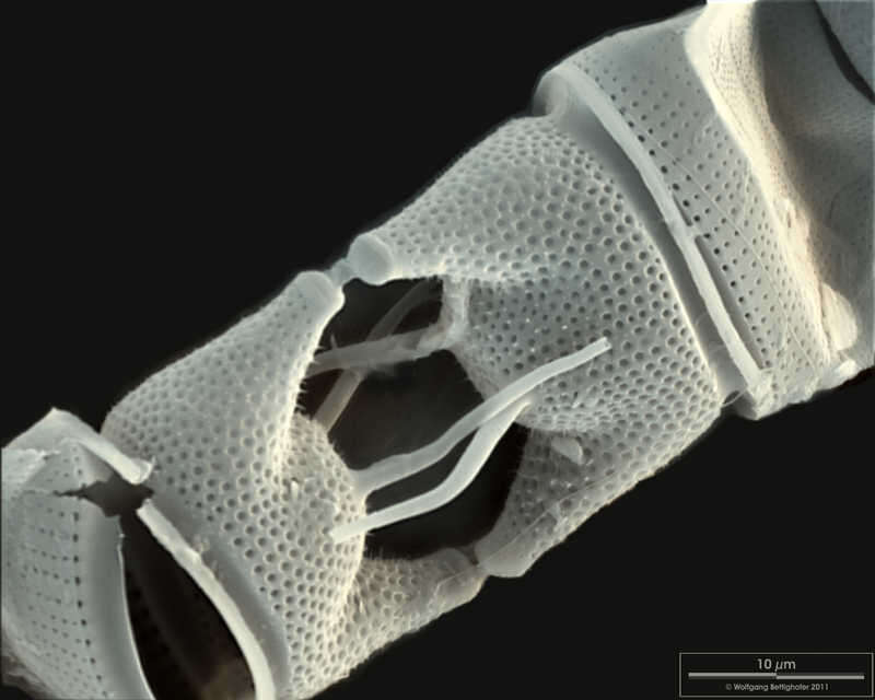
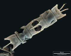
Scale bar indicates 10 µm. Sample from North Sea near Heligoland (spring diatom bloom). Use of SEM equipment courtesy of Lab Dr. Karl-Heinz Schäffner, Solingen, Germany.
-
Pera, Faro, Portugal
-
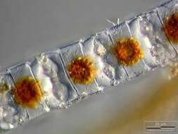
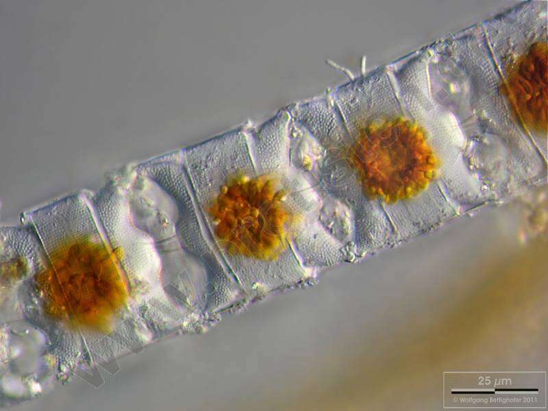
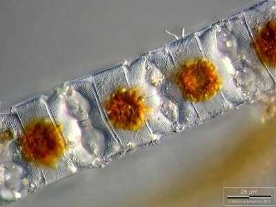
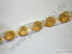
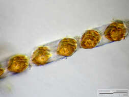
Depth-of-focus image exhibiting structure of the frustules. Chloroplasts are concentrated in cell centers due to long lasting exposition with microscope's illumination. Scale bar indicates 25 m. The image was built up using several photomicrographic frames with manual stacking technique. Sample from North Sea near Heligoland (spring diatom bloom). Images were taken using Zeiss Universal with Olympus C7070 CCD camera.For more look at
www.protisten.de/english/gallery_main/gallery_main.htmlFor high-resolution images please ask postmaster@protisten.de.
-
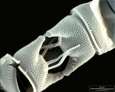
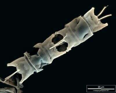
Scale bar indicates 25 µm. Sample from North Sea near Heligoland (spring diatom bloom). Use of SEM equipment courtesy of Lab Dr. Karl-Heinz Schäffner, Solingen, Germany.
-
Odontella aurita.Scale bar indicates 10 m. Sample from North Sea near Heligoland (spring diatom bloom). Use of SEM equipment courtesy of Lab Dr. Karl-Heinz Schffner, Solingen, Germany. For more look at
www.protisten.de/english/gallery_main/gallery_main.htmlFor high-resolution images please ask postmaster@protisten.de.
-
Depth-of-focus image exhibiting structure of the frustules. Chloroplasts are concentrated in cell centers due to long lasting exposition with microscope's illumination. Scale bar indicates 25 µm. The image was built up using several photomicrographic frames with manual stacking technique. Sample from North Sea near Heligoland (spring diatom bloom). Images were taken using Zeiss Universal with Olympus C7070 CCD camera.
-
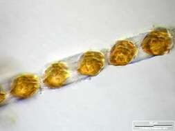
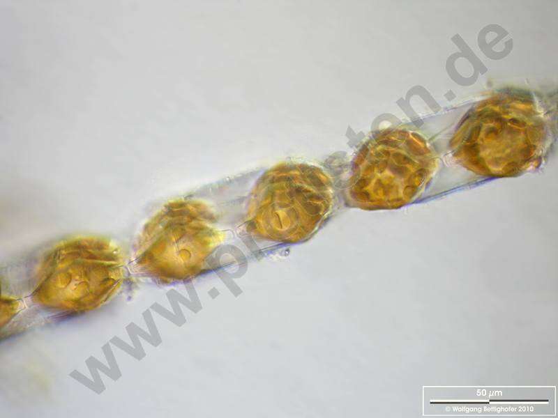
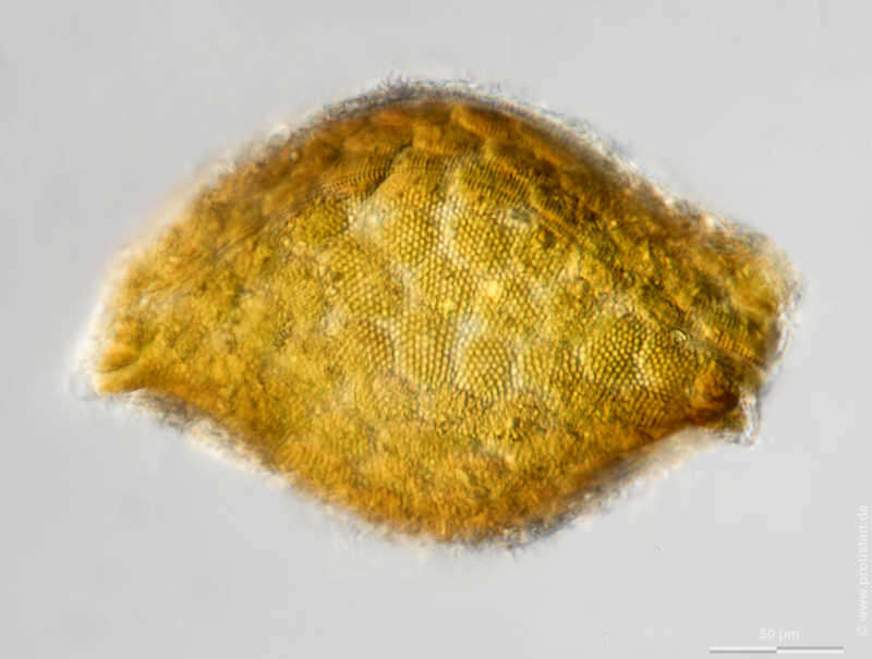
Odontella aurita.Depth-of-focus image exhibiting structure of the frustules (apical view). Oblique light. Scale bar indicates 50 m. The image was built up using several photomicrographic frames with manual stacking technique. Sample from North Sea near Heligoland (spring diatom bloom). Images were taken using Zeiss Universal with Olympus C7070 CCD camera.For more look at
www.protisten.de/english/gallery_main/gallery_main.htmlFor high-resolution images please ask postmaster@protisten.de.
-
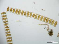
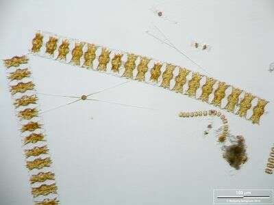
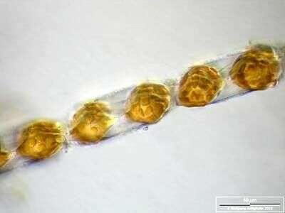
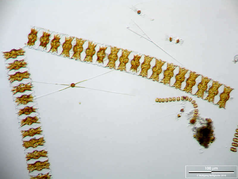
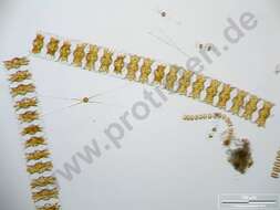
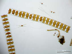
Long chains of Odontella aurita accompanied by Chaetoceros danicus and Thalassiosira nordenskjoeldii. Scale bar indicates 100 µm. The image was built up using several photomicrographic frames with manual stacking technique. Sample from North Sea near Heligoland (spring diatom bloom). Images were taken using Zeiss Universal with Olympus C7070 CCD camera.
-
Long chains of Odontella aurita accompanied by Chaetoceros danicus and Thalassiosira nordenskjoeldii. Scale bar indicates 100 m. The image was built up using several photomicrographic frames with manual stacking technique. Sample from North Sea near Heligoland (spring diatom bloom). Images were taken using Zeiss Universal with Olympus C7070 CCD camera.For more look at
www.protisten.de/english/gallery_main/gallery_main.htmlFor high-resolution images please ask postmaster@protisten.de.
-
Depth-of-focus image exhibiting structure of the frustules (apical view). Oblique light. Scale bar indicates 50 µm. The image was built up using several photomicrographic frames with manual stacking technique. Sample from North Sea near Heligoland (spring diatom bloom). Images were taken using Zeiss Universal with Olympus C7070 CCD camera.
-
-
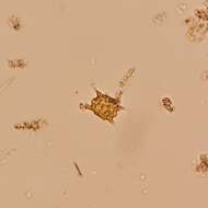
Description: English: Odontella mobiliensis preserved by lugol solution. A field sample from Long Island Sound. Date: 17 September 2013. Source:
flickr. Author: NOAA Photo Library.
-
Richard A. Ingebrigtsen, Department of Arctic and Marine Biology, University of Tromsø
Wikimedia Commons
-
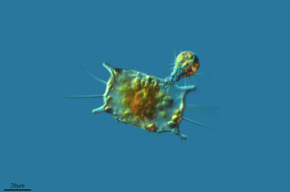
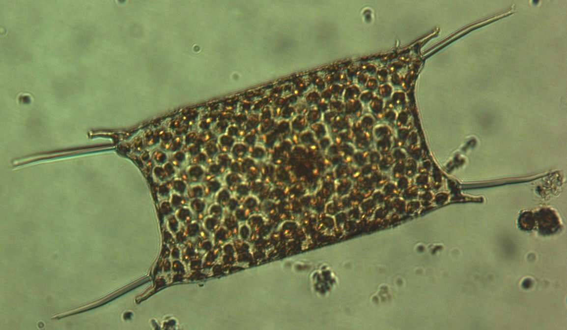
Odontella rhombus Sample from North Sea near Heligoland (spring diatom bloom). Scale bar indicates 50 µm.The image was built up using several photomicrographic frames with manual stacking technique. Images were taken using Zeiss Axioplan with MFT camera Olympus OM-D E-M5 II.Image under Creative Commons License V 3.0 (CC BY-NC-SA). Place name: North Sea around Heligoland Latitude: 54.186311 Longitude: 7.895034 Multiebenen-Abbildung, manuell gestapelt. Der Messbalken markiert eine Länge von 50 µm. Probe aus der Nordsee vor Helgoland in der Zeit der Frühjahrsblüte. Mikrotechnik: Zeiss Axioplan Kamera: Olympus OM-D E-M5 II.Creative Commons License V 3.0 (CC BY-NC-SA). For permission to use of (high-resolution) images please contact postmaster@protisten.de.
-

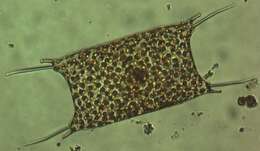
Odontella aurita Depth-of-focus image exhibiting structure of the frustules. Chloroplasts are concentrated in cell centers due to long lasting exposition with microscope's illumination. Scale bar indicates 25 µm. The image was built up using several photomicrographic frames with manual stacking technique. Sample from North Sea near Heligoland (spring diatom bloom). Images were taken using Zeiss Universal with Olympus C7070 CCD camera.Image under Creative Commons License V 3.0 (CC BY-NC-SA). Place name: North Sea around Heligoland Latitude: 54.186311 Longitude: 7.895034 Die Multiebenen-Abbildung zeigt die Schalenstruktur. Die Chloroplasten haben sich nach einiger Zeit intensiver Beleuchtung in der Zellmitte nahe dem Kern gesammelt. Der Messbalken markiert eine Länge von 25 µm. Probe aus der Nordsee vor Helgoland in der Zeit der Frühjahrsblüte. Mikrotechnik: Zeiss Universal, Kamera: Olympus C7070.Creative Commons License V 3.0 (CC BY-NC-SA). For permission to use of (high-resolution) images please contact postmaster@protisten.de.
-
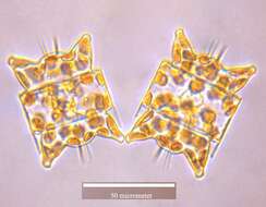
Odontella aurita Depth-of-focus image exhibiting structure of the frustules (apical view). Oblique light. Scale bar indicates 50 µm. The image was built up using several photomicrographic frames with manual stacking technique. Sample from North Sea near Heligoland (spring diatom bloom). Images were taken using Zeiss Universal with Olympus C7070 CCD camera.Image under Creative Commons License V 3.0 (CC BY-NC-SA). Place name: North Sea around Heligoland Latitude: 54.186311 Longitude: 7.895034 Die Multiebenen-Abbildung zeigt die Schalenstruktur (Apikalsicht). Multiebenen-Abbildung, manuell gestapelt. Der Messbalken markiert eine Länge von 50 µm. Probe aus der Nordsee vor Helgoland in der Zeit der Frühjahrsblüte. Mikrotechnik: Zeiss Universal, Kamera: Olympus C7070.Creative Commons License V 3.0 (CC BY-NC-SA). For permission to use of (high-resolution) images please contact postmaster@protisten.de.
-
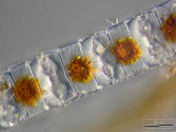
Odontella aurita Long chains of Odontella aurita accompanied by Chaetoceros danicus and Thalassiosira nordenskjoeldii. Scale bar indicates 100 µm. The image was built up using several photomicrographic frames with manual stacking technique. Sample from North Sea near Heligoland (spring diatom bloom). Images were taken using Zeiss Universal with Olympus C7070 CCD camera.Image under Creative Commons License V 3.0 (CC BY-NC-SA). Place name: North Sea around Heligoland Latitude: 54.186311 Longitude: 7.895034 Lange Zellketten von Odontella aurita zusammen mit Chaetoceros danicus und Thalassiosira nordenskjoeldii. Multiebenen-Abbildung, manuell gestapelt. Der Messbalken markiert eine Länge von 100 µm. Probe aus der Nordsee vor Helgoland in der Zeit der Frühjahrsblüte. Mikrotechnik: Zeiss Universal, Kamera: Olympus C7070.Creative Commons License V 3.0 (CC BY-NC-SA). For permission to use of (high-resolution) images please contact postmaster@protisten.de.
-
Odontella aurita Scale bar indicates 10 µm. Sample from North Sea near Heligoland (spring diatom bloom). Use of SEM equipment courtesy of Lab Dr. Karl-Heinz Schäffner, Solingen, Germany. Place name: North Sea around Heligoland Latitude: 54.186311 Longitude: 7.895034 Der Messbalken markiert eine Länge von 10 µm. Probe aus der Nordsee vor Helgoland in der Zeit der Frühjahrsblüte. Die Aufnahme entstand im Labor Dr. Karl-Heinz Schäffner, Solingen.Creative Commons License V 3.0 (CC BY-NC-SA). For permission to use of (high-resolution) images please contact postmaster@protisten.de.
-
Odontella aurita Scale bar indicates 25 µm. Sample from North Sea near Heligoland (spring diatom bloom). Use of SEM equipment courtesy of Lab Dr. Karl-Heinz Schäffner, Solingen, Germany. Place name: North Sea around Heligoland Latitude: 54.186311 Longitude: 7.895034 Der Messbalken markiert eine Länge von 25 µm. Probe aus der Nordsee vor Helgoland in der Zeit der Frühjahrsblüte. Die Aufnahme entstand im Labor Dr. Karl-Heinz Schäffner, Solingen.Creative Commons License V 3.0 (CC BY-NC-SA). For permission to use of (high-resolution) images please contact postmaster@protisten.de.