-
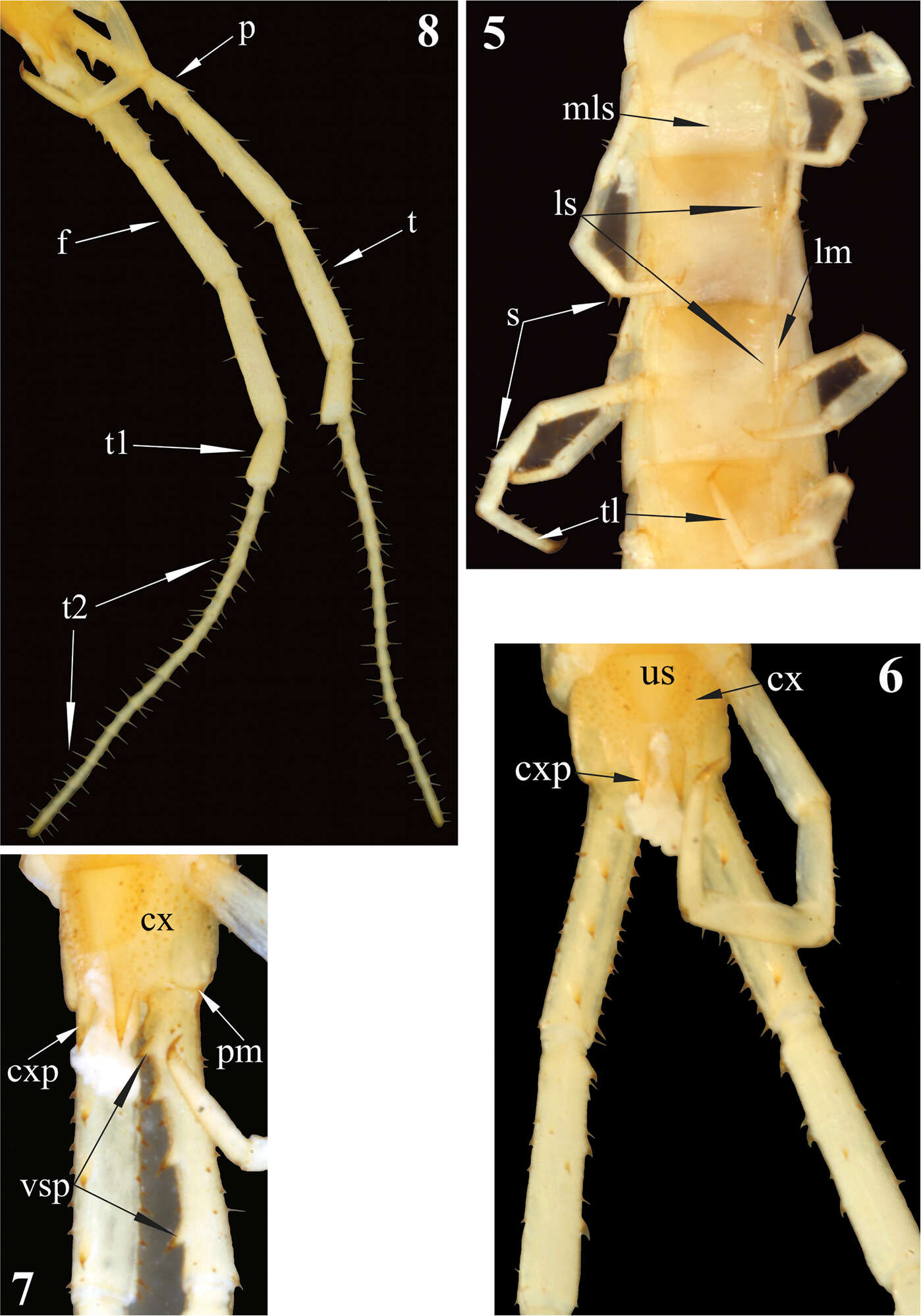
Figure 1–4.1 Rhysida celeris from Ecuador. Cephalic plate 2 Forcipular Coxosternum 3 Tooth plates 4 Forcipular trochanteroprefemur process. Scale bars 1 mm.
-
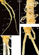
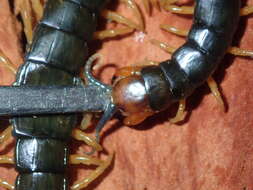
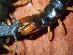
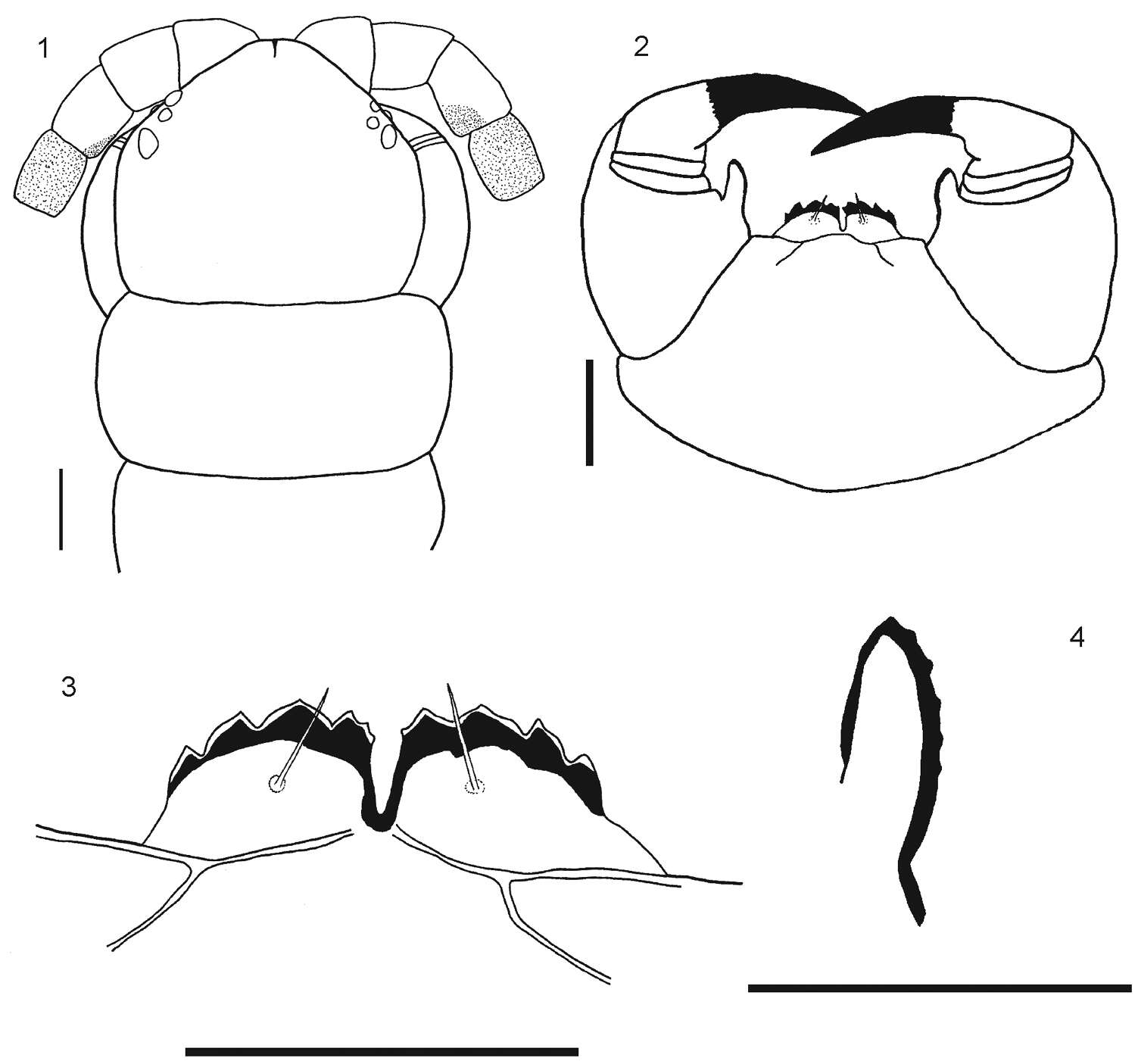
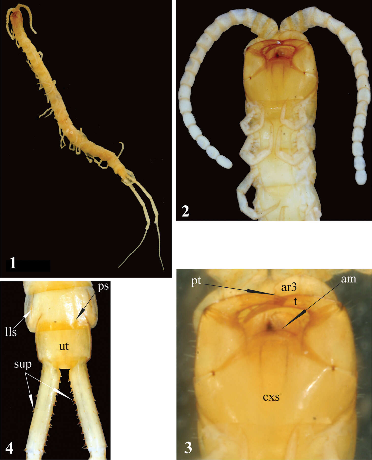
Figures 1–4.Newportia stoevi,sp. n. 1 Habitus 2 Head and anterior segments, ventral view 3 Forcipular segment, ventral view 4 Tergites 22 and 23 and prefemora of ultimate legs, dorsal view; (pt) – pretarsus of second maxilla, (ar3) – article 3 of telopodite of second maxilla, (cxs) – forcipular coxosternite, (am) – anterior margin of coxosternite, (t) – tarsungulum, (ps) – paramedian sutures, (lls) – lateral longitudinal sutures, (ut) – tergite of ultimate leg-bearing segment, (sup) – spurs of ultimate prefemur.
-
Figure 5–8. 5 Tergites 11, 12 and 13 6 Sternites 4, 5 and 6 7 Tergite 21 8 Segment 21 showing sternite 21 and coxopleuron. Scale bars 1 mm.
-
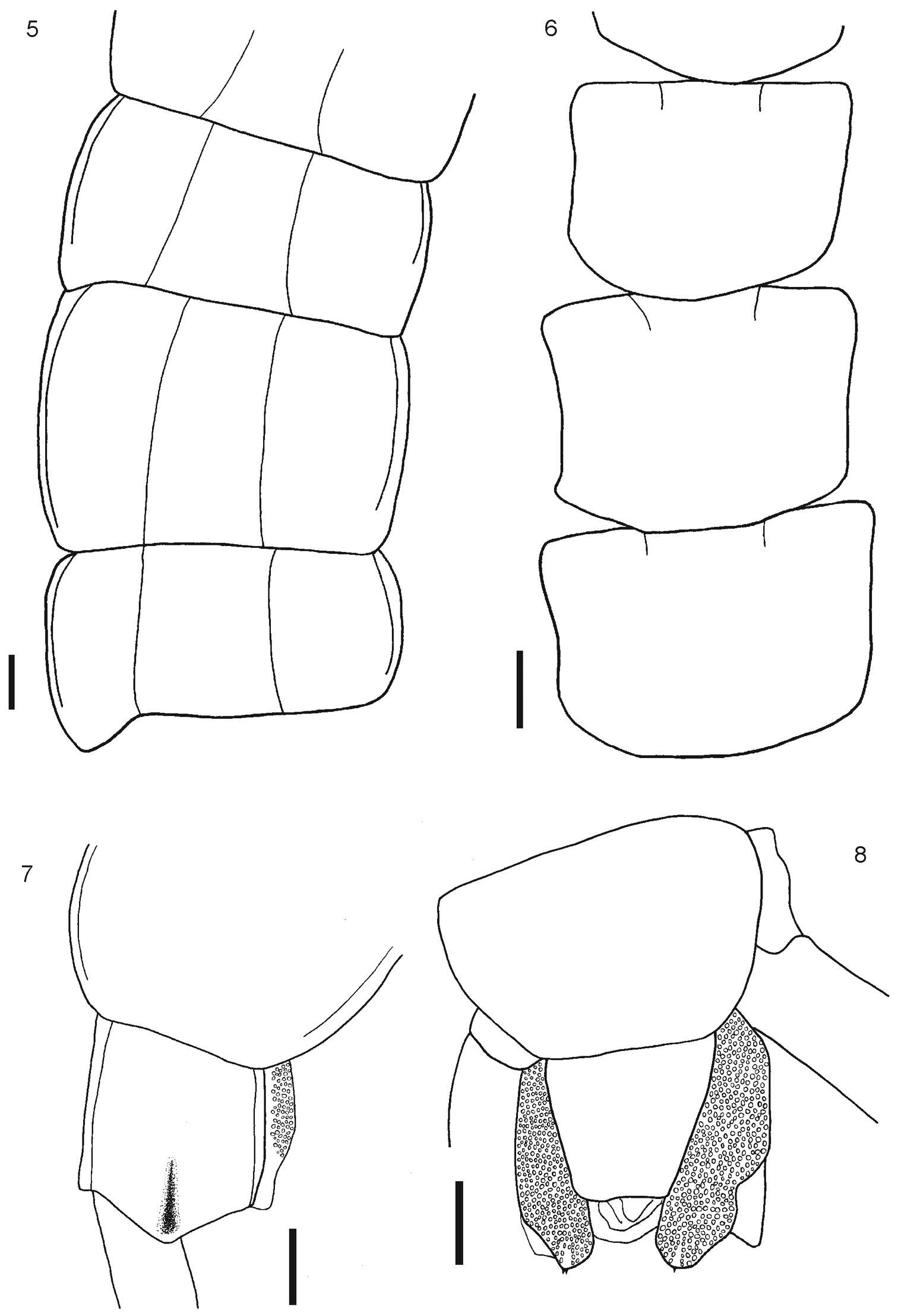
Figures 5–8.Newportia stoevi,sp. n. 5 Segments and midbody legs, ventral view 6 Posterior body end, ventral view 7 Left side of ultimate leg-bearing segment and prefemora of ultimate legs, ventro-lateral view 8 Ultimate legs, ventro-lateral view; (mls) – median longitudinal sulcus, (ls) – lateral sutures, (lm) – lateral margination, (s) – setae, (tl) – monoarticulated tarsus of locomotory leg, (us) – sternite of ultimate leg-bearing segment, (cx) – coxopleuron, (cxp) – coxopleural process, (pm) – posterior margin of pleuron of ultimate leg-bearing segment, (vsp) – ventral spinous processes of ultimate prefemur, (p) – prefemur, (f) – femur, (t) – tibia, (t1) – tarsus 1, (t2) – tarsus 2.
-
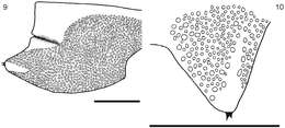
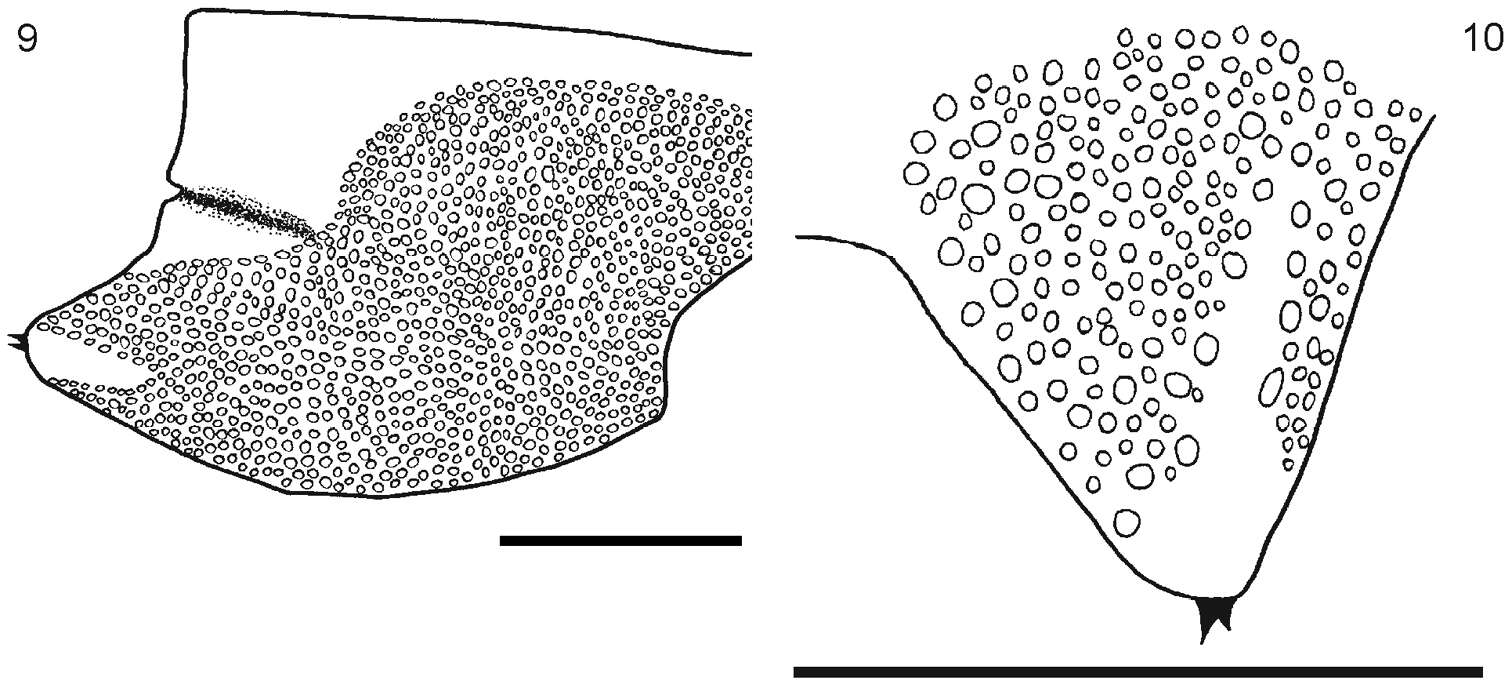
Figure 9–10. 9 Segment 21 showing the coxopleuron 10 Detail of the terminal part of the coxopleuron showing the spines. Scale bar 1 mm.
-
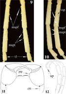
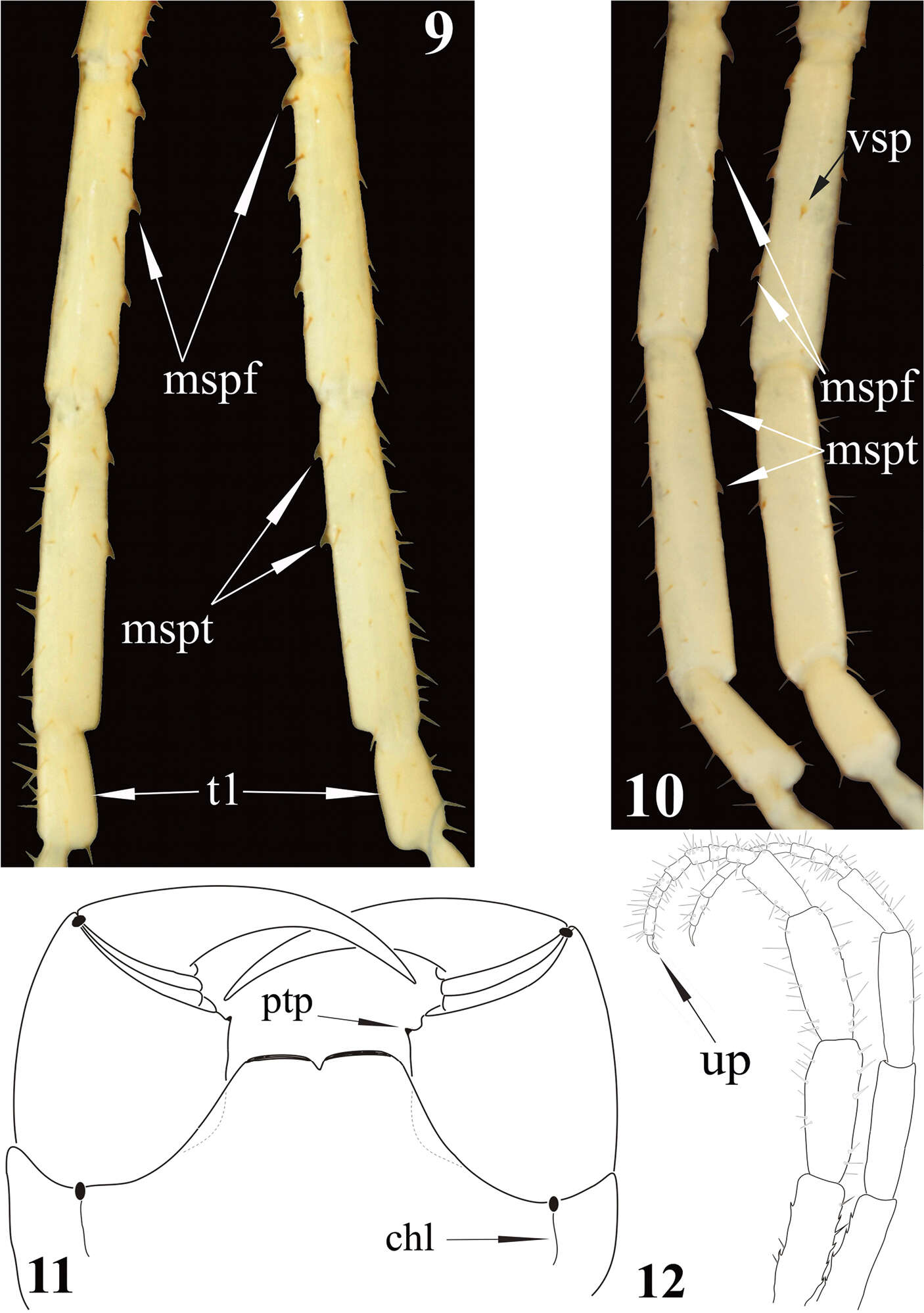
Figures 9–12.Newportia stoevi,sp. n. 9 Femora, tibiae and tarsi 1 of ultimate legs, dorsal view 10 Femora, tibiae and tarsi 1 of ultimate legs, ventral view; Newportia divergens Chamberlin, 1922 11 Forcipular segment, ventral view (after Schileyko and Minelli 1998); Newportia unguifer Chamberlin, 1921 12 Ultimate legs, dorso-lateral view (after Schileyko and Minelli 1998); (mspf) – medial spinous processes of ultimate femur, (mspt) – medial spinous processes of ultimate tibia, (vsp) – ventral spinous process of ultimate femur, (t1) – tarsus 1, (up) – ultimate pretarsus, (chl) – chitin-lines, (ptp) – process of trochanteroprefemur.
-
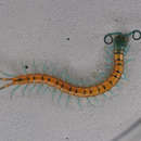
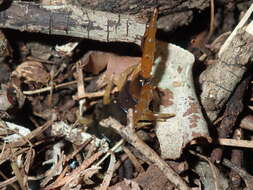
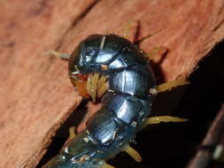
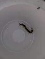
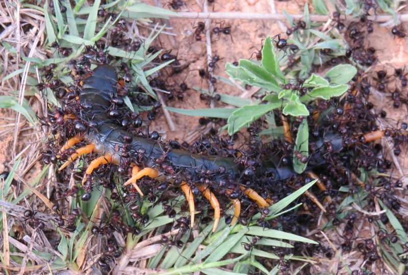
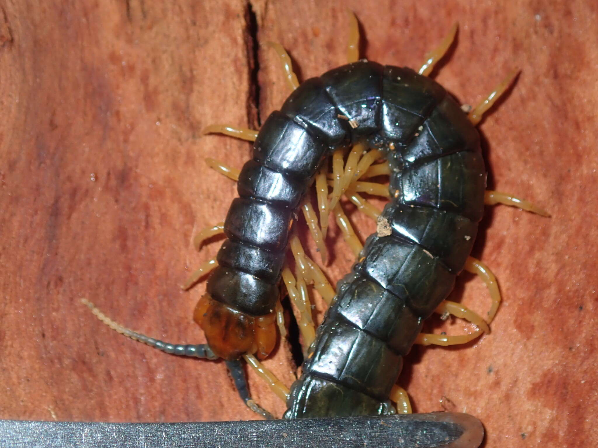
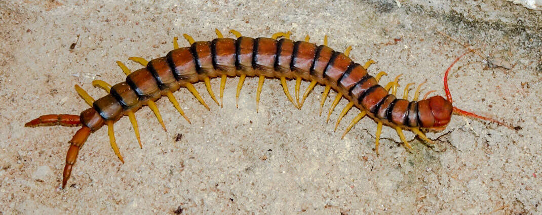
Being attacked by ants. Tentative i.d.
-
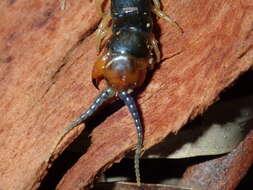
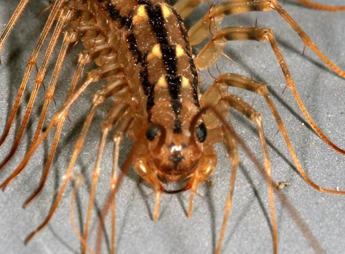
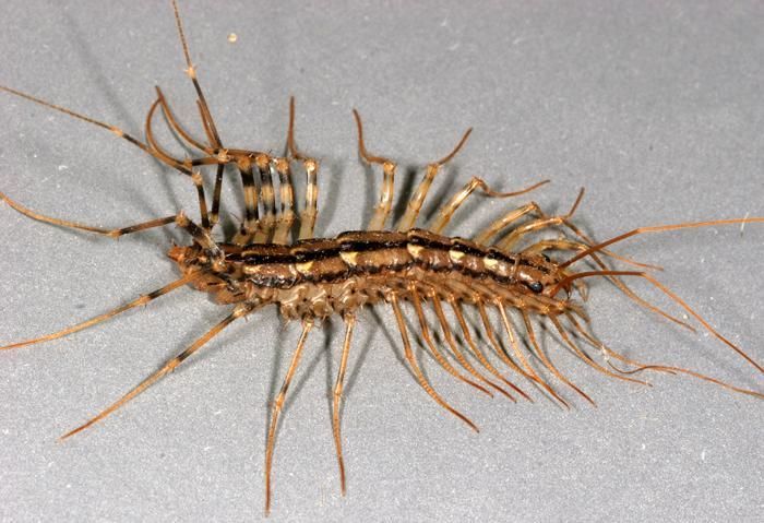
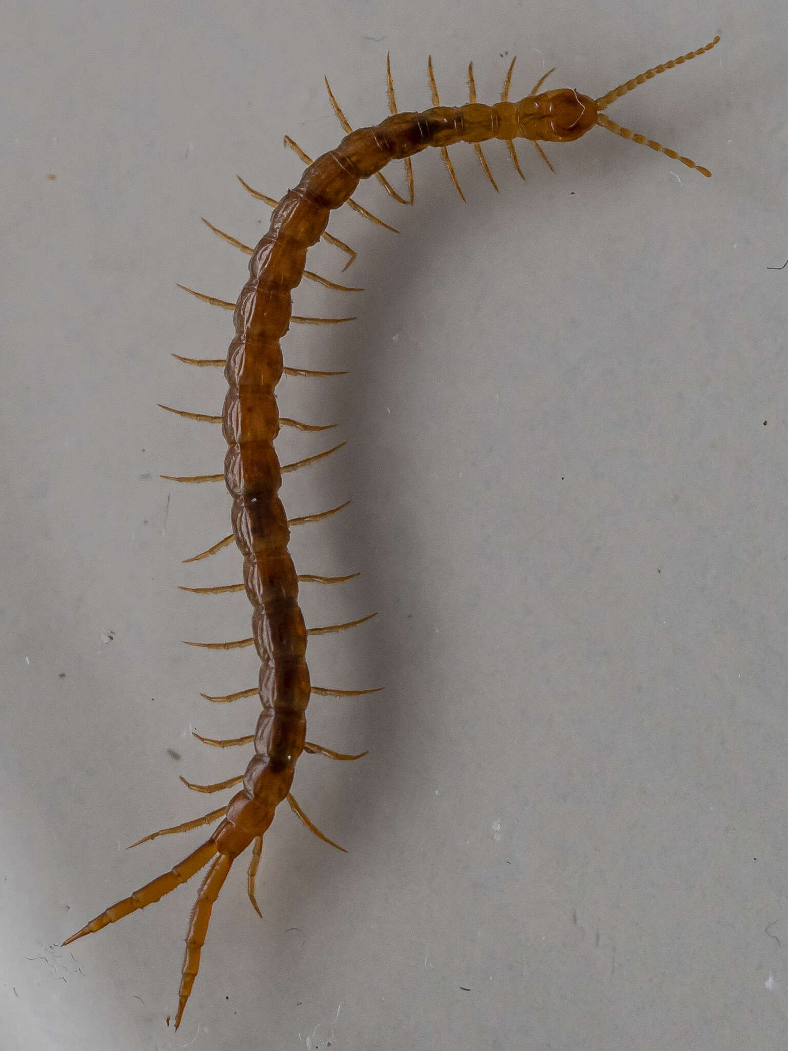
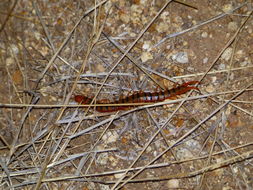
Tentative i.d.
-
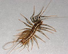
Photographed by Harvard University, Dept. of Environmental Health and Safety entomologist/environmental biologist, Dr. Gary Alpert, this 2006 image depicted a anterosuperior view of a common house centipede, Scutigera coleoptrata. See PHIL 9823, and 9824 for two additional views of this animal. Each segment of a centipedes body possesses a single pair of legs. Emanating from the head is a pair of sensorial antennae, and its three pairs of mouthparts, consisting of paired mandibulae with teeth, maxillae, and palps. Emanating from just behind its head, and visible in this image, is this arthropods first pair of legs which has evolved into a pair of fangs. Although a rare occurrence, when it does bite its victims, the centipede ejects its venom, which is not generally toxic, although it may cause extreme pain to humans.Created: 2006
-
Photographed by Harvard University, Dept. of Environmental Health and Safety entomologist/environmental biologist, Dr. Gary Alpert, this 2006 image depicted a dorsoposterior view of a common house centipede, Scutigera coleoptrata. Youll note that a number of its legs are missing. However, these appendages may easily be detached if grasped by an enemy, and will continue to wriggle, in order to distract predators. See PHIL 9823, and 9825 for two additional views of this animal. Each segment of a centipedes body possesses a single pair of legs. Emanating from the head is a pair of sensorial antennae, and its three pairs of mouthparts, consisting of paired mandibulae with teeth, maxillae, and palps. Located behind its head, this arthropods first pair of legs has evolved into a pair of fangs. Although a rare occurrence, when it does bite its victims, the centipede ejects its venom, which is not generally toxic, although it may cause extreme pain to humans.Created: 2006
-
Photographed by Harvard University, Dept. of Environmental Health and Safety entomologist/environmental biologist, Dr. Gary Alpert, this 2006 image depicted a dorsolateral view of a common house centipede, Scutigera coleoptrata. Youll note that a number of its legs are missing. However, these appendages may easily be detached if grasped by an enemy, and will continue to wriggle, in order to distract predators. See PHIL 9824, and 9825 for two additional views of this animal. Each segment of a centipedes body possesses a single pair of legs. Emanating from the head is a pair of sensorial antennae, and its three pairs of mouthparts, consisting of paired mandibulae with teeth, maxillae, and palps. Located behind its head, this arthropods first pair of legs has evolved into a pair of fangs. Although a rare occurrence, when it does bite its victims, the centipede ejects its venom, which is not generally toxic, although it may cause extreme pain to humans.Created: 2006
-
-
-
-
-
-
-
-
-
-
-
-
-