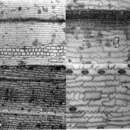
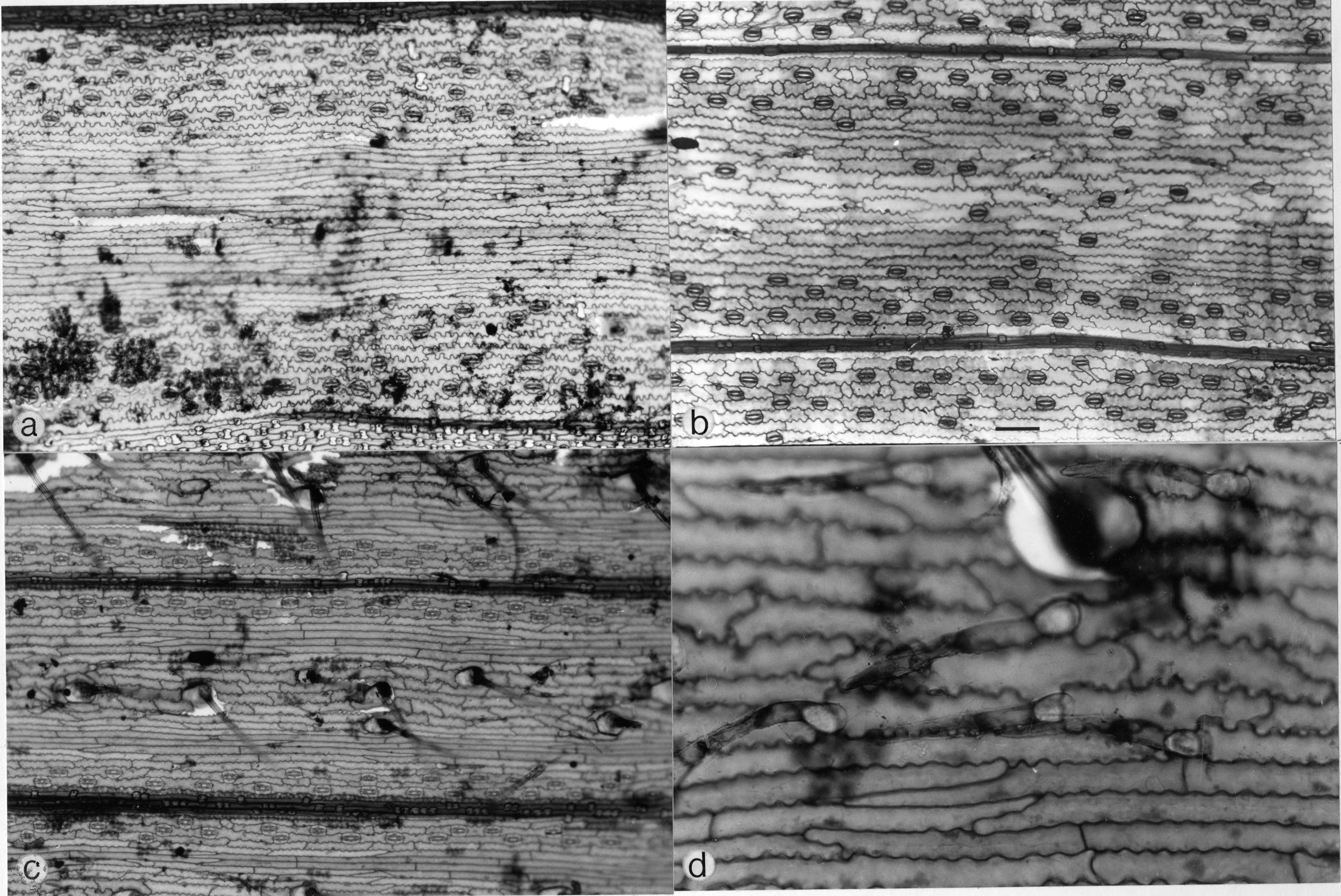
Leaf epidermides of Streptochaeta species: a,b, adaxial view of S. sodiroana showing (a) bulliform cells (below) and (b) structure of costal and isolated intercostal silica cells; c, adaxial view of S. angustifofia showing poorly differentiated bulliform cells; d, abaxial, S. spicata subsp. spicata, showing bicellular microhairs (arrows). (a, b based on Soderstrom 1205, Costa Rica; c based on Soderstrom and Sucre 1969, Brazil; d based on Soderstrom and Calderon 1861, Brazil.) Scale bar: a = 100 µm; b,d = 40 µm; c = 12a0 µm.
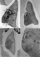
Median sagittal sections, embryos of Streptochaeta and Anomochloa: a, S. spicata subsp. spicata (Soderstrom and Sucre 1896, Brazil), showing poorly developed cleft between scutellum and coleorhiza (arrow); b, S. sodiroana (Calderon 2096, Panama), showing overlapping margins of coleoptile (arrow); c, A, marantoidea (dos Santos el al. 3880, Brazil), showing massive scutellum; d, detail of c showing abaxial position of cleft between coleorhiza and scutellum (arrow). Abbreviations: cp = coleoptile; cr = coleorhiza; ep = epiblast; If = first embryonic leaf; ra = radicle; sc = scutellum; vt = vascular trace. Scale bar: a,b,d = 100 µm; c = 25 µm.
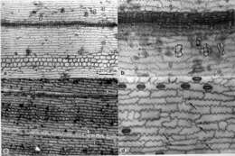
Abaxial epidermides of Streptochaeta species: a, S. sodiroana, showing elongate mid-intercostal long cells; b, S. spicata subsp. spicafa, showing shorter mid-intercostal long cells with more sinuous walls; c,d, S. angustifolia, showing (c) prominent mid-intercostal cilia and (4 bicellular microhairs. (a based on Soderstrom 1205, Costa Rica; b based on Soderstrom and Calderon 1861, Brazil; c, d based on Soderstrom and Sucre 1969, Brazil.) Scale bar: a = 60 µm: b = 70 p; c = 80 µm; d = 20 µm.