-
Wayne N. Mathis, Alessandra Rung, Marion Kotrba
Zookeys
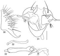
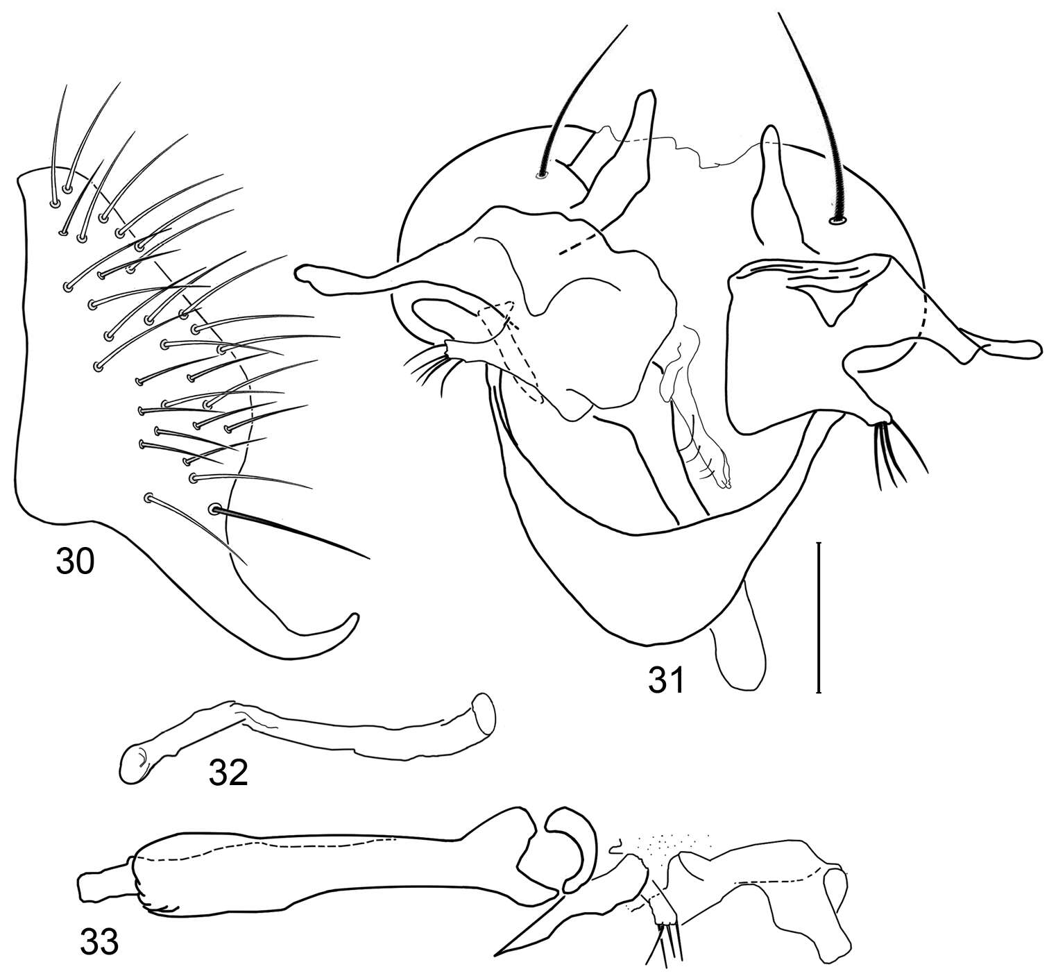
Figures 30–33.Illustrations of Planinasus miradorus sp. n. (male). 30 epandrium, surstylus, , lateral view 31 epandrium, hypandrium, and internal structures of male terminalia, ventral view 32 ejaculatory apodeme, lateral view 33 internal structures of male terminalia, lateral view. Scale bar = 0.1 mm.
-
Wayne N. Mathis, Alessandra Rung, Marion Kotrba
Zookeys
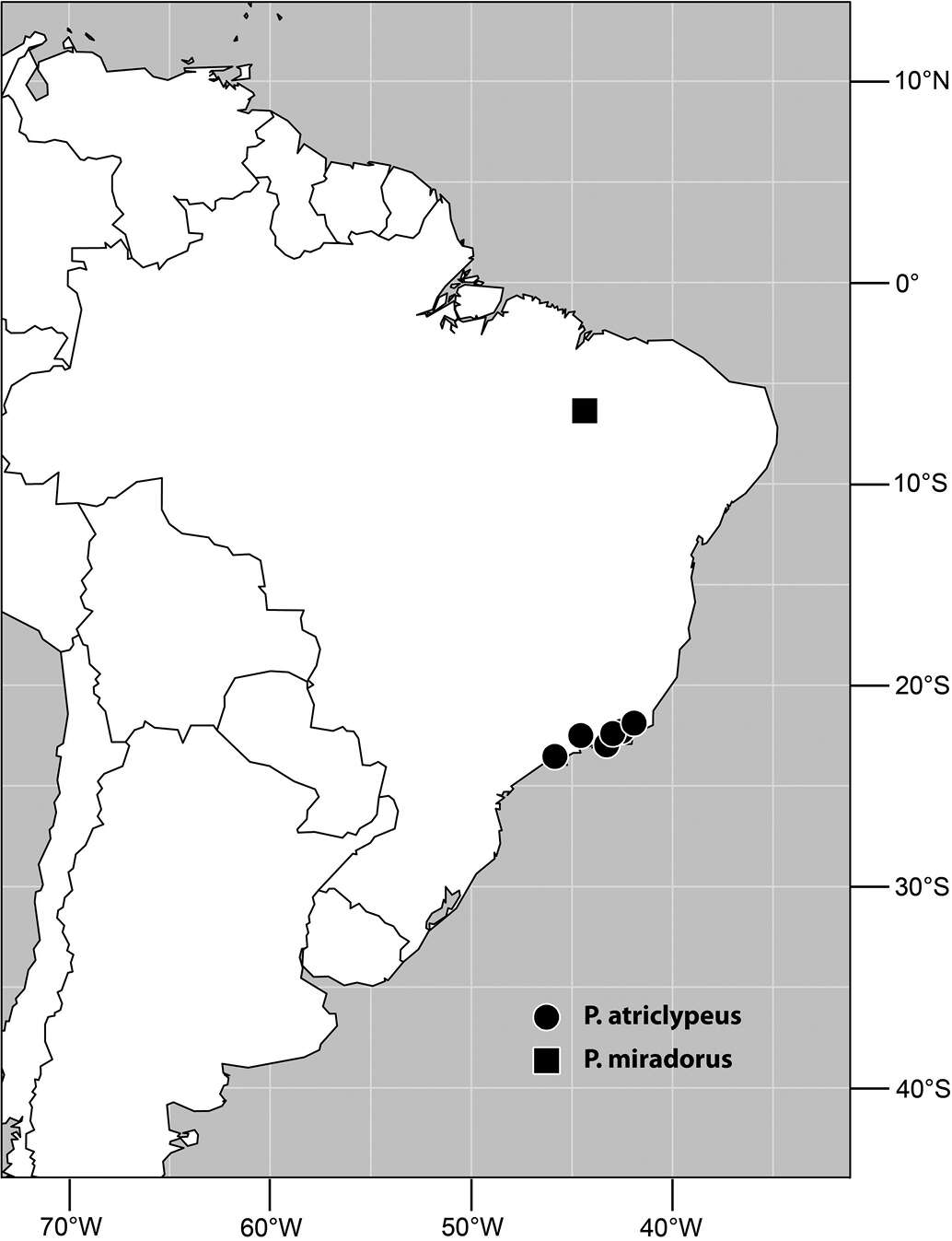
Figure 34.Distribution of Planinasus miradorus sp. n. (square) and Planinasus atriclypeus (dots).
-
Wayne N. Mathis, Alessandra Rung, Marion Kotrba
Zookeys
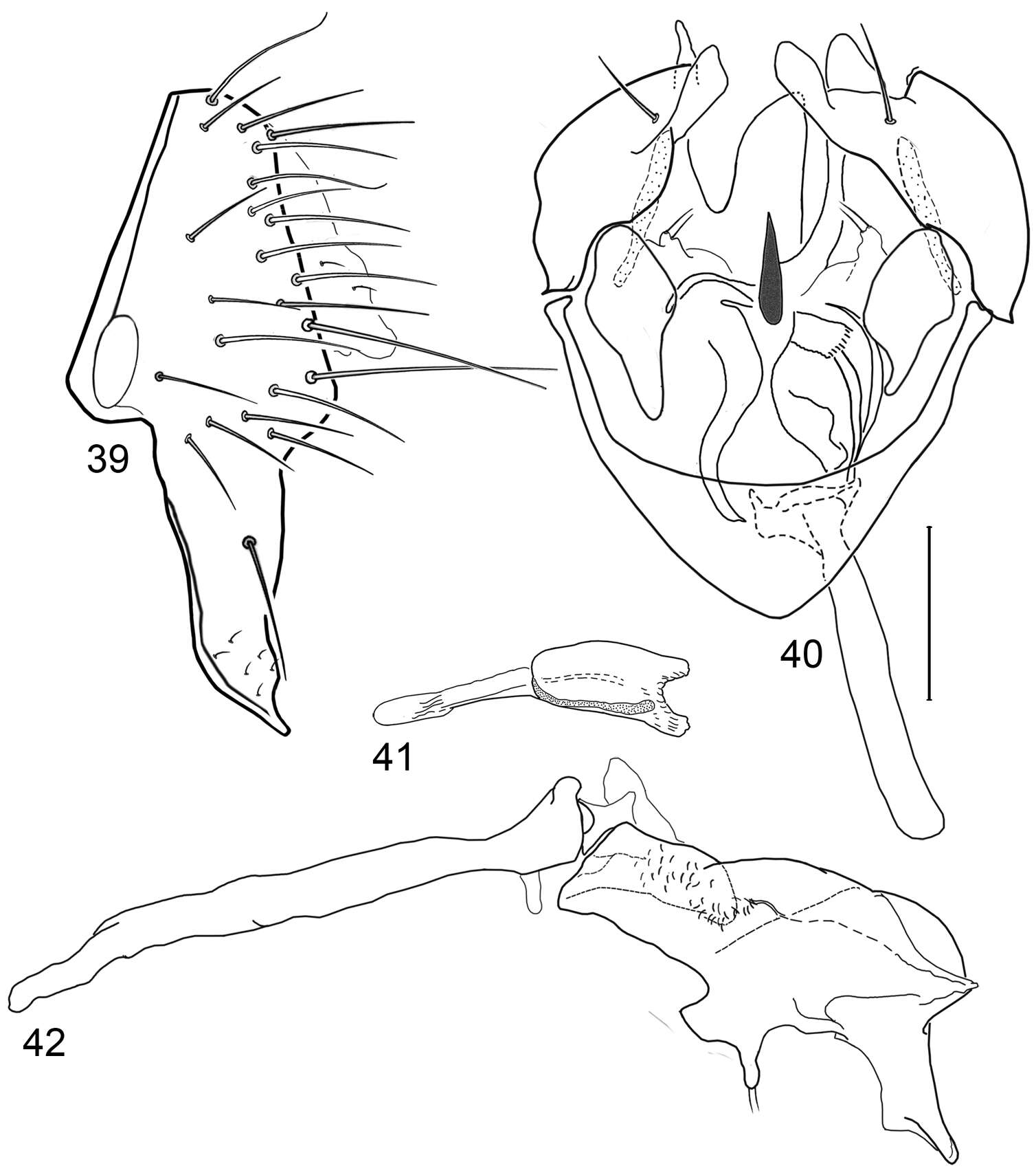
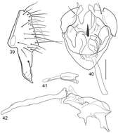
Figures 39–42.Illustrations of Planinasus tobagoensis sp. n. (male). 39 epandrium, surstylus, lateral view 40 structures of internal male terminalia, ventral view 41 ejaculatory apodeme, lateral view 42 internal structures of male terminalia, lateral view. Scale bar = 0.1 mm.
-
Wayne N. Mathis, Alessandra Rung, Marion Kotrba
Zookeys
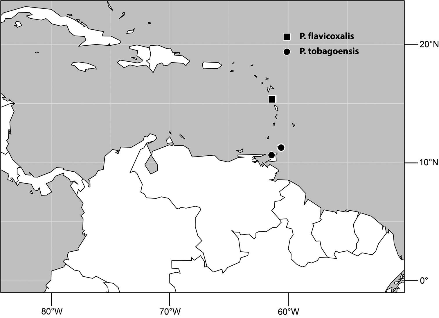
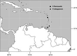
Figure 43.Distribution of Planinasus tobagoensis sp. n. (dots) and Planinasus flavicoxalis sp. n. (square).
-
Wayne N. Mathis, Alessandra Rung, Marion Kotrba
Zookeys
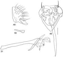
Figures 48–51.Illustrations of Planinasus xanthops sp. n. (male). 48 epandrium, surstylus, lateral view 49 structures of internal male terminalia, ventral view 50 ejaculatory apodeme, lateral view 51 phallus, phallapodeme, pre- and postgonite, lateral view. Scale bar = 0.1 mm.
-
Wayne N. Mathis, Alessandra Rung, Marion Kotrba
Zookeys
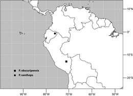
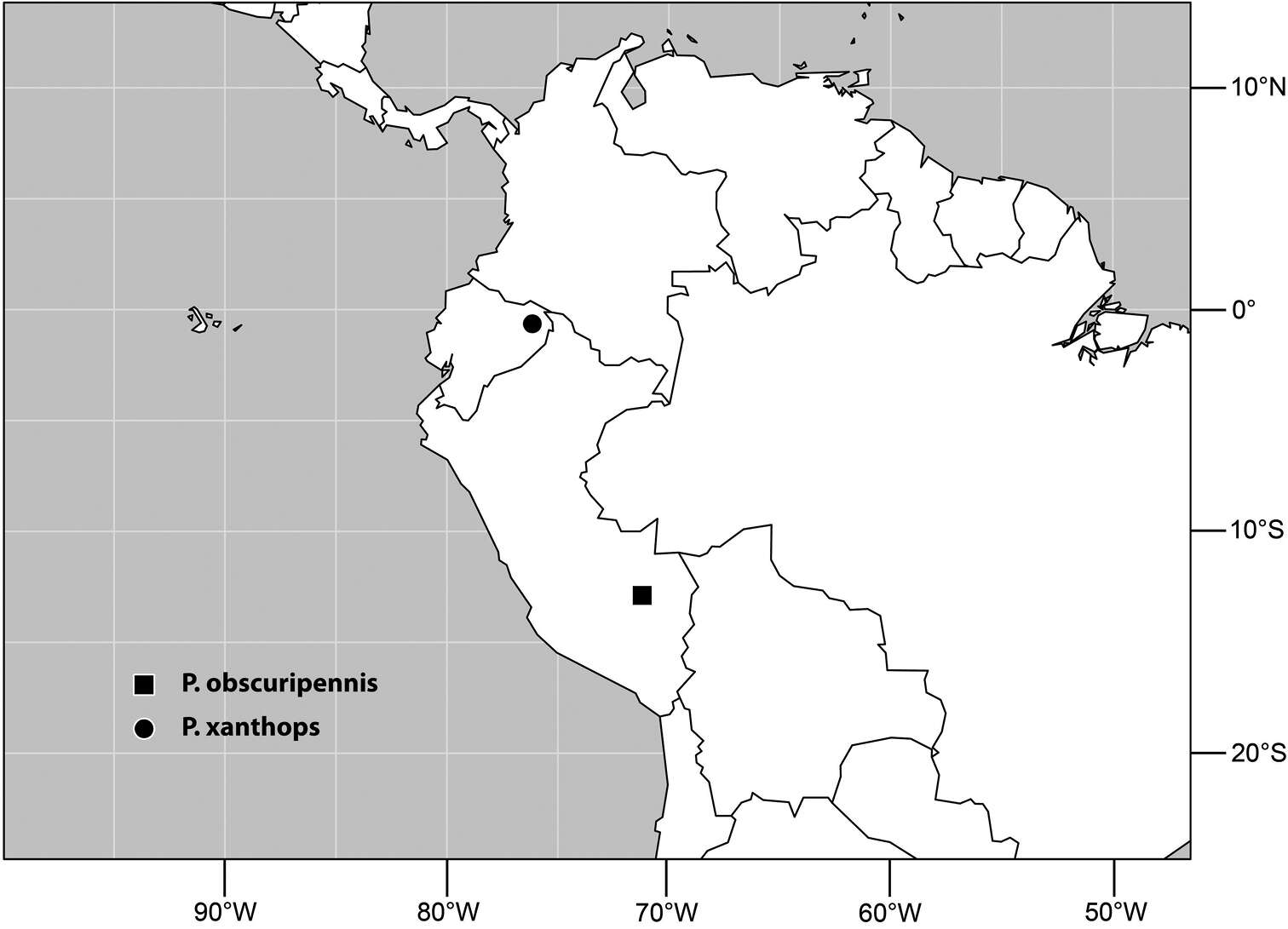
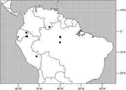
Figure 52.Distribution of Planinasus xanthops sp. n. (dot) and Planinasus obscuripennis sp. n. (square).
-
Wayne N. Mathis, Alessandra Rung, Marion Kotrba
Zookeys
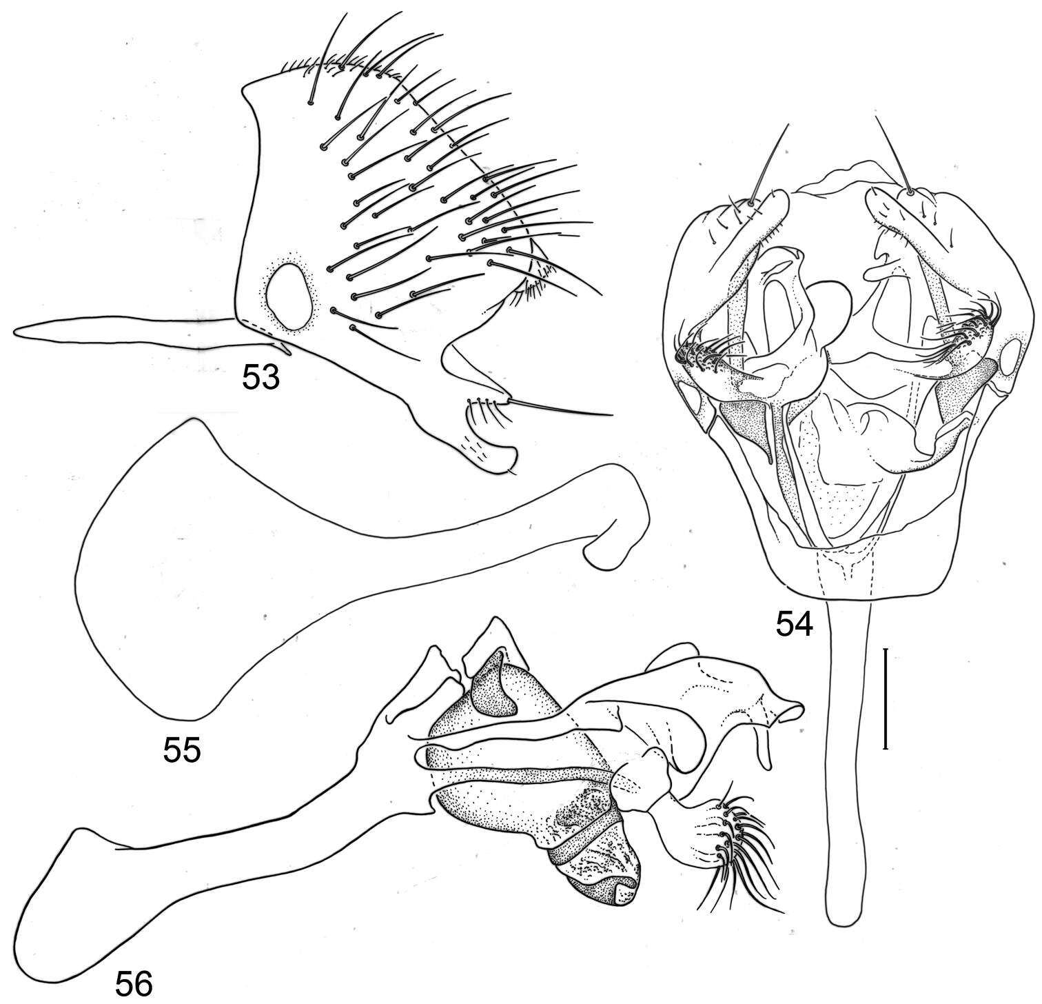
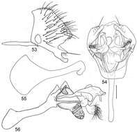
Figures 53–56.Illustrations of Planinasus argentifacies sp. n. (male). 53 epandrium, surstylus, and hypandrium, lateral view 54 structures of internal male terminalia, ventral view 55 ejaculatory apodeme, lateral view 56 internal structures of male terminalia, lateral view. Scale bar = 0.1 mm.
-
Wayne N. Mathis, Alessandra Rung, Marion Kotrba
Zookeys
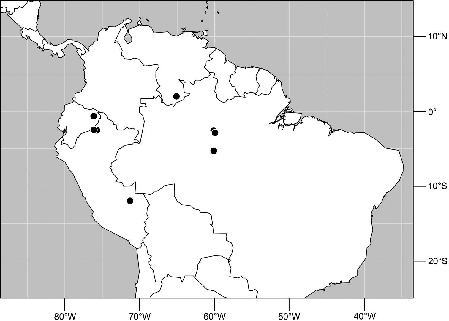
Figure 57.Distribution of Planinasus argentifacies sp. n.
-
Wayne N. Mathis, Alessandra Rung, Marion Kotrba
Zookeys
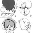
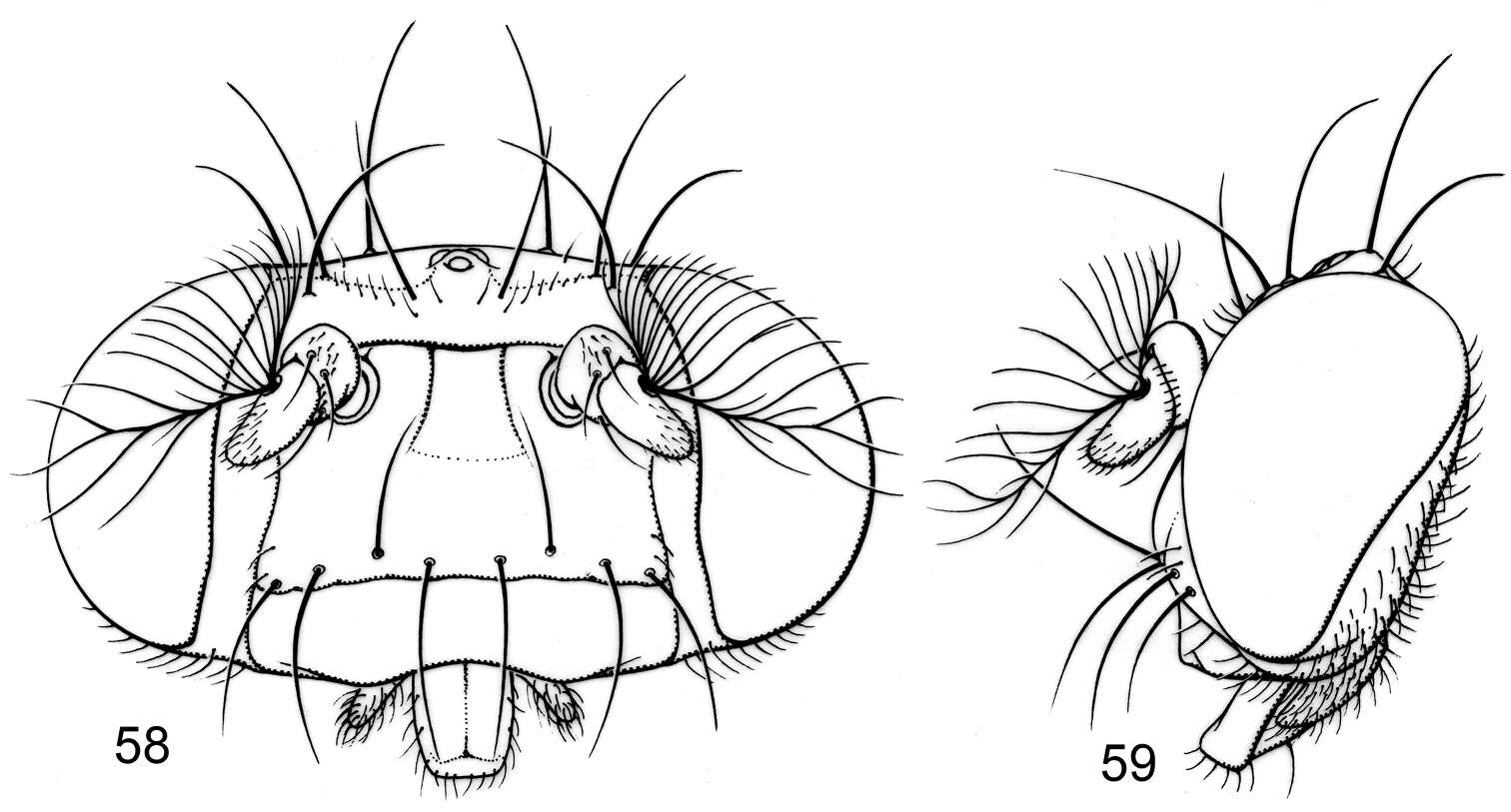
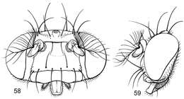
Figures 58–59.Illustrations of Planinasus insulanus sp. n. (male). 58 head, anterior view 59 same, lateral view.
-
Wayne N. Mathis, Alessandra Rung, Marion Kotrba
Zookeys
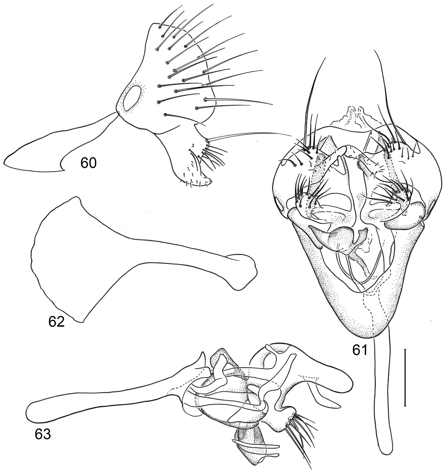
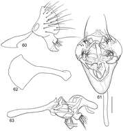
Figures 60–63.Illustrations of Planinasus insulanus sp. n. (male). 60 epandrium, surstylus, lateral view 61 structures of internal male terminalia, ventral view 62 ejaculatory apodeme, lateral view 63 internal structures of male terminalia, lateral view. Scale bar = 0.1 mm.
-
Wayne N. Mathis, Alessandra Rung, Marion Kotrba
Zookeys
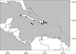
Figure 64.Distribution of Planinasus insulanus sp. n.
-
Wayne N. Mathis, Alessandra Rung, Marion Kotrba
Zookeys
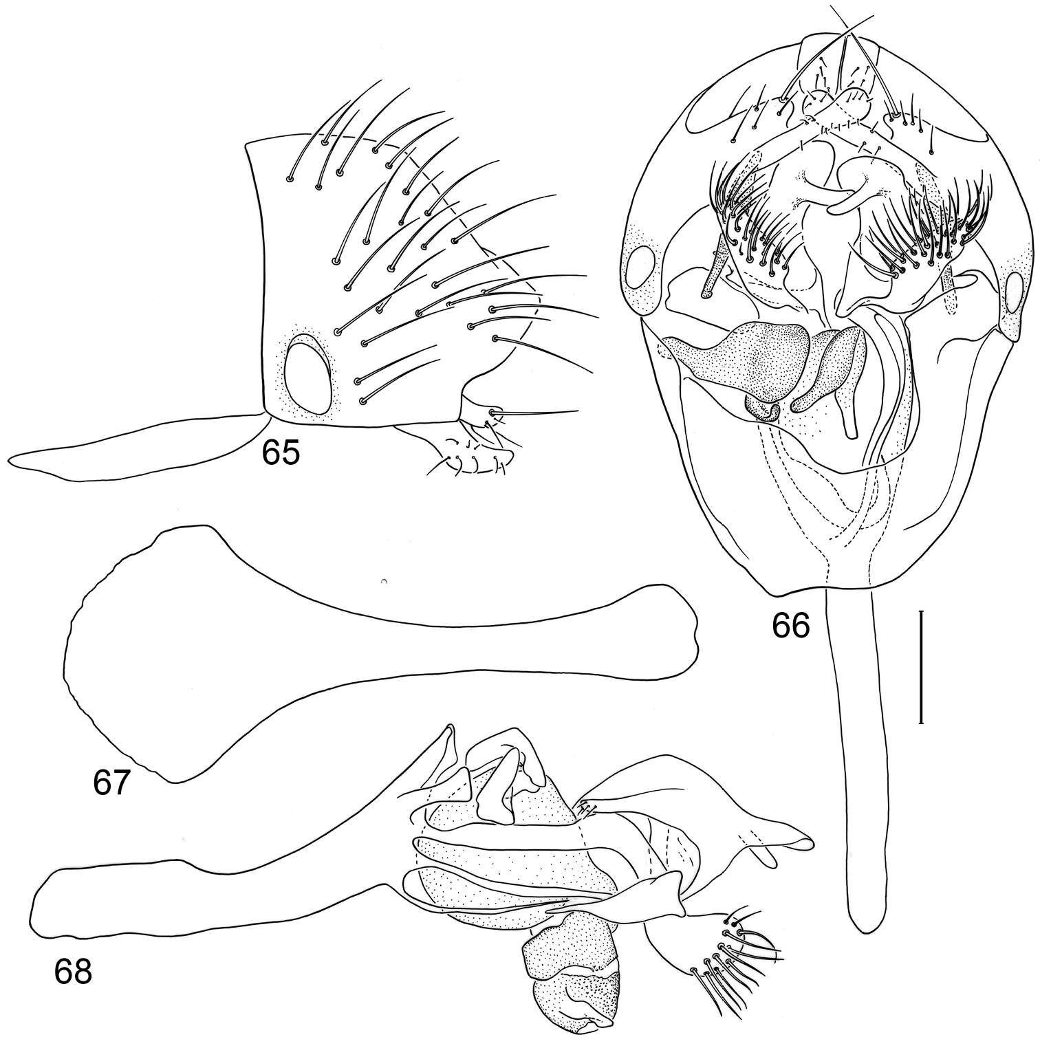
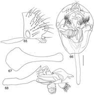
Figures 65–68.Illustrations of Planinasus nigritarsus sp. n. (male). 65 epandrium, surstylus, lateral view 66 structures of internal male terminalia, ventral view 67 ejaculatory apodeme, lateral view 68 internal structures of male terminalia, lateral view. Scale bar = 0.1 mm.
-
Wayne N. Mathis, Alessandra Rung, Marion Kotrba
Zookeys
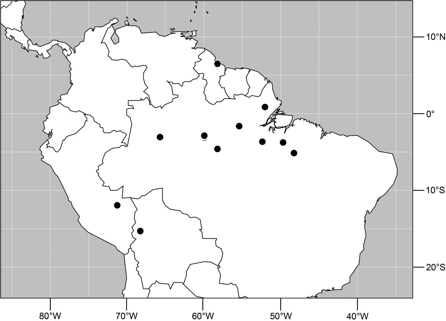
Figure 69.Distribution of Planinasus nigritarsus sp. n.
-
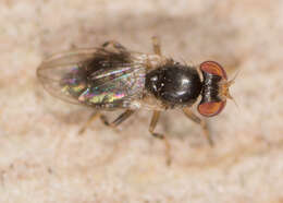
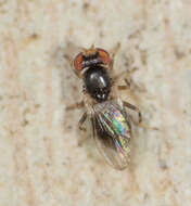
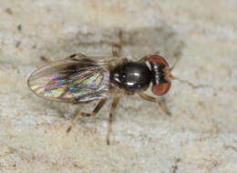
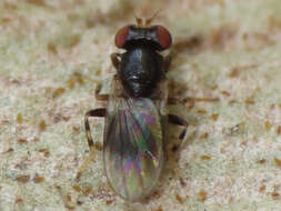
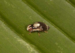
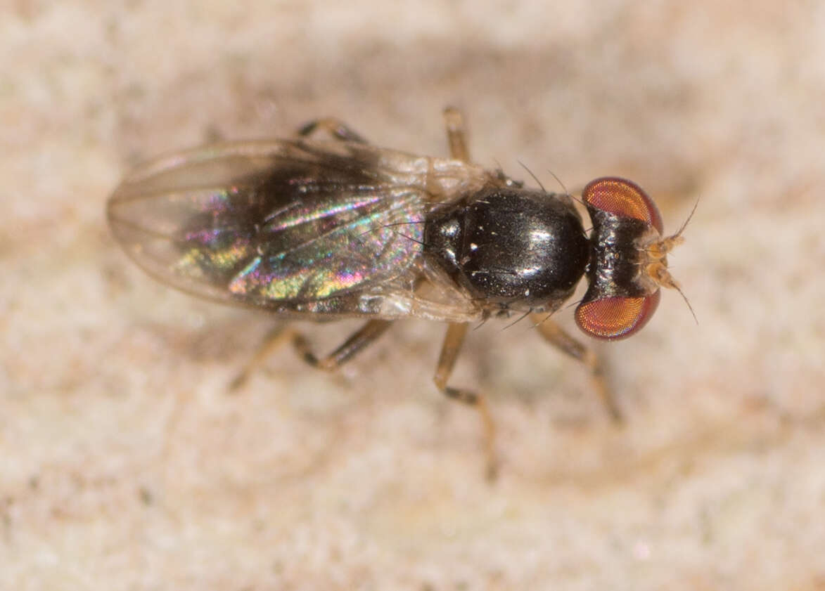
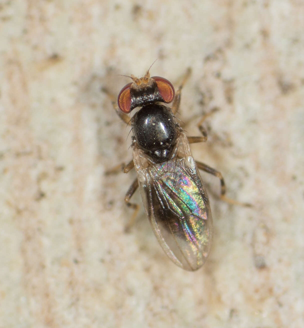
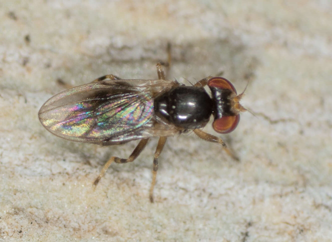
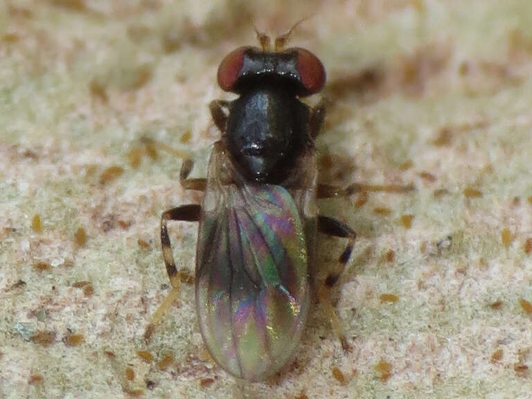
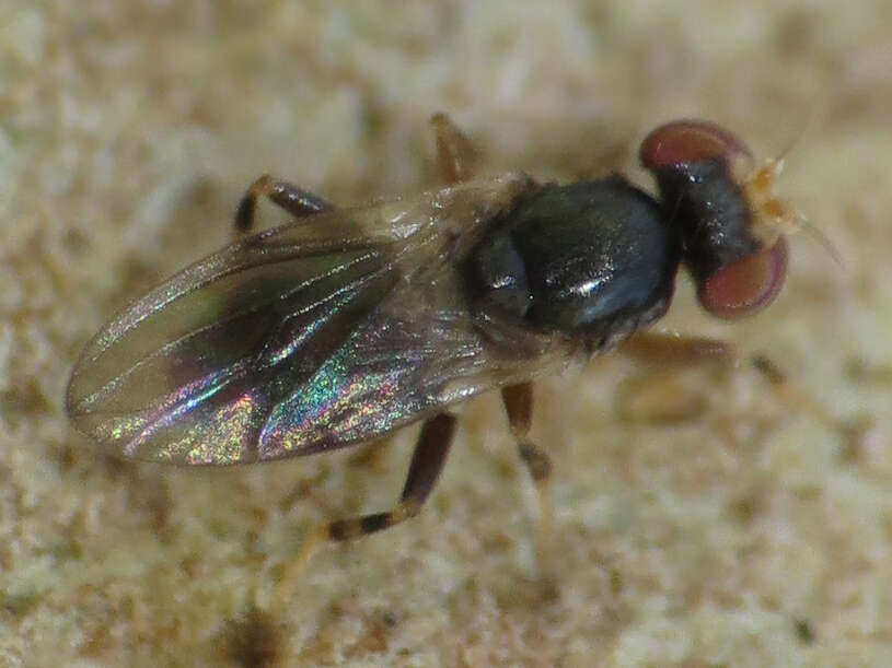
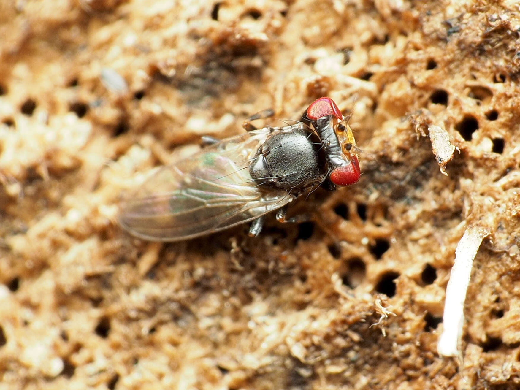
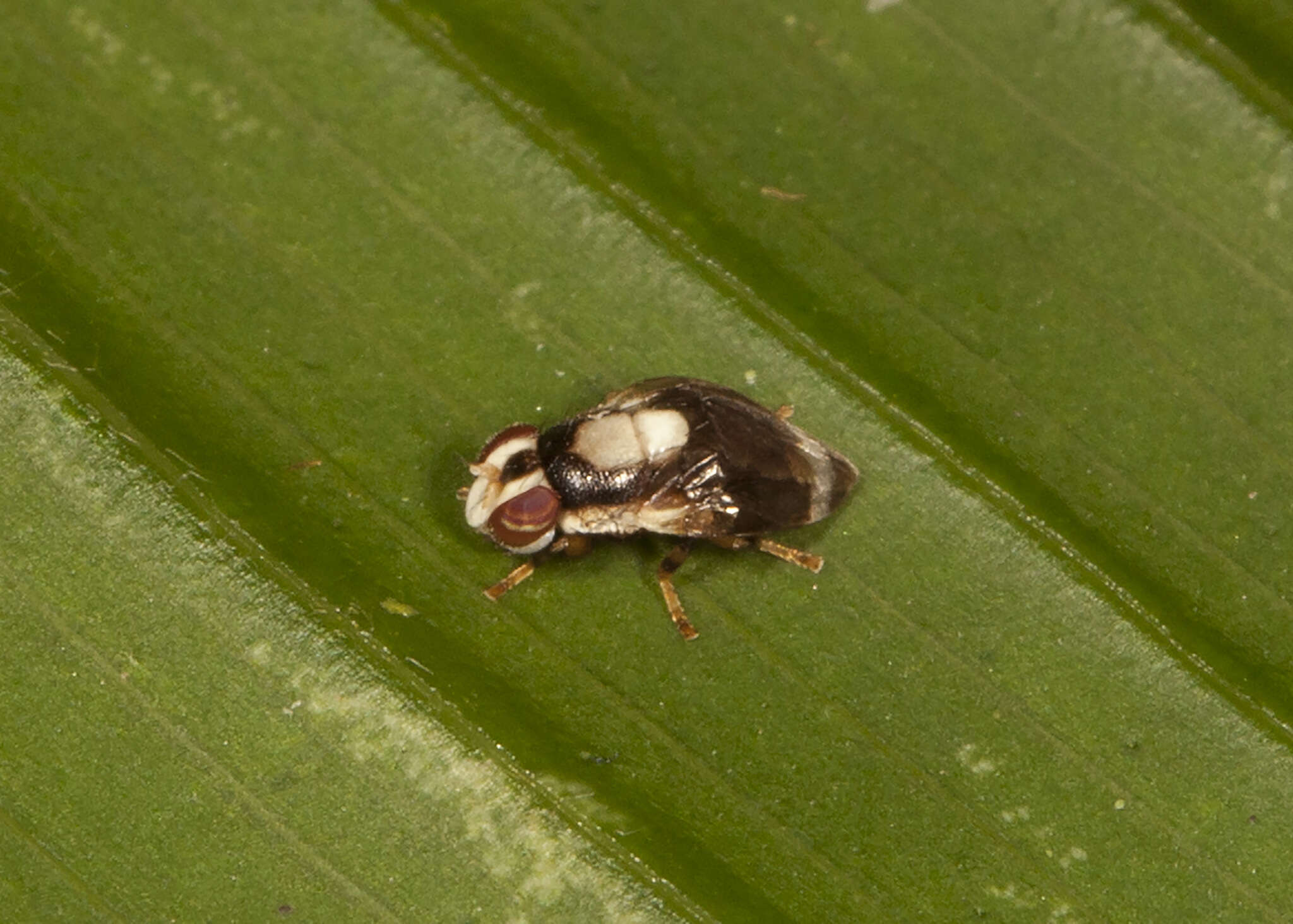
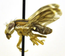
undet. Pericelididae, Costa Rica: Santa Rosa NP
-
-
-
-
-
-
-
-
-
-