Toxicity
provided by Harmful Phytoplankton Project
Producer of dinophysistoxin-1 (DTX1) (Lee et al. 1989).
- bibliographic citation
- Guide to UK Coastal Planktonic Ciliates © 2001 DJS Montagnes, University of Liverpool http://www.liv.ac.uk/ciliate/
- author
- David J.S. Montagnes
Distribution
provided by Harmful Phytoplankton Project
Tends not to form blooms. Widely distributed around the world though most abundant in warmer regions. Has been identified in both the Mediterranean and the North Sea.
- Larsen, J. and O. Moestrup 1992. Potentially toxic phytoplankton. 2. Genus Dinophysis (Dinophyceae). In: J.A. Lindley (ed), ICES Identification Leaflets for Plankton. ICES, Copenhagen, 180: 1-12.
- Moita, M.T. and M.A. de M. Sampayo 1993. Are there cysts in the genus Dinophysis? In: T.J. Smayda and Y. Shimizu (eds), Toxic Phytoplankton Blooms in the Sea, Elsevier, Amsterdam: 153-157.
- Lee J.S., lgarashi T., Fraga S., Dahl E., Hovgaard P. & Yasumoto T. 1989. Determination of diarrhetic shellfish toxins in various dinoflagellate species. J. Appl. Phycol. 1: 147-152.
- bibliographic citation
- Guide to UK Coastal Planktonic Ciliates © 2001 DJS Montagnes, University of Liverpool http://www.liv.ac.uk/ciliate/
- author
- David J.S. Montagnes
Comprehensive Description
provided by Harmful Phytoplankton Project
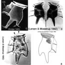
Dinophysis tripos is an armoured, marine, planktonic dinoflagellate. Cells are very distinctive thanks to the presence of two posterior projections. The ventral projection is longer than the dorsal one. The projections are often toothed (i.e. they have small spines on the tip). Like other Dinophysis species they have a large hypotheca and a small cap like epitheca. Cells are laterally compressed. The left sulcal list (LSL) is large and often reticulated. D. tripos is a photosynthetic species with chloroplasts (Larsen & Moestrup 1992).
- bibliographic citation
- Guide to UK Coastal Planktonic Ciliates © 2001 DJS Montagnes, University of Liverpool http://www.liv.ac.uk/ciliate/
- author
- David J.S. Montagnes
Diagnostic Description
provided by Harmful Phytoplankton Project
The epitheca is much smaller than the hypotheca although both are made up of four plates. On the epitheca the cingulum is narrow with two large lists (wings); anterior cingular list (ACL) and posterior cingular list (PCL), oriented anteriorly. The ACL is supported by many ribs. The LSL is also highly visible and supported by three ribs. The sulcus is made up of several irregularly shaped plates. The flagellar pore is located in the sulcal area. The hypotheca contains two posterior projections; the short dorsal and long ventral. The ventral margin is straight but the dorsal margin is concave below the cingulum and then convex continuing down to the dorsal projection (Larsen & Moestrup 1992).
- bibliographic citation
- Guide to UK Coastal Planktonic Ciliates © 2001 DJS Montagnes, University of Liverpool http://www.liv.ac.uk/ciliate/
- author
- David J.S. Montagnes
Ecology
provided by NMNH Marine Dinoflagellates
Dinophysis tripos is a planktonic species commonly found in neritic, estuarine and oceanic waters (Steidinger & Tangen 1996). No blooms for this species have been reported (Larsen & Moestrup 1992).
- bibliographic citation
- Faust, Maria A. and Rose A. Gulledge. Identifying Harmful Marine Dinoflagellates. Smithsonian Contributions from the United States National Herbarium, volume 42: 1-144 (including 48 plates, 1 figure and 1 table).
Habitat and Locality
provided by NMNH Marine Dinoflagellates
D. tripos is widely distributed in tropical and temperate waters, and occasionally is found in colder regions (Larsen & Moestrup 1992; Taylor et al. 1995; Steidinger & Tangen 1996).
- bibliographic citation
- Faust, Maria A. and Rose A. Gulledge. Identifying Harmful Marine Dinoflagellates. Smithsonian Contributions from the United States National Herbarium, volume 42: 1-144 (including 48 plates, 1 figure and 1 table).
Morphology and Structure
provided by NMNH Marine Dinoflagellates
D. tripos is a photosynthetic species with chloroplasts (Fig. 2). D. diegensis, a smaller form very similar in morphology to D. tripos with a reduced hypothecal process, is suspected to be a gamete of the latter species (Moita & Sampayo 1993).
- bibliographic citation
- Faust, Maria A. and Rose A. Gulledge. Identifying Harmful Marine Dinoflagellates. Smithsonian Contributions from the United States National Herbarium, volume 42: 1-144 (including 48 plates, 1 figure and 1 table).
Nomenclatural Types
provided by NMNH Marine Dinoflagellates
Holotype: Dinophysis tripos Gourret, 1883: 114, plate 3, fig. 53
Type Locality: Mediterranean Sea: Gulf of Marseille, France
- bibliographic citation
- Faust, Maria A. and Rose A. Gulledge. Identifying Harmful Marine Dinoflagellates. Smithsonian Contributions from the United States National Herbarium, volume 42: 1-144 (including 48 plates, 1 figure and 1 table).
Remarks
provided by NMNH Marine Dinoflagellates
Many authors consider Phalacroma to be synonymous with Dinophysis (Steidinger & Tangen 1996).
- bibliographic citation
- Faust, Maria A. and Rose A. Gulledge. Identifying Harmful Marine Dinoflagellates. Smithsonian Contributions from the United States National Herbarium, volume 42: 1-144 (including 48 plates, 1 figure and 1 table).
Reproduction
provided by NMNH Marine Dinoflagellates
D. tripos reproduces asexually by binary fission. Moita and Sampayo (1993) speculate that sexual reproduction, with sexual dimorphism, is part of the life cycle for this species.
- bibliographic citation
- Faust, Maria A. and Rose A. Gulledge. Identifying Harmful Marine Dinoflagellates. Smithsonian Contributions from the United States National Herbarium, volume 42: 1-144 (including 48 plates, 1 figure and 1 table).
Species Comparison
provided by NMNH Marine Dinoflagellates
Dinophysis tripos can be confused with D. caudata; some cells of D. caudata, bearing a short hypothecal process, can superficially resemble D. tripos. However, D. tripos can be distinguished by the presence of two posterior projections (Larsen & Moestrup 1992; Steidinger & Tangen 1996).
- bibliographic citation
- Faust, Maria A. and Rose A. Gulledge. Identifying Harmful Marine Dinoflagellates. Smithsonian Contributions from the United States National Herbarium, volume 42: 1-144 (including 48 plates, 1 figure and 1 table).
Species Overview
provided by NMNH Marine Dinoflagellates
Dinophysis tripos is an armoured, marine, planktonic dinoflagellate species. It is a toxic species common in warm temperate to tropical waters.
- bibliographic citation
- Faust, Maria A. and Rose A. Gulledge. Identifying Harmful Marine Dinoflagellates. Smithsonian Contributions from the United States National Herbarium, volume 42: 1-144 (including 48 plates, 1 figure and 1 table).
Synonyms
provided by NMNH Marine Dinoflagellates
Dinophysis caudata var. tripos (Gourret) Gail, 1950
- bibliographic citation
- Faust, Maria A. and Rose A. Gulledge. Identifying Harmful Marine Dinoflagellates. Smithsonian Contributions from the United States National Herbarium, volume 42: 1-144 (including 48 plates, 1 figure and 1 table).
Taxonomic Description
provided by NMNH Marine Dinoflagellates
Species in this genus are laterally compressed with a small, cap-like epitheca and a much larger hypotheca (dorso-ventral depth of epitheca is 1/3 to 1/2 hypotheca). The shape of the cell in lateral view is the most important criterion used for identification (Taylor et al. 1995).
D. tripos is a very distinctive species. Cells are large, anterio-posteriorly elongated and asymmetrical with two posterior hypothecal projections; a longer ventral process and a shorter dorsal one (Figs. 1-4). The V-shaped processes are often toothed on their posterior ends (small knob-like spines) (Fig. 1). The well developed left sucal list (LSL) widens posteriorly and is often reticulated (Figs. 1-3) (Larsen & Moestrup 1992; Taylor et al. 1995; Steidinger & Tangen 1996).
The thick thecal plates are heavily areolated (Fig. 1). Cell size ranges: 90-125 µm in length and 50-60 µm in dorso-ventral width (Larsen & Moestrup 1992; Taylor et al. 1995).
- bibliographic citation
- Faust, Maria A. and Rose A. Gulledge. Identifying Harmful Marine Dinoflagellates. Smithsonian Contributions from the United States National Herbarium, volume 42: 1-144 (including 48 plates, 1 figure and 1 table).
Thecal Plate Description
provided by NMNH Marine Dinoflagellates
The small epitheca is made up of four plates. The cingulum is narrow with two well developed lists, anterior cingular list (ACL) and posterior cingular list (PCL), oriented anteriorly (Figs. 1-4). The ACL is supported by many ribs (Figs. 1, 4). The wide ACL forms a narrow, funnel-like structure obscuring the epitheca on the bottom. The sulcus is comprised of several irregularly shaped plates. The flagellar pore is housed in the sulcal area. The prominent wide LSL has a straight margin and is supported by three ribs (Figs. 1-4) (Larsen & Moestrup 1992; Taylor et al. 1995; Steidinger & Tangen 1996). The hypotheca, with four large plates, comprises the majority of the cell. It is long, narrowing into two tapered or pointed posterior projections: one short and dorsal, and one longer and ventral (Figs. 1-3). The dorsal projection is sometimes seen with a narrow list connecting two daughter cells during cell division (Fig. 3). The ventral margin of the hypotheca is straight or slightly undulate. The dorsal margin is concave below the cingulum and then convex continuing down to the dorsal projection (Figs. 1, 2) (Larsen & Moestrup 1992; Taylor et al. 1995; Steidinger & Tangen 1996).
- bibliographic citation
- Faust, Maria A. and Rose A. Gulledge. Identifying Harmful Marine Dinoflagellates. Smithsonian Contributions from the United States National Herbarium, volume 42: 1-144 (including 48 plates, 1 figure and 1 table).
Toxicity
provided by NMNH Marine Dinoflagellates
D. tripos is associated with diarrhetic shellfish poisoning (DSP) events; it produces Dinophysistoxin-1 (DTX1)(Lee et al. 1989).
- bibliographic citation
- Faust, Maria A. and Rose A. Gulledge. Identifying Harmful Marine Dinoflagellates. Smithsonian Contributions from the United States National Herbarium, volume 42: 1-144 (including 48 plates, 1 figure and 1 table).