-
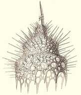
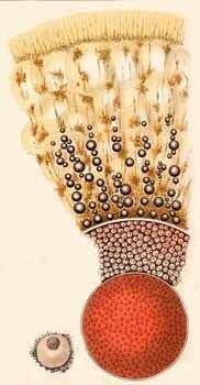
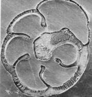
Calocyclas monumentum.
-
-
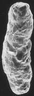
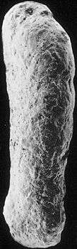
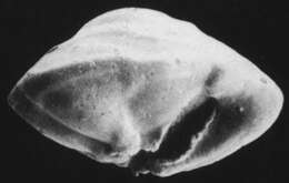
Cristellaria siddalliana.
-
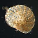
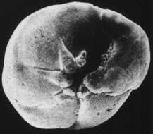
Truncatulina ungeriana.
-
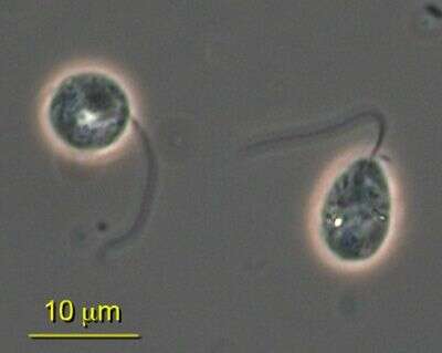
Cyranomonas is a biflagellated colourless protist of unknown affinities. Cells are ovoid, 4.5 - 5 µm long, dorso-ventrally flattened, and somewhat flexible. The anterior part is depressed of concave. Two thickened flagella emerge from the right side of the cell and are not acronematic. The anterior flagellum is about the length of the cell and flickers stiffly forwards. The posterior flagellum inserts to the left of the anterior flagellum, is 1.5 the length of the cell, and trails behind the cell. The nucleus is located anteriorly. The cells contain small food materials. The cells glide slowly with the anterior flagellum. C. australis is currently the only species in the genus, which has so far only been reported from Australia.
-
Collected on a mudflat in Sandebukta, Oskofjord, Norway. Image courtesy of Elisabeth Alve, University of Oslo. Originally published in J. Foram. Res. 16: 261-284; used with permission.
-
ATCC culture 50636.
-
The outer surface of the test is much smoother on this specimen than in most members of its species. Image courtesy of Elisabeth Alve, University of Oslo. Originally published in J. Foram. Res. 16: 261-284; used with permission.
-
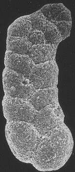
The typical chamber arrangement of this genus (planispiral in the early chambers, at the base, and biserial later) is evident here. Image courtesy of Elisabeth Alve, University of Oslo. Originally published in J. Foram. Res. 16: 261-284; used with permission.
-
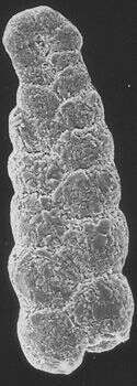
This individual shows an unusual bending of the early chambers. Individual collected in Saanich Inlet, Vancouver Island, British Columbia. Image courtesy of R. Timothy Patterson, Carleton University. This image first appeared in J. Foram. Res. 28:201-219 and is used with permission.
-
Individual collected in Saanich Inlet, Vancouver Island, British Columbia. This species was most common in deep water in the center of the inlet. Image courtesy of R. Timothy Patterson, Carleton University. This image first appeared in J. Foram. Res. 28:201-219 and is used with permission.
-
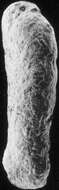
Fossil (Badenian) specimen, from Nussdorff, Austria. Image courtesy of Stefan Revets. This image first appeared in Hansen and Revets, J. Foram. Res. 22:166-180 (1992) and is used with permission.
-
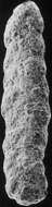
Polished and etched horizontal section through test. Image courtesy of Stefan Revets. This image first appeared in Hansen and Revets, J. Foram. Res. 22:166-180 (1992) and is used with permission.
-
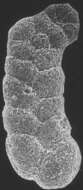
Image courtesy of Stefan Revets. This image first appeared in Hansen and Revets, J. Foram. Res. 22:166-180 (1992) and is used with permission.
-
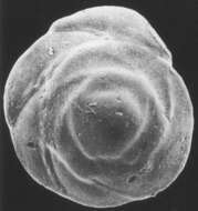
Image courtesy of Stefan Revets. This image first appeared in Hansen and Revets, J. Foram. Res. 22:166-180 (1992) and is used with permission.
-
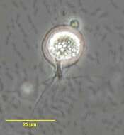
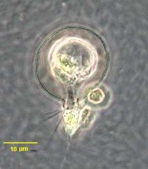
in vivo portrait of the small shelled amoeba of uncertain affinity, Apogromia pagei (ERTL, 1984).Collected from a rain barrel in Boise, Idaho. April 2006. Phase contrast.
-
in vivo portrait of the small shelled amoeba of uncertain affinity, Apogromia pagei (ERTL, 1984).Collected from a rain barrel in Boise, Idaho. April 2006. Phase contrast.
-
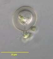
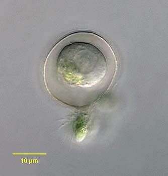
in vivo portrait of the small shelled amoeba of uncertain affinity, Apogromia pagei (ERTL, 1984).Collected from a rain barrel in Boise, Idaho. April 2006. DIC.
-
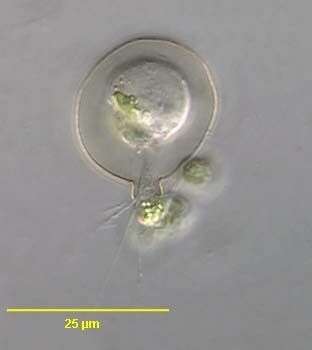
in vivo portrait of the small shelled amoeba of uncertain affinity, Apogromia pagei (ERTL, 1984).Collected from a rain barrel in Boise, Idaho. April 2006.
-
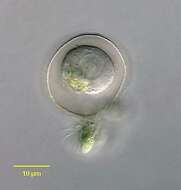
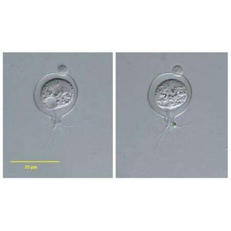
View of the contractile vacuole in diastole (left) and at the end of systole (right) in the small shelled amoeba of uncertain affinity, Apogromia pagei (ERTL, 1984).Collected from a rain barrel in Boise, Idaho. April 2006. Phase contrast.
-
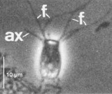
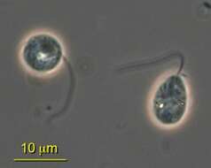
Small heterotrophic flagellate of about 15 µm which has four anteriorly directed flagella and bears posterior axopods with extrusomes. Flagella and axopods arise from a centroplast situated on the anterior face of the nucleus. Two free-living species from freshwater habitats which feed on bacteria. Phase contrast.
-
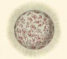
Section of cell with central capsule, associated black pigment, central nucleus, and calymma. Inset is an endoplasmic vacuole.
-
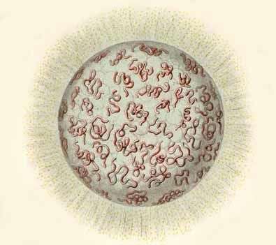
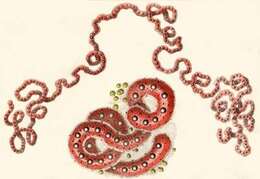
The central capsules are worm-like. With oil droplets, nuclei, small pigment spots and yellow symbiotic algae.
-
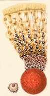
Living coenobium, with serpentine central capsules. Numerous yellow algal cells are scattered among the radial pseudopodia.