-
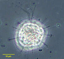
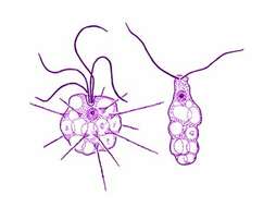
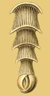
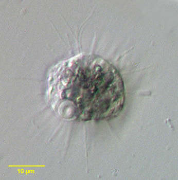
Tetradimorpha, a dimorphid helioflagellate. Four weakly oscillating flagella are seen emerging from the cell at the one o clock position. Axopodia with extrusomes radiate around the circumference of the cell. The axopodia may be completely withdrawn into the cell when it is disturbed. Although the size of these individuals is smaller than that described for T. pterbica (65-95 microns) the circumferential distribution of axopodia and the complete withdrawal of axopods and the shorter flagella are consistent with descriptions of T. pterbica. What appears to be the spherical dense axoplast (the termination of axopodial axonemes) is seen in the anterior of the cell between the flagellar insertions and the nucleus. From temporary rainwater pool near Boise, Idaho. Oblique illumination.
-
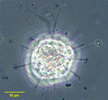
Tetradimorpha, a dimorphid helioflagellate. Four weakly oscillating flagella are seen emerging from the cell at the top of the cell. Axopodia with extrusomes radiate around the circumference of the cell. The axopodia may be completely withdrawn into the cell when it is disturbed. Although the size of these individuals is smaller than that described for T. pterbica (65-95 microns) the circumferential distribution of axopodia and the complete withdrawal of axopods and the shorter flagella are consistent with descriptions of T. pterbica. From temporary rainwater pool near Boise, Idaho. Phase contrast illumination
-
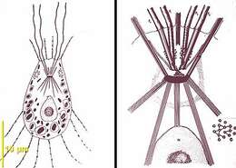
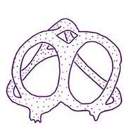
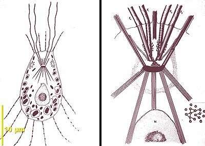
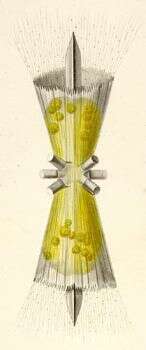
Tetradimorpha diagram based on light microscopy and transmission electron microscopy showing the four anterior flagella linked to the axosplast and the posterior axopods originating from microtubules arising from the axosplast, microtubules of the axopods are not regularly arranged like in Dimorpha (from G. Brugerolle and J.-P. Mignot).
-
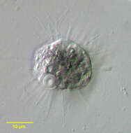
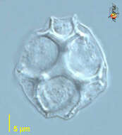
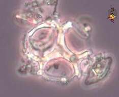
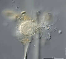
Tetradimorpha marina Fenchel et al., 1995. This heliozoan-like organism is almost spherical and most cells measured between 15 and 20 microns in diameter. The spherical nucleus is situated towards one side adjacent to a depression in the cell surface, from the bottom of which either two or four flagella arise. Cells with four flagella might represent early division stages, but there was no correlation between the number of flagella and cell diameter. About twenty axopodia with extrusomes radiate from the cell. The cell are packed with spherical food vacuoles containing bacteria and the surrounding cytoplasm is filled with rod-shaped (~ 1 microns) organelles or endosymbiotic bacteria. The cells are capable of absorbing the axopodia and of transforming into a slug-like flagellated form.
-
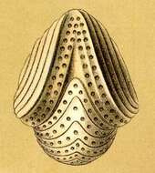
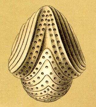
Hermesinum adriaticum Zacharias, 1906. The skeleton of this species is asymmetrical and difficult to describe. The skeleton is 48-50 microns long, 20 microns wide at its widest point and 12 microns high.
-
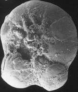
Ebria (ee-bree-a) A small group of marine flagellates, although may be abundant, and large numbers have been assoicated with fish kills. They deposit complex siliceous endoskeletons within the cells. As you can see. Differential interference contrast.
-
Ebria (ee-bree-a) A small group of marine flagellates, although may be abundant, and large numbers have been assoicated with fish kills. They deposit complex siliceous endoskeletons within the cells and (as in this case) the skeleton may be found on its own. The ebriids have been implicated in fish kills. Phase contrast.
-
Ebria tripartita (Shumann, 1867) Lemmermann, 1899. Specimens of this species are almost spherical in shape and average 24 microns in diameter.
-
-
-
-
-
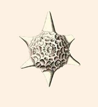
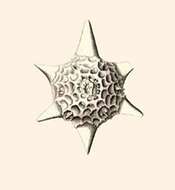
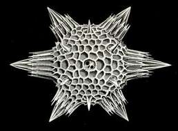
Hexaconus serratus.
-
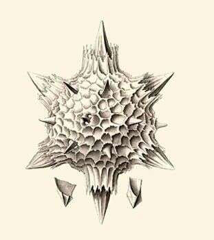
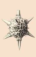
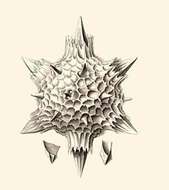
Haeckel says: Lower left- Central base of an equatroial spine. Lower right: Central base of a tropical spine.
-
-
Vertebralina mucronata.
-
Orbiculina adunca.
-
Cristellaria calcar.
-
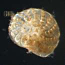
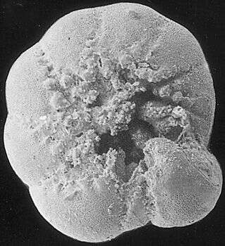
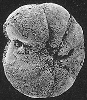
Foraminiferans living in polluted environments often show alterations in the morphology of their tests. This individual, isolated from a site in Norway which is contaminated with heavy metals, has protuberances on two of its chambers (bottom), which distort the characteristic coiling pattern of the test. Image courtesy of Dr. Elisabeth Alve, University of Oslo. Citation: Alve, E. Benthic foraminifera reflecting pollution. Journal of Foraminiferal Research 21:1-19.
-
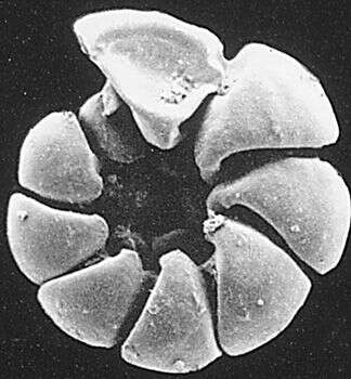
Foraminiferans living in polluted environments often show alterations in the morphology of their tests. This individual, isolated from a site in Norway which is contaminated with heavy metals, exhibits reduced chamber size in its second-to-last chamber, at bottom. Image courtesy of Dr. Elisabeth Alve, University of Oslo. Citation: Alve, E. Benthic foraminifera reflecting pollution. Journal of Foraminiferal Research 21:1-19.
-
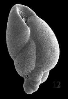
This specimen, a dead test, has been in an environment that stripped the calcium carbonate away; only the organic lining remains. Image courtesy of Elisabeth Alve, University of Oslo. Originally published in J. Foram. Res. 16: 261-284; used with permission.
-
This dead test has had some of the calcium carbonate leached from it, giving the test surface a rough appearance. Image courtesy of Elisabeth Alve, University of Oslo. Originally published in J. Foram. Res. 16: 261-284; used with permission.
-
This species is an "opportunistic" marine taxon, which readily invades nearshore environments if conditions allow. Sample collected at Hamble Estuary, Hampshire, England. Image courtesy of Elisabeth Alve, University of Oslo. Originally published in the Journal of Foraminiferal Research 31:1; used with permission.
-
Scale bars indicate 100 µm (1), 50 µm (3–5).Five images.Please click on < or > on the image edges or on the dots at the bottom edge of the images to browse through the slides!Place name: Tropical freshwater aquarium Latitude: 54.3018013 Longitude: 10.07120132Microscope Zeiss Axioplan, camera Olympus OM-D M5 MKII.© Wolfgang Bettighofer,images under Creative Commons License V 3.0 (CC BY-NC-SA).For permission to use of (high resolution) images please contact
postmaster@protisten.de.For further information about the image, please click here:
Link to protisten.de page