-
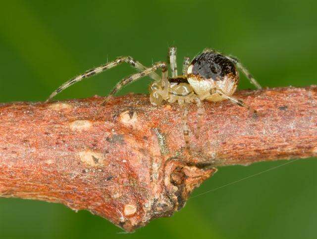
Asker, Akershus, Norge
-
Asker, Akershus, Norge
-
Hadsund Syd
-
Østrup Skov
-
Glatved Strand nord
-
Kattinge ved Roskilde
-
Døstrup ved Hobro, Jylland, Danmark
-
Åleklinta Öland
-
Asker, Akershus, Norge
-
Asker, Akershus, Norge
-
Asker, Akershus, Norge
-
Asker, Akershus, Norge
-
Asker, Akershus, Norge
-
Sangstrup/Karlby Klint
-
Kenneth F. Haynes and Kenneth V. Yeargan
EOL staff
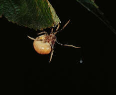
Mastophora stowei
-
Figures 1–6.Chrysso bifurca sp. n., 1–4 male holotype 1 body, dorsal view 2 abdomen, lateral view 3 male left palp, ventral view 4 same, prolateral view 5–6 female paratype 5 epigynum, ventral view 6 vulva, dorsal view. Scale bars: 0.5 mm (1–2); 0.1 mm (3–6).
-
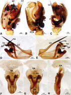
Figure 26.Theridiosoma vimineum sp. n., male holotype. A Right pedipalp, prolateral view B Right pedipalp, retrolateral view C Embolic division of right pedipalp, retrolateral view D Right pedipalp, ventral view. Co conductor; EA embolic apophysis; MA median apophysis; PC paracymbium; T tegulum. Scale bars: A as B.
-
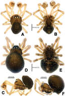
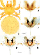
Figure 1.Mysmena wawuensis sp. n., male holotype (A–C) and female paratype (D–F). A–F Habitus. A, D dorsal view B, E ventral view C, F lateral view.
-
Ning Sun, Yuri M. Marusik, Lihong Tu
Zookeys
Figure 2.Acanoides beijingensis sp. n. A male palp, prolateral B male palp, prolateral, with embolic division removed C male palp, retrolateral D embolic division, ventral E embolic division, dorsal F epigynum, ventral G epigynum, dorsal H epigynum, lateral. CG copulatory groove; CO copulatory opening; DP dorsal plate; EA extensible area of epigynal basal part; EM embolic membrane; EP embolus proper; FG fertilization groove; FiG Fickert’s gland; LC lamella characteristica; MP median plate; P paracymbium; PCA proximal cymbial apophysis; R radix; S spermathecae; TA terminal apophysis; TH thumb of embolus; VP ventral plate. [Scale bars: mm].
-
Yucheng Lin, Shuqiang Li, Peter Jäger
Zookeys
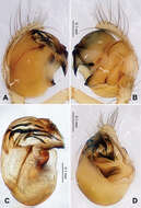
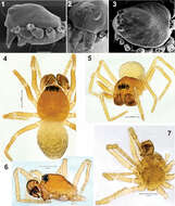
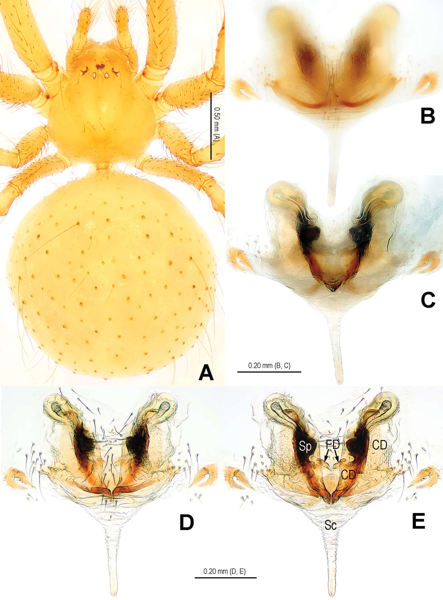
Figure 17.Chthonopes thakekensis sp. n., holotype female. A Habitus, dorsal B Epigyne (untreated), ventral C Vulva, dorsal D Epigyne (lactic acid-treated), ventral E Vulva, dorsal. CD = copulatory duct; FD = fertilization duct; Sc = scape; Sp = spermathecae.
-
Carles Ribera, Mert Elverici, Kadir Boğaç Kunt, Recep Sulhi Özkütük
Zookeys
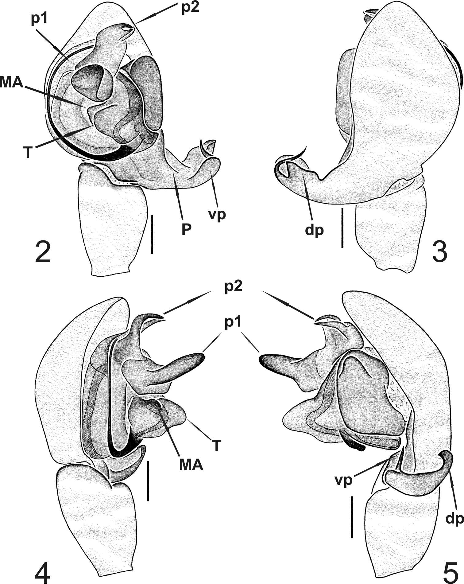
Figures 2–5.Typhlonesticus gocmeni sp. n. male palp. 2 ventral view 3 dorsal view 4 prolateral view 5 retrolateral view. Abbreviations: T = tegulum, MA = median apophysis, p1 = process 1 of TTA, p2 = process 2 of TTA, P = paracymbium, vp = ventral process of paracymbium, dp = dorsal process of paracymbium. Scale bars 0.1 mm.
-
Yuri M. Marusik, Sergei Zonstein
Zookeys
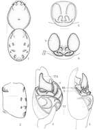
Figures 1–7.Prosoma and habitus of Synaphris wunderlichi sp. n. 1–3 prosoma with removed legs and palps, lateral, frontal and dorsal 4–5 habitus, dorsal and frontal 6–7 prosoma, lateral and ventral. Scale = 0.1 mm if not otherwise stated.
-
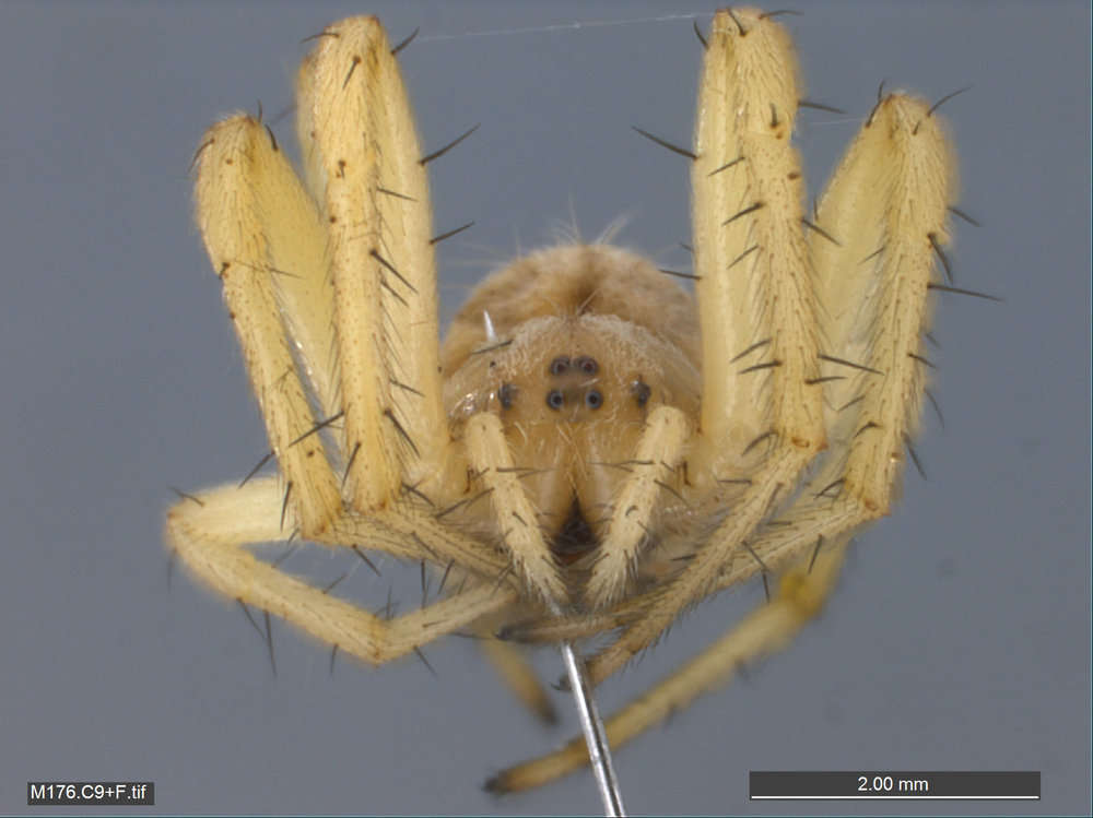
Gratiot Co., Michigan
-
All Biocode files are based on field identifications to the best of the researcher’s ability at the time.