-
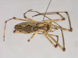
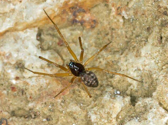
Asker, Akershus, Norge
-
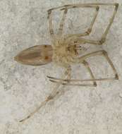
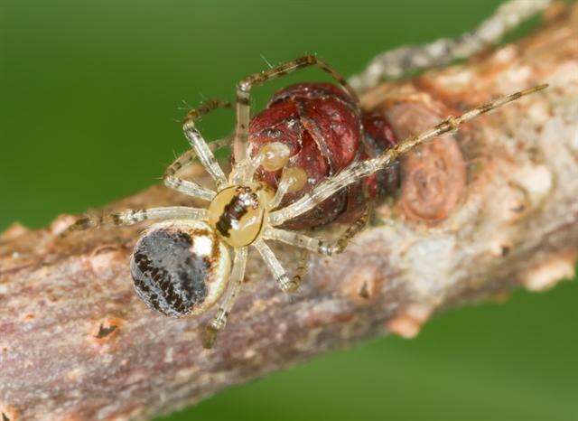
Store Klinteskov Møn
-
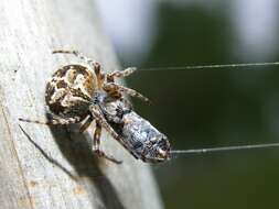
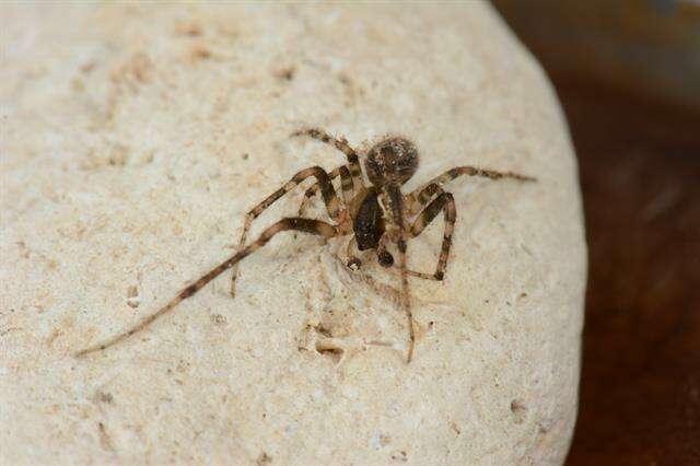
Asker, Akershus, Norge
-
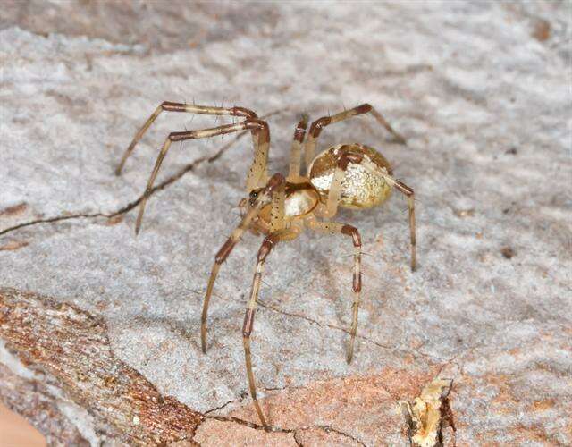
Asker, Akershus, Norge
-
Sangstrup/Karlby Klint
-
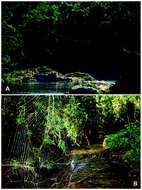
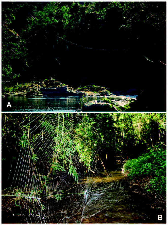
Webs of Caerostris darwini spanning Madagascar streams and rivers, with longest bridgelines exceeding 10 meters
-
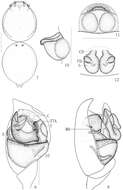
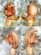
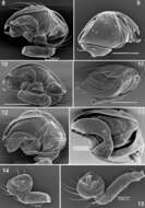
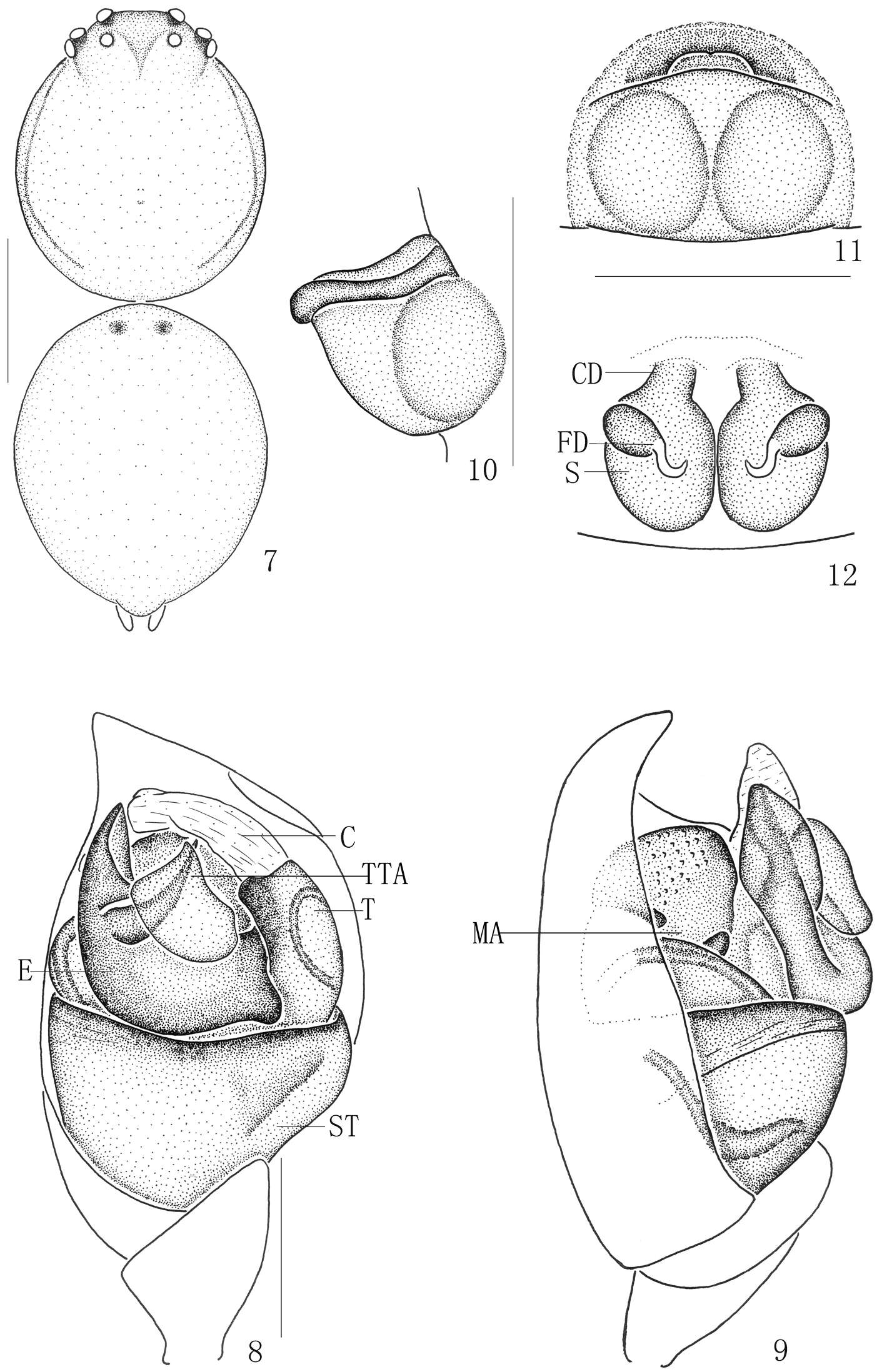
Figures 7–12.Chrysso bicuspidata sp. n., 7–9 female holotype 7 body, dorsal view 8 male left palp, ventral view 9 same, prolateral view 10–12 female paratype 10 epigynum, lateral view 11 same, ventral view 12 vulva, dorsal view. Scale bars: 0.5 mm (7); 0.1 mm (8–12).
-
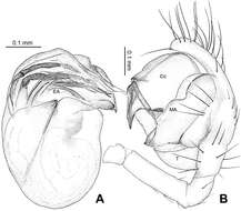
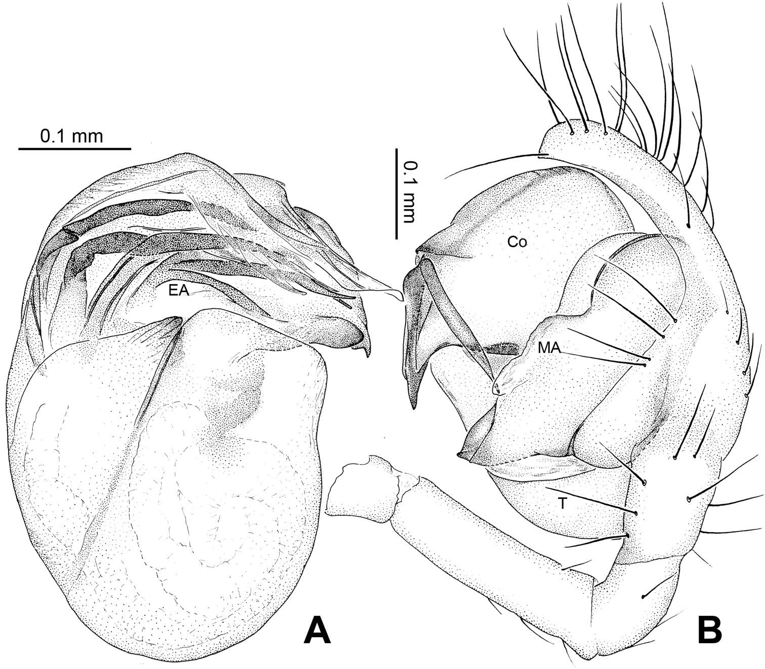
Figure 27.Theridiosoma vimineum sp. n., male holotype. A Embolic division of right pedipalp, retrolateral view B Right pedipalp, prolateral view. Co conductor; EA embolic apophysis; MA median apophysis; T tegulum.
-
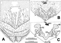
Figure 7.Mysmena wawuensis sp. n., female paratype. A Epigynum, ventral view B Epigynum (lactic acid-treated), ventral view C Vulva (cleared), dorsal view. Abbrs.: AB accessory bursa; CD copulatory duct; FD fertilization duct; S spermatheca; Sp scape.
-
Ning Sun, Yuri M. Marusik, Lihong Tu
Zookeys
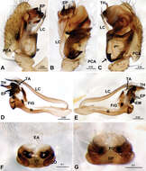
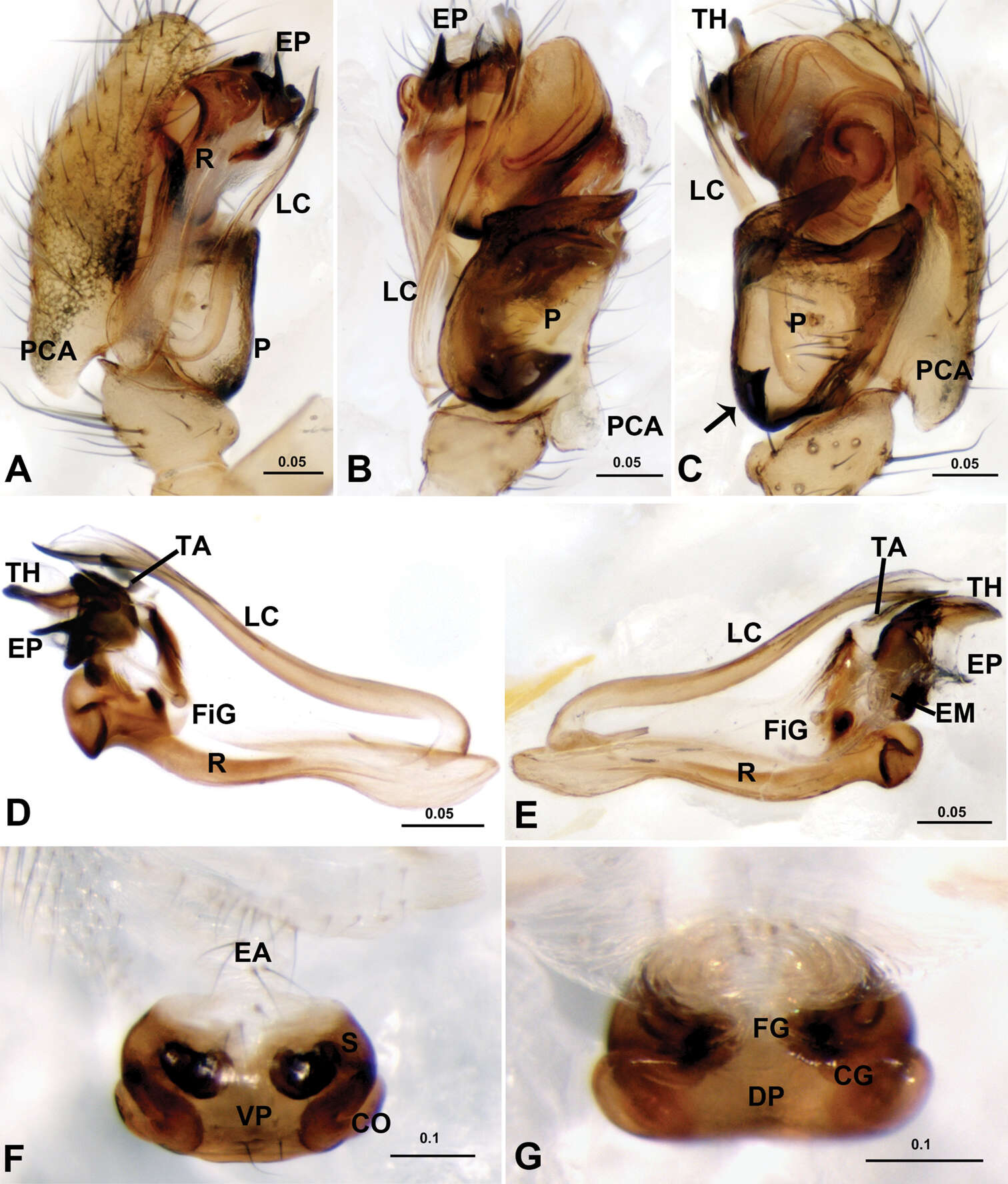
Figure 3.Acanoides hengshanensis. A male palp, prolateral B male palp, ventral C male palp, retrolateral, arrow indicates pointed tooth on posterolateral margin D embolic division, ventral E embolic division, dorsal F epigynum, ventral G epigynum, dorsal. CG copulatory groove; CO copulatory opening; DP dorsal plate; EA extensible area of epigynal basal part; EM embolic membrane; EP embolus proper; FG fertilization groove; FiG Fickert’s gland; LC lamella characteristica; P paracymbium; PCA proximal cymbial apophysis; R radix; S spermatheca; TA terminal apophysis; TH thumb of embolus; VP ventral plate. [Scale bars: mm].
-
Yucheng Lin, Shuqiang Li, Peter Jäger
Zookeys
Figure 18.Chthonopes thakekensis sp. n., holotype female. A Epigyne (untreated), ventral B Ditto (lactic acid-treated), ventral E Vulva (lactic acid-treated), dorsal. CD = copulatory duct; FD = fertilization duct; Sc = scape; Sp = spermathecae.
-
Carles Ribera, Mert Elverici, Kadir Boğaç Kunt, Recep Sulhi Özkütük
Zookeys
Figures 6–9.Typhlonesticus gocmeni sp. n. male palp. 6 ventral view 7 dorsal view 8 retrolateral view 9 prolateral view. Abbreviations: T = tegulum, MA = median apophysis, p1 = process 1 of TTA, p2 = process 2 of TTA, vp = ventral process of paracymbium, dp = dorsal process of paracymbium. Scale bars 0.5 mm.
-
Yuri M. Marusik, Sergei Zonstein
Zookeys
Figures 8–15. Scanning electron microphotographs of the male palp of Synaphris wunderlichi sp. n. (8–11, 14), Synaphris lehtineni (12, 15) and Synaphris orientalis (13). 8, 12–13 prolateral 9 retrolateral 10 caudal 11 anterior 14–15 palp with removed bulbus showing femur-tibia, anterior. Scale = 0.1 mm if not otherwise stated.
-
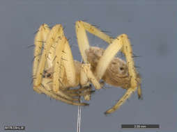
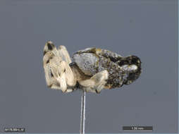
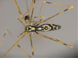
All Biocode files are based on field identifications to the best of the researcher’s ability at the time.
-
All Biocode files are based on field identifications to the best of the researcher’s ability at the time.
-
All Biocode files are based on field identifications to the best of the researcher’s ability at the time.
-
All Biocode files are based on field identifications to the best of the researcher’s ability at the time.
-
All Biocode files are based on field identifications to the best of the researcher’s ability at the time.
-
Læsten Bakker, Jylland, Danmark
-
Midtsjælland, Denmark
-
Asker, Akershus, Norway
-
Frijsenborg Dyrehave
-
Asker, Akershus, Norge
-
Asker, Akershus, Norge