Comprehensive Description
provided by Smithsonian Contributions to Botany
Vellozia phalocarpa Pohl
SPECIMENS EXAMINED.—Anderson et al. 35855.
SURFACE VIEW.—Hairs: absent. Epidermis: adaxial cells rounded to square, few almost triangular; thin walled; abaxial cells rectangular, few square shaped, thin walled. Stomata: paracytic, 24 × 12 μm; present on abaxial surface.
TRANSVERSE SECTION OF LAMINA.—Dorsiventral; V-shaped with small median adaxial groove. Extreme margins of lamina turned slightly inversely. Adaxial surface slightly undulating; abaxial surface furrowed one-third to one-half thickness of blade. Epidermis: cells on adaxial and abaxial surfaces rounded; wall thickened. Adaxial epidermis interspersed with strands of sclerenchyma. Subjacent to epidermis and strands of fibers occur two to three layers of parenchyma cells. Cuticle: markedly thick and slightly ridged on both surfaces. Stomata: present in abaxial furrows; virtually no substomatal chamber observed; somewhat associated with small projections in furrow. Mesophyll: three to four layers of palisade cells changing abruptly into a compactly arranged spongy tissue of small, rounded cells. Two to three layers of large translucent cells radially arranged above vascular bundles and furrows. Large translucent cells occur above midvein. Vascular bundles: 26; few commissural bundles observed. One to two large vessels present in each vascular bundle. Two phloem units lying laterally in flanges of Y-shaped abaxial girder. Adaxial cap present. One to two layers of sclerenchyma strands between abaxial epidermis and a layer of parenchyma cells subjacent to spongy mesophyll. Bundle sheath present on each vascular bundle. Crystals: present. Tannins: some observed.
- bibliographic citation
- Ayensu, Edward S. 1974. "Leaf Anatomy and Systematics of New World Velloziaceae." Smithsonian Contributions to Botany. 1-125. https://doi.org/10.5479/si.0081024X.15
Comprehensive Description
provided by Smithsonian Contributions to Botany
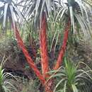
Vellozia glabra Mikan
Vellozia glabra Mikan, Delect. Fl. & Faun. Brasil. fasc. 2, 1820.—Sprengel, Syst. Veg., 3:338, 1826.—Schultes f. in Roemer & Schultes, Syst., 7:293, 1826.—L. B. Smith, Contr. U.S. Nat. Herb., 35:260, 1962.—Ayensu, Smithsonian Contr. Bot., 15:24, pls. 24f–g, 25a, 1974.—L. B. Smith, Taxon, 24(4):474, 1975.
Vellozia capsulis scapisque glabris Vandelli, Fl. Lusit. & Brasil. Spec., 33, pl. 2, 1788.
Vellozia phalocarpa Pohl, Pl. Brasil., 1:123, pl. 98, 1827.—Seubert in Martius, Fl. Bras., 3(1):76, 1847 [type: in stands,
dry mountains, Serra de São Felis, Goiás, Brazil, October 1819, Pohl s n (W n v)].
TYPE.—Description and illustration. No specimen known.
DISTRIBUTION.—Brazil: Minas Gerais: Jaboticatubas.
- bibliographic citation
- Smith, Lyman B. and Ayensu, Edward S. 1976. "A Revision of American Velloziaceae." Smithsonian Contributions to Botany. 1-172. https://doi.org/10.5479/si.0081024X.30
Comprehensive Description
provided by Smithsonian Contributions to Botany
Vellozia glabra Mikan
SPECIMENS EXAMINED.—Maguire et al. 44622; Maguire et al. 49048; Duarte 8355-Mathos 464.
SURFACE VIEW.—Hairs: on abaxial furrows. Epidermis: cells square to rectangular; thin walled. Stomata: paracytic, 21 × 12 μm; present mainly on abaxial surface.
TRANSVERSE SECTION OF LAMINA.—Dorsiventral; V-shaped with very small median adaxial groove; very slight inversion at margins. Adaxial surface almost smooth; abaxial surface furrowed about one-half thickness of blade. Epidermis: adaxial and abaxial cells square to rectangular; walls thicker on inner tangential side. Subjacent to epidermis occur two to three layers of parenchyma cells interspersed with one to two layers of sclerenchyma strands. Below parenchyma and sclerenchyma layers is a distinct layer of large translucent parenchyma cells. Cuticle: slightly thicker on adaxial side; slightly ridged on both surfaces. Stomata: present in furrows and on abaxial surface; small substomatal chamber present; protected by hairs in furrows. Mesophyll: definite palisade tissue of three layers abruptly changing into compact spongy tissue; two to three layers of translucent palisade cells arranged radially above vascular bundles and furrows. Three distinct layers of large translucent palisade cells in midvein region. Vascular bundles: 16–46; no commissural bundles observed. One to two large vessels in each vascular bundle, mostly one. Two phloem units lying laterally in flanges of Y-shaped abaxial girder. Each vascular bundle has an adaxial cap. Some sclerenchyma strands occur between abaxial epidermis and abaxial parenchyma layer. Bundle sheath completely surrounds each vascular bundle; few large translucent cells may appear at abaxial end of vascular bundle. Abaxial furrows contain many epidermal extensions. Crystals: none observed. Tannins: few present.
- bibliographic citation
- Ayensu, Edward S. 1974. "Leaf Anatomy and Systematics of New World Velloziaceae." Smithsonian Contributions to Botany. 1-125. https://doi.org/10.5479/si.0081024X.15