-
Dmitri V. Logunov, Yuri M. Marusik
Zookeys
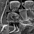
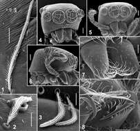
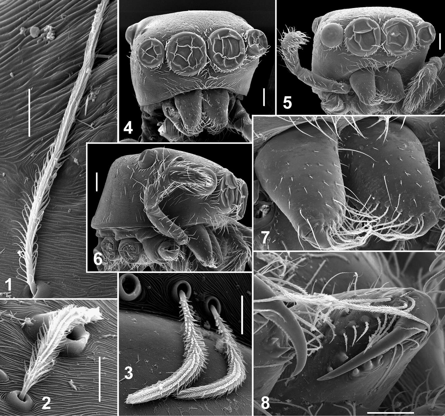
Figures 1–8.Somatic characters of Eupoa lehtineni sp. n. 1–3 plumose scales on female carapace. 4 male carapace, frontal view 5 female carapace, frontal view 6 ditto, lateral view 7 female chelicerae, frontal view 8 female fang and cheliceral teeth. Scale bars: 10 μm (1–3), 50 μm (7–8), 0.1 mm (4–6).
-
Dmitri V. Logunov, Yuri M. Marusik
Zookeys
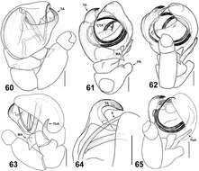
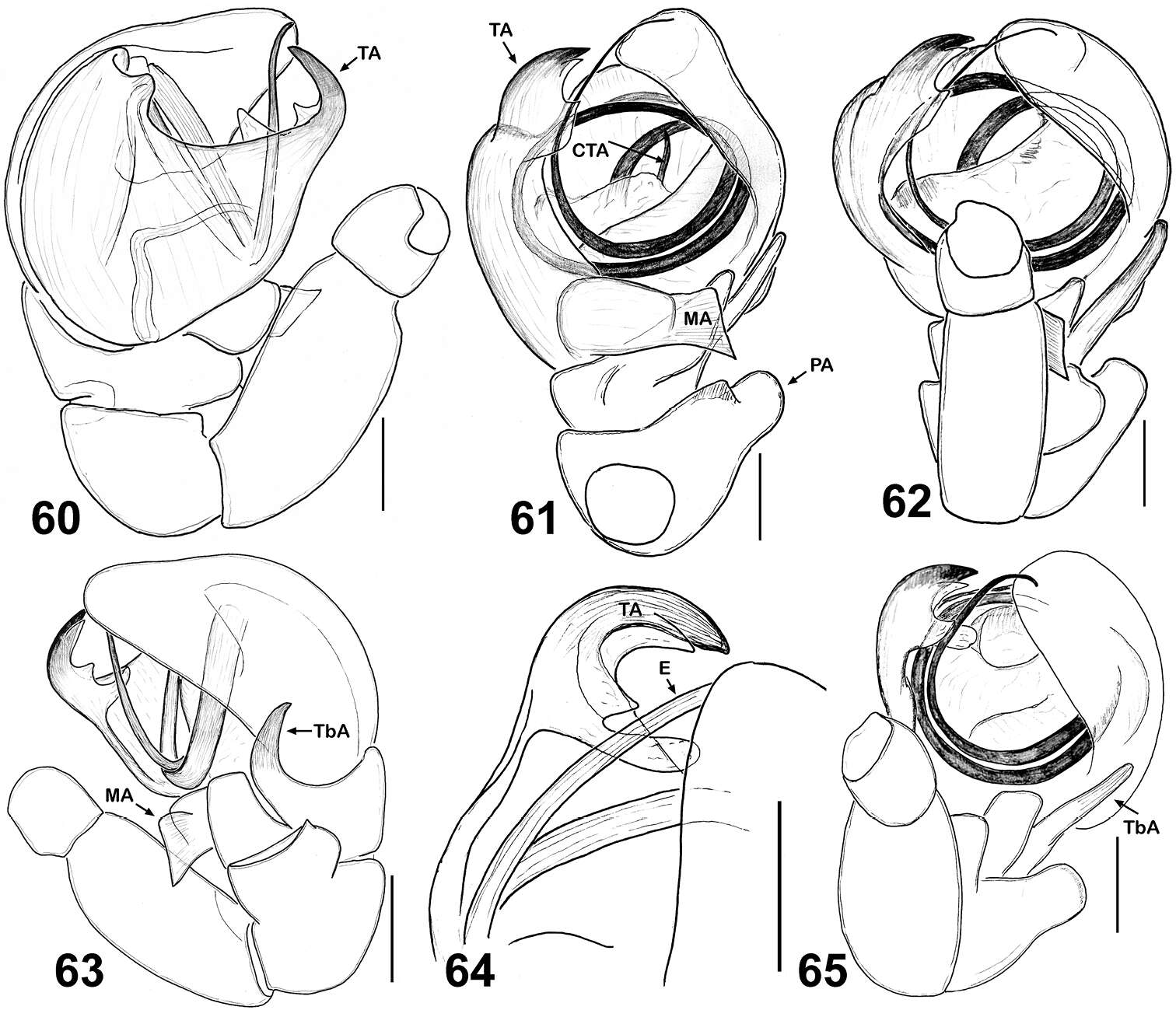
Figures 60–65.Copulatory organs of Eupoa lobli sp. n. (the holotype). 60 male palp, median view 61–62, 65 ditto, ventral view 63 ditto, retrolateral view 64 tegular apophysis, retrolateral view. Abbreviations as explained in ‘Material and methods’. Scale bars: 0.1 mm.
-
Dmitri V. Logunov, Yuri M. Marusik
Zookeys
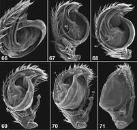
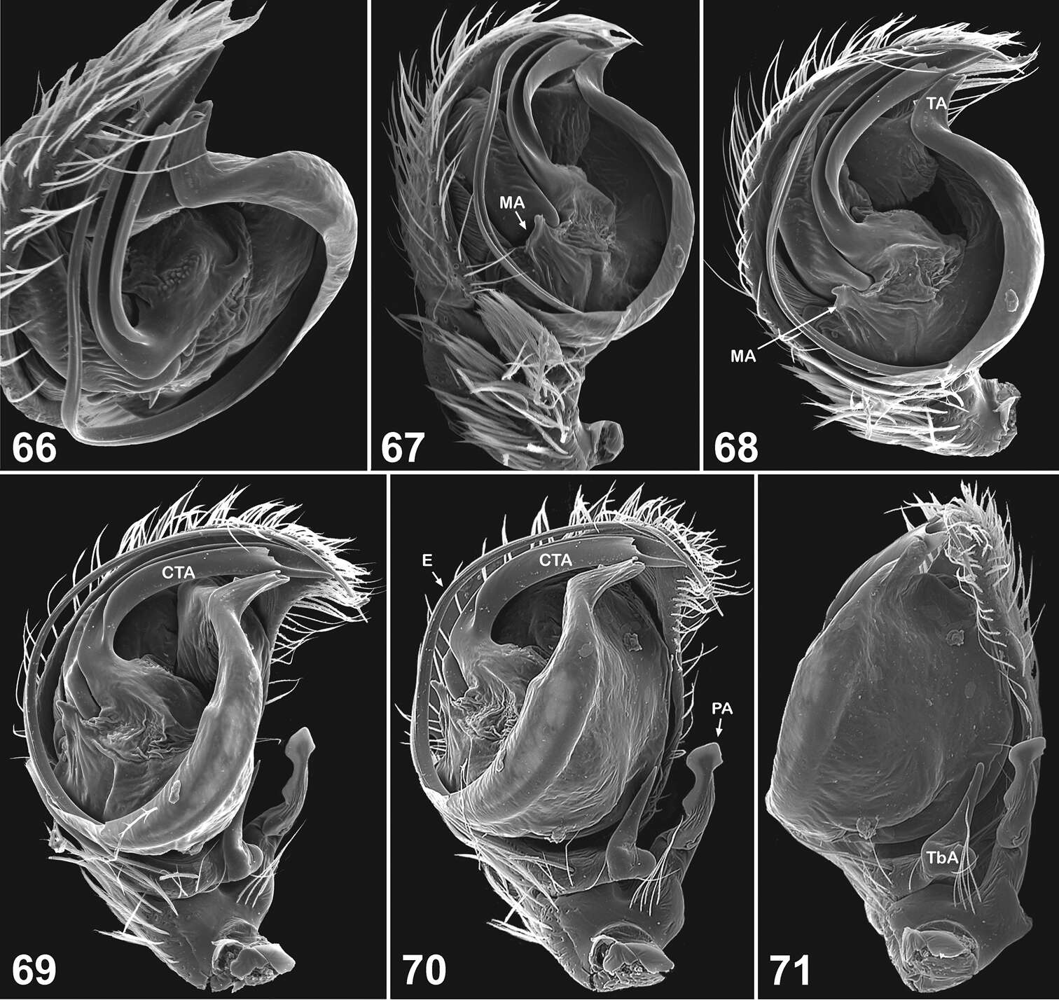
Figures 66–71.Copulatory organs of Eupoa pappi sp. n. (the holotype). 66 male palp, apical view 67–68 ditto, median view 69 ditto, ventral view 70–71 ditto, retrolateral view. Abbreviations as explained in ‘Material and methods’.
-
Dmitri V. Logunov, Yuri M. Marusik
Zookeys
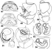
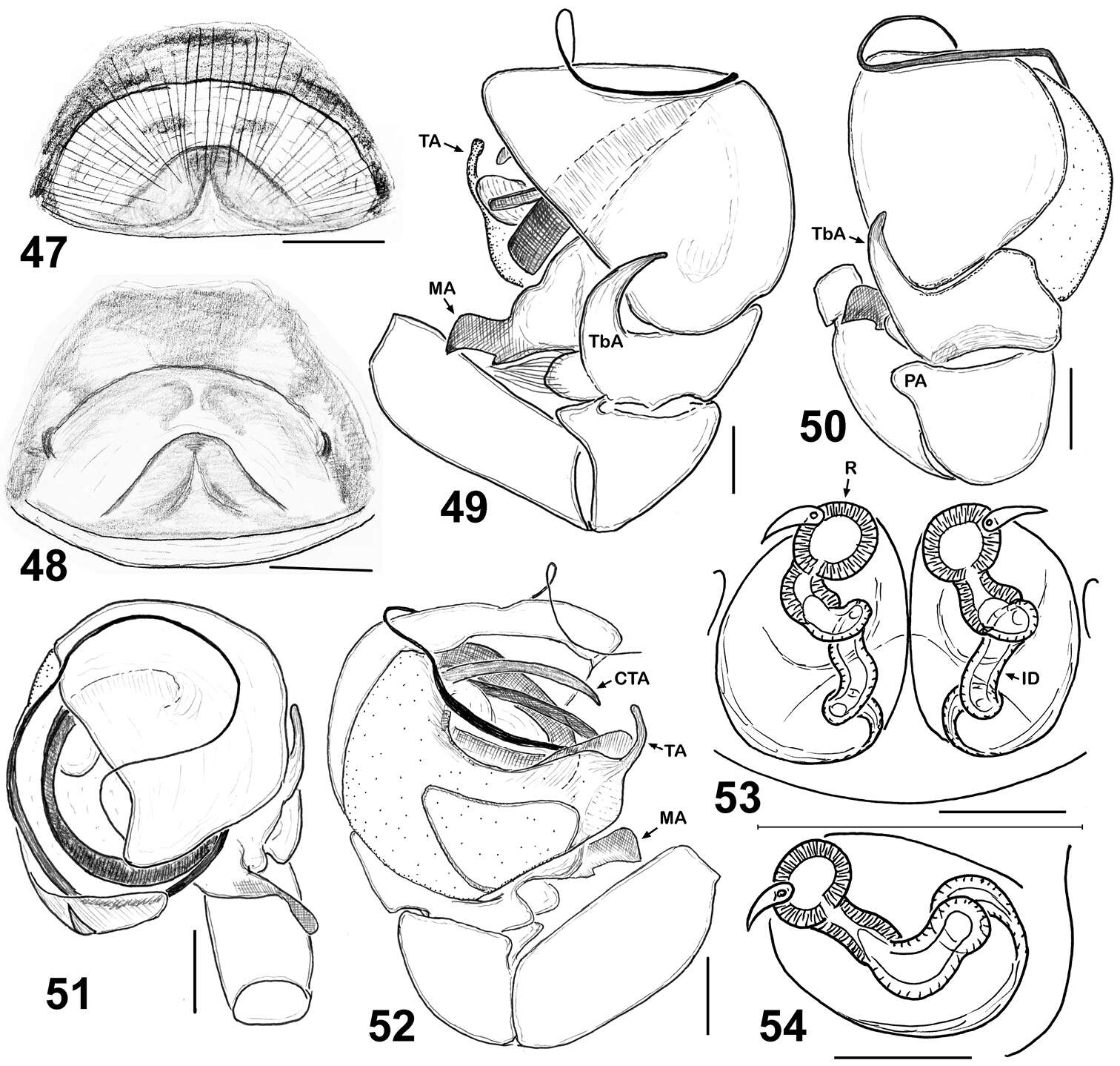
Figures 47–54.Copulatory organs of Eupoa lehtineni sp. n. 47–48 epigyne, ventral view 49 male palp, retrolateral view 50 ditto, dorsal view 51 ditto, apical view 52 ditto, median view 53–54 vulva, dorsal view. Abbreviations as explained in ‘Material and methods’. Specimens: 49–53 – India; 47–48, 54– Viet-Nam. Scale bars: 50 μm (27, 29), 0.1 mm (25–26, 28, 30–32).
-
Dmitri V. Logunov, Yuri M. Marusik
Zookeys
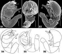
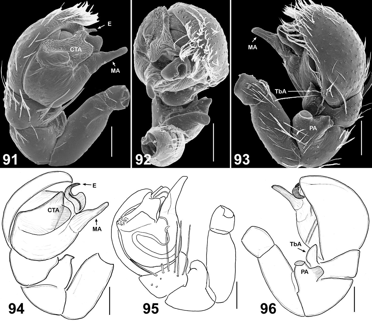
Figures 91–96.Copulatory organs of Eupoa pulchella sp. n. (91–93, the holotype; 94–96, ♂ paratype). 91, 94–95 male palp, median view 92 ditto, apical view 93, 96 ditto, retrolateral view. Abbreviations as explained in ‘Material and methods’. Scale bars: 0.1 mm.
-
Dmitri V. Logunov, Yuri M. Marusik
Zookeys
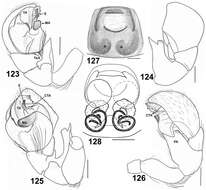
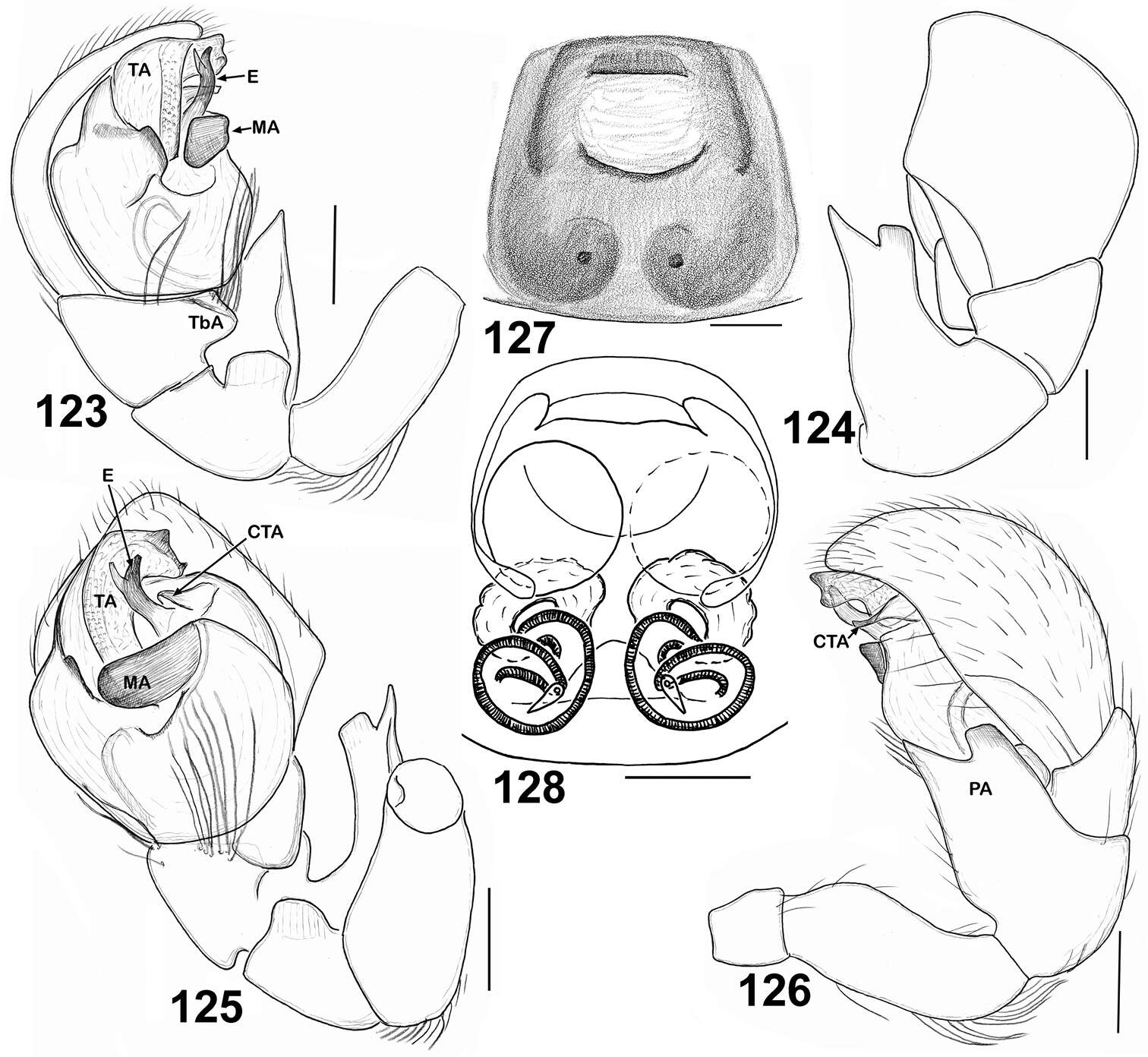
Figures 123–128.Copulatory organs of Eupoa yunnanensis from Laos. 123 male palp, median view 124 ditto, dorsal view 125 ditto, ventral view 126 ditto, retrolateral view 127 epigyne, verntral view 128 ditto, dorsal view. Abbreviations as explained in ‘Material and Methods’. Scale bars: 0.1 mm.
-
Dmitri V. Logunov, Yuri M. Marusik
Zookeys
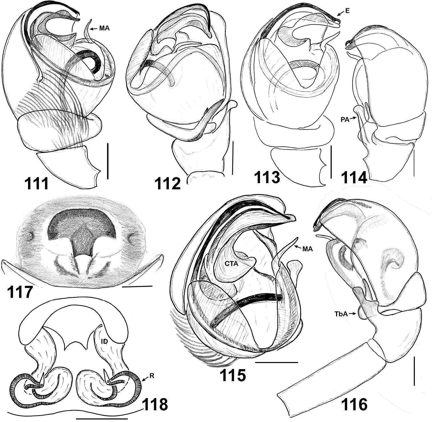
Figures 111–118.Copulatory organs of Eupoa thailandica sp. n. (♂ and ♀ paratypes). 111, 113 male palp, median view 112 ditto, ventral view 114 male palp, dorsal view 115 ditto, ventral view 116 ditto, retrolateral view 117 epigyne, ventral view 118 vulva, dorsa view. Abbreviations as explained in ‘Material and methods’. Scale bars: 0.1 mm.
-
Dmitri V. Logunov, Yuri M. Marusik
Zookeys
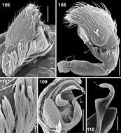
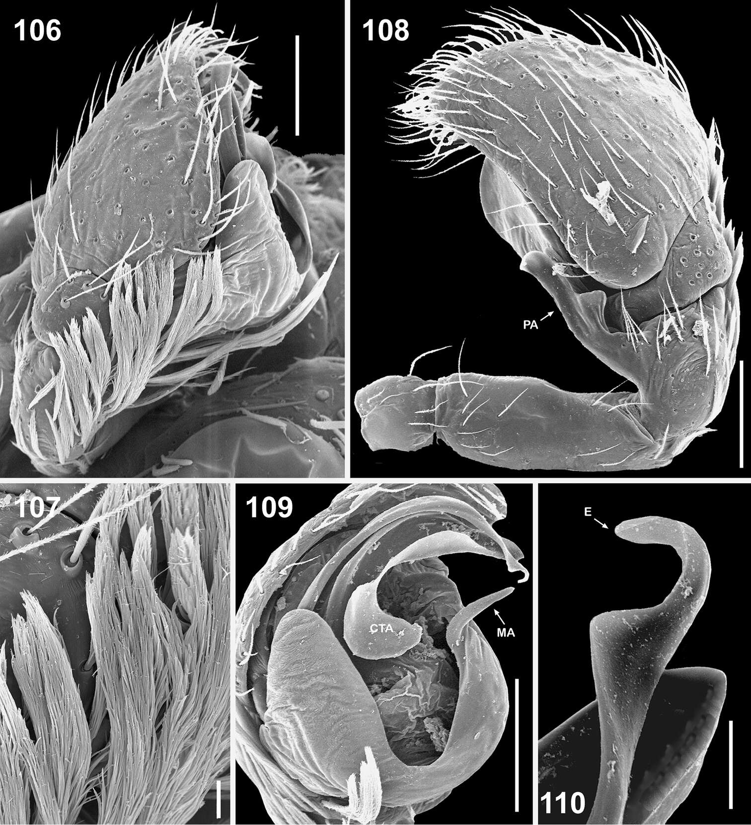
Figures 106–110.Copulatory organs of Eupoa thailandica sp. n. (♂ paratype). 106 male palp, median view 107 bunches of white hairs at the base of cymbium, median view 108 male palp, retrolateral view 109 ditto, ventral view 110 embolic tip, apical view. Abbreviations as explained in ‘Material and methods’. Scale bars: 10 μm (107, 110), 0.1 mm (106, 108–109).
-
Dmitri V. Logunov, Yuri M. Marusik
Zookeys
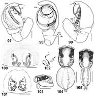
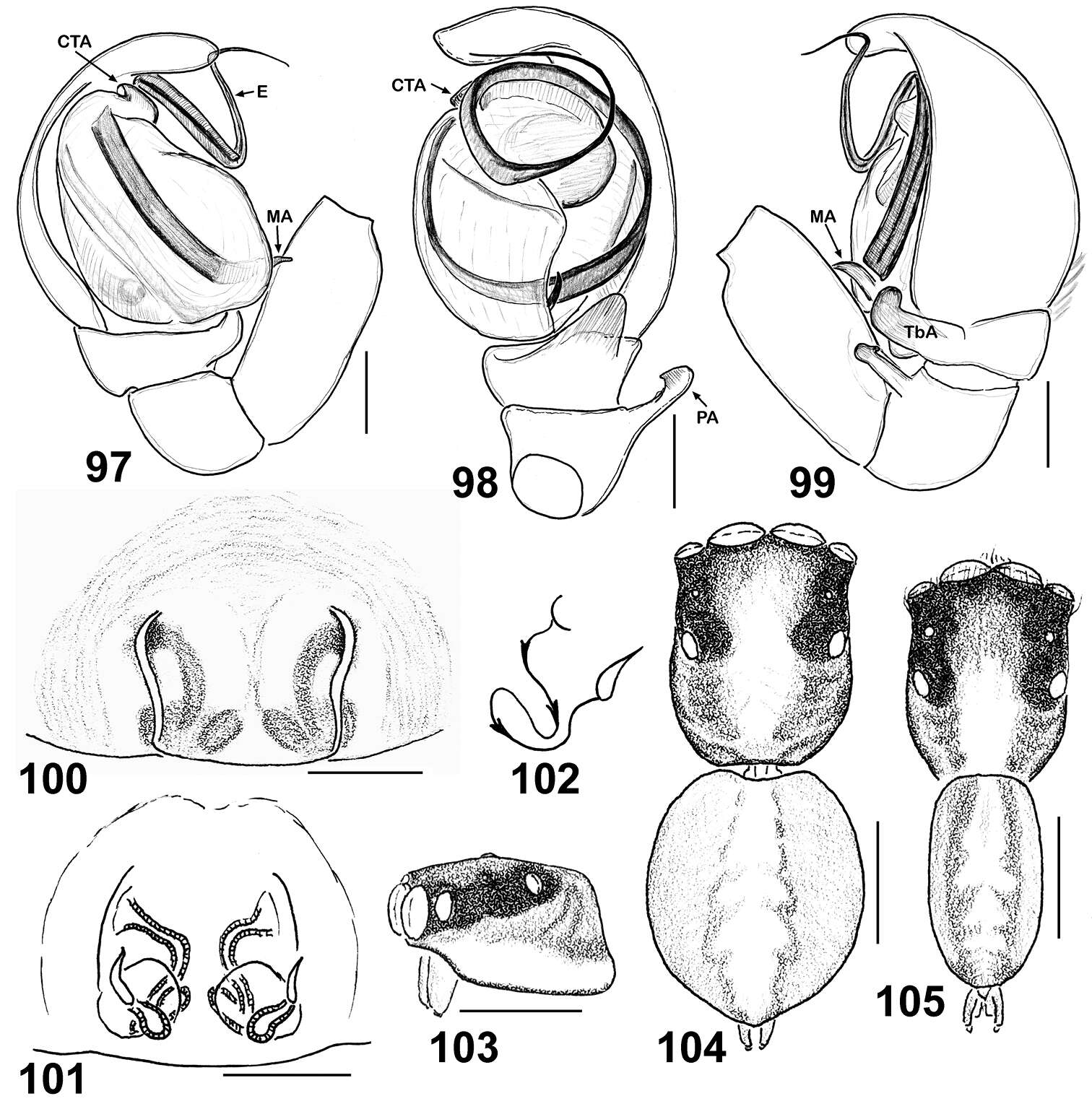
Figures 97–105.Copulatory organs and somatic characters of Eupoa schwendingeri sp. n. (♂ and ♀ paratypes). 97 male palp, median view 98 ditto, ventral view 99 ditto, retrolateral view 100 epigyne, ventral view 101 vulva, dorsal view 102 diagrammatic course of insemination ducts 103 female carapace, lateral view 104 female body, dorsal view 105 male body, dorsal view. Abbreviations as explained in ‘Material and methods’. Scale bars: 0.1 mm (97–101), 0.25 mm (103–105).
-
Dmitri V. Logunov, Yuri M. Marusik
Zookeys
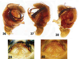
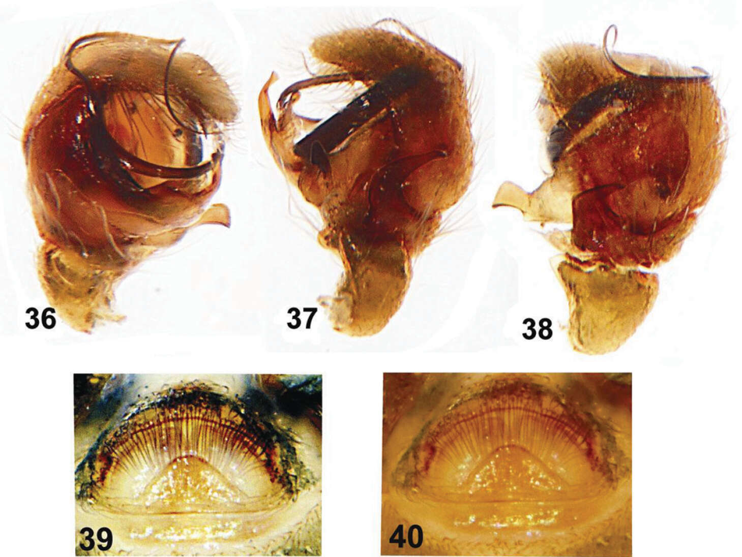
Figures 36–40.Copulatory organs of Eupoa lehtineni sp. n. from India. 36 male palp, median view 37–38 ditto, retrolateral view 39–40 epigyne, ventral view.
-
Dmitri V. Logunov, Yuri M. Marusik
Zookeys
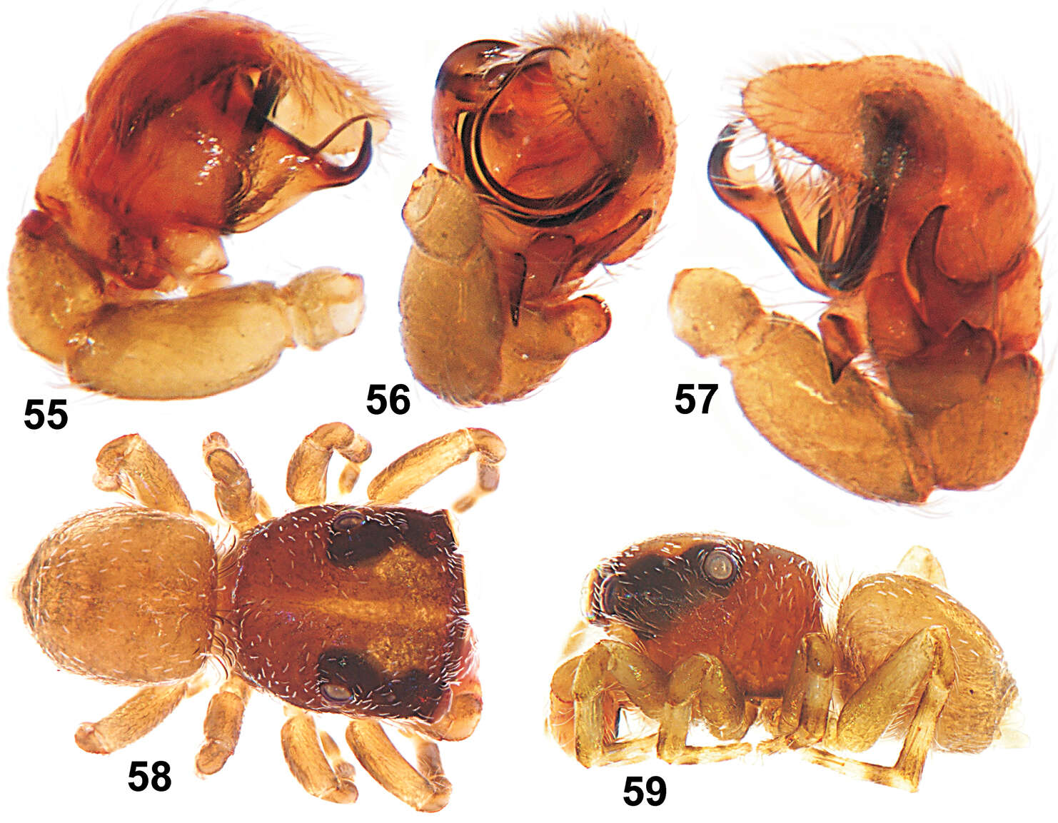
Figures 55–59.Copulatory organs and somatic characters of Eupoa lobli sp. n. (the holotype). 55 male palp, median view 56 ditto, apical view 57 ditto, retrolateral view 58 male general appearance, dorsal view 59 ditto, lateral view.
-
Dmitri V. Logunov, Yuri M. Marusik
Zookeys
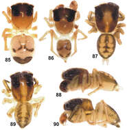
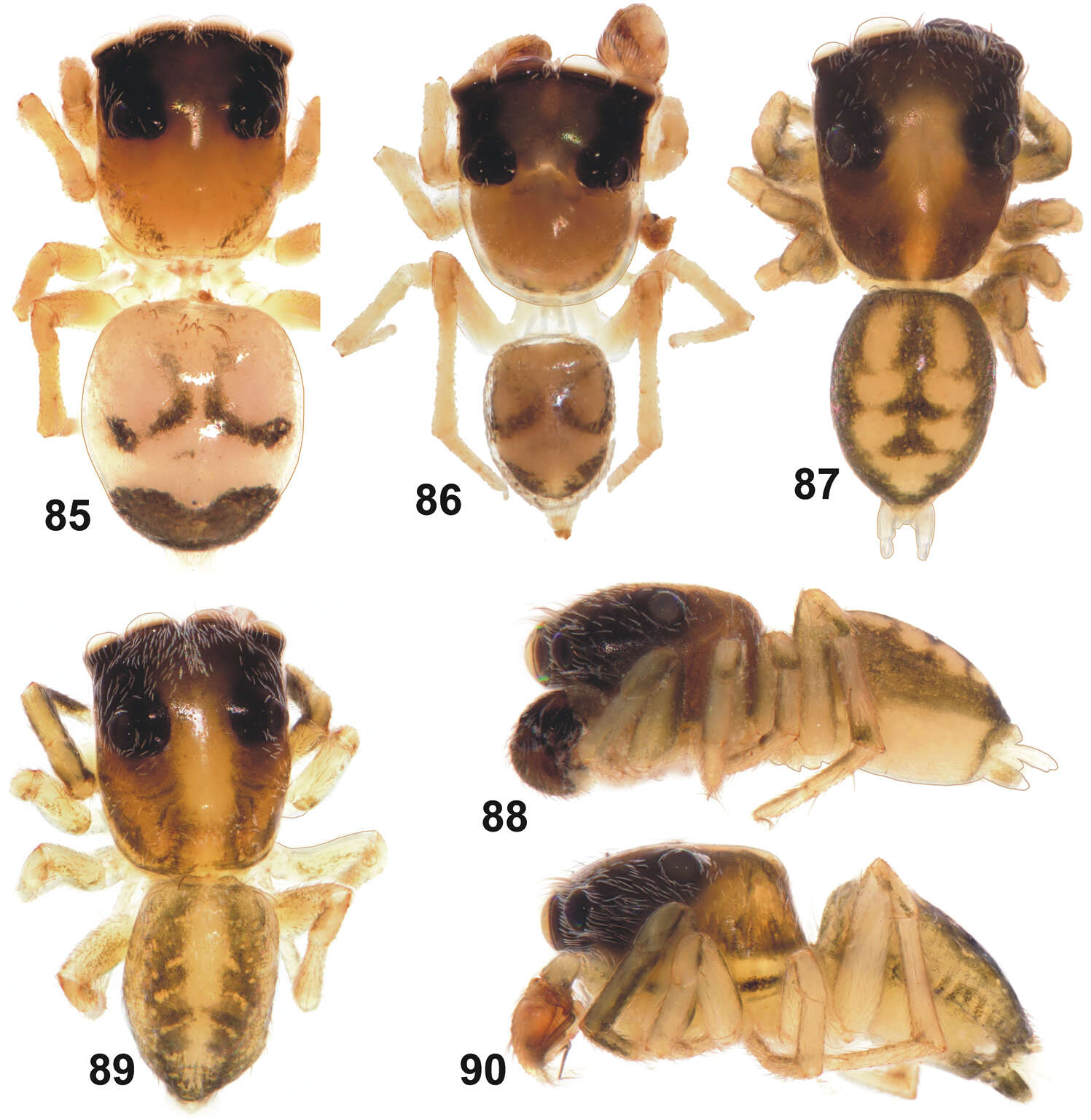
Figures 85–90.General appearance of Eupoa pulchella sp. n. (87–88, ♂ paratype), Eupoa schwendingeri sp. n. (♂ holotype, 89–90) and Eupoa thailandica sp. n. (♀ and ♂ paratypes, 85–86). 85 female body, dorsal view 86–87, 89 male body, dorsal view 88, 90 ditto, lateral view.
-
Dmitri V. Logunov, Yuri M. Marusik
Zookeys
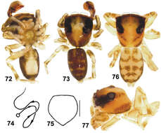
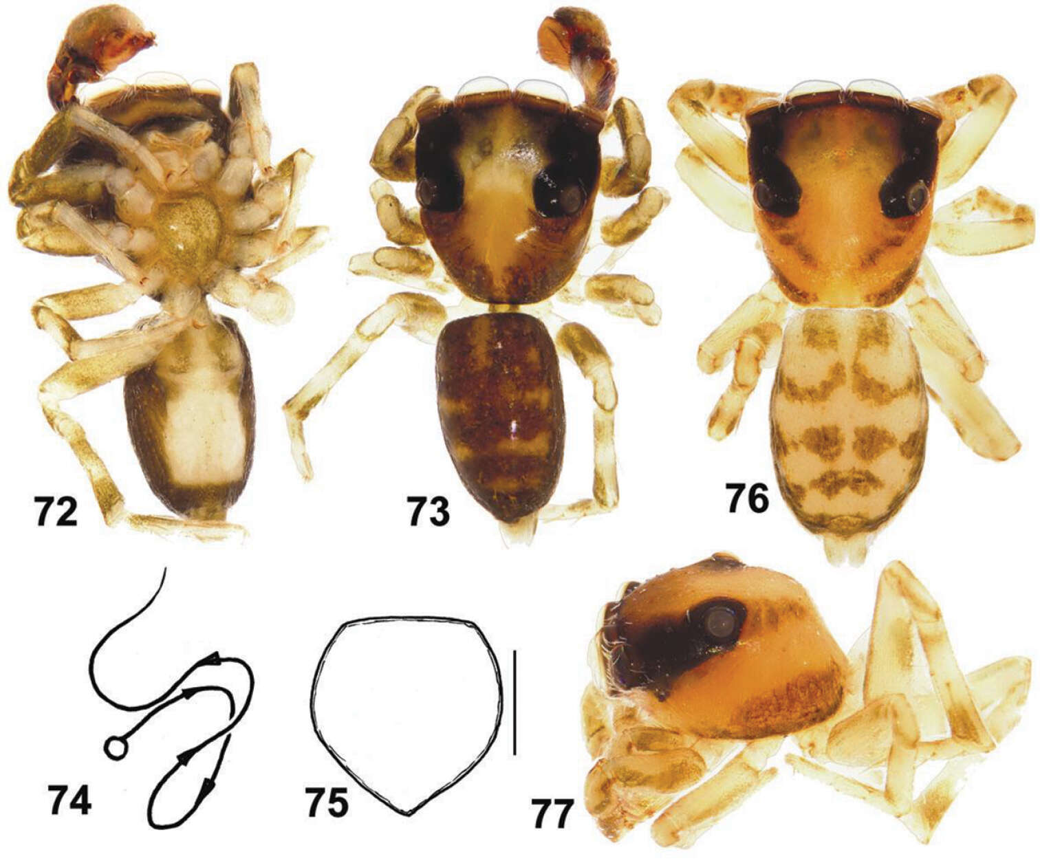
Figures 72–77.General appearance and somatic characters of Eupoa prima (♂ holotype and ♀ allotype). 60 male body, ventral view 73 ditto, dorsal view 74 diagrammatic course of the embolar path 75 female sternum, ventral view 76 female body, dorsal view 77 female carapace, lateral view. Scale bars: 0.25 mm (76).
-
Dmitri V. Logunov, Yuri M. Marusik
Zookeys
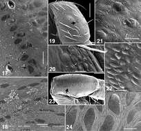
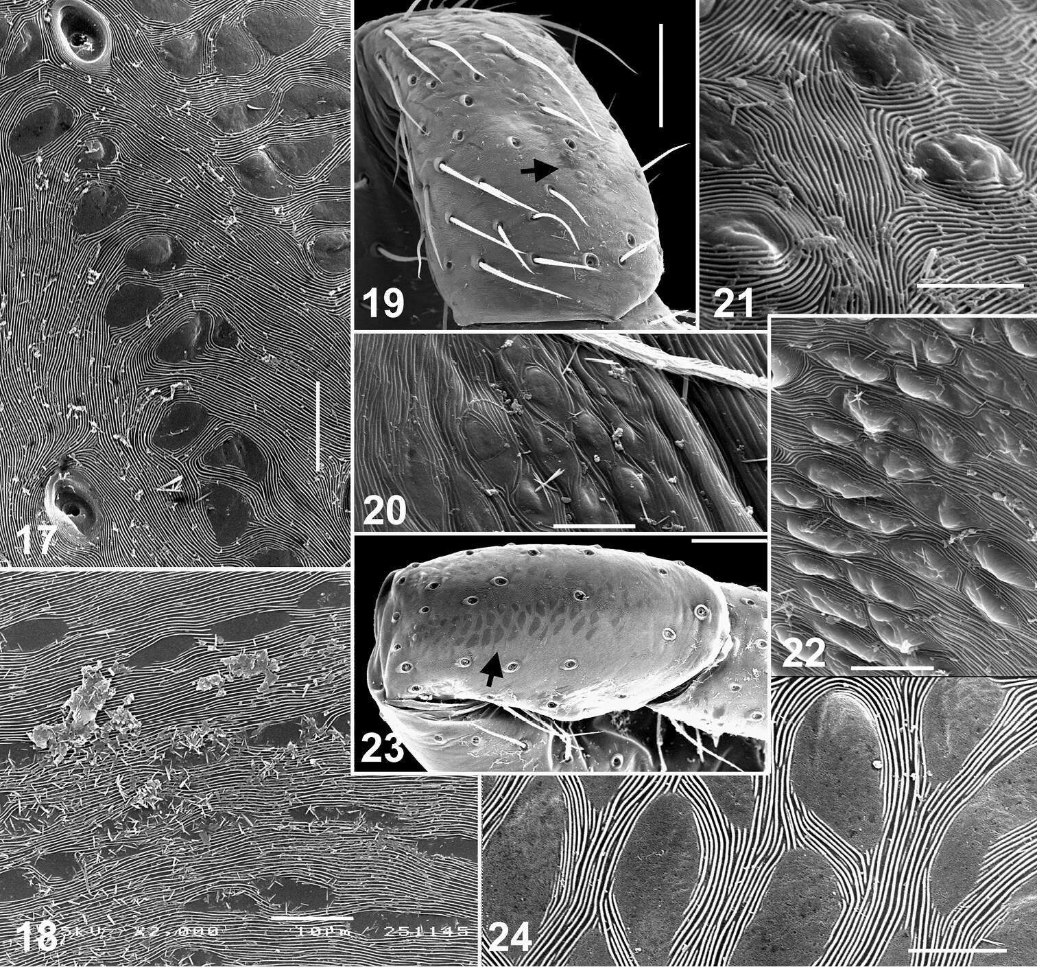
Figures 17–24.Skin structures of Eupoa lehtineni sp. n. (17, 19–22) and Eupoa thailandica sp. n. (18, 23, 24). 17 male patella I, dorsal view 18, 20, 22 female carapace, lateral view 19, 21, 23–24 female patella I, dorsal view. Scale bars: 5 μm (21, 24), 10 μm (17–18, 20, 22), 50 μm (19, 23).
-
Dmitri V. Logunov, Yuri M. Marusik
Zookeys
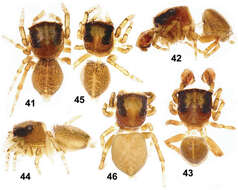
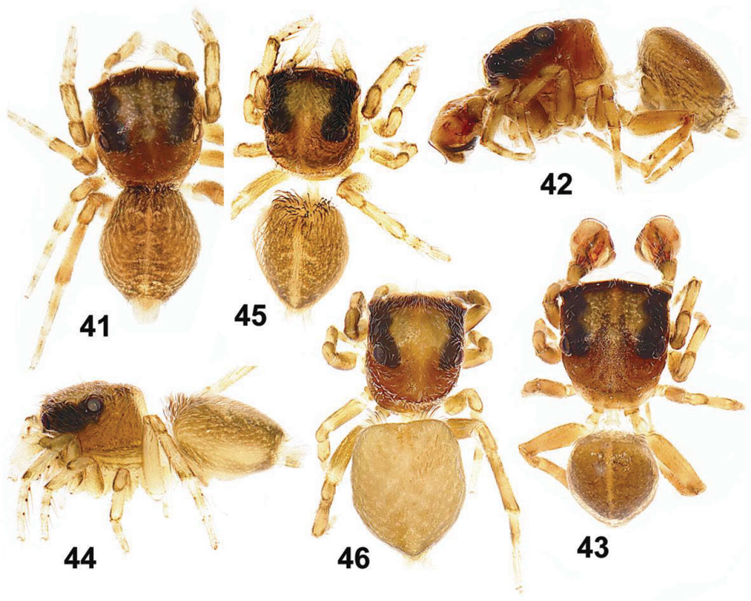
Figures 41–46.General appearance of Eupoa lehtineni sp. n. 41, 45–46 females, dorsal view 42 male, lateral view 43 ditto, dorsal view 44 female, lateral view. Specimens: 41–43 – India; 44–46 – Viet-Nam.
-
Dmitri V. Logunov, Yuri M. Marusik
Zookeys
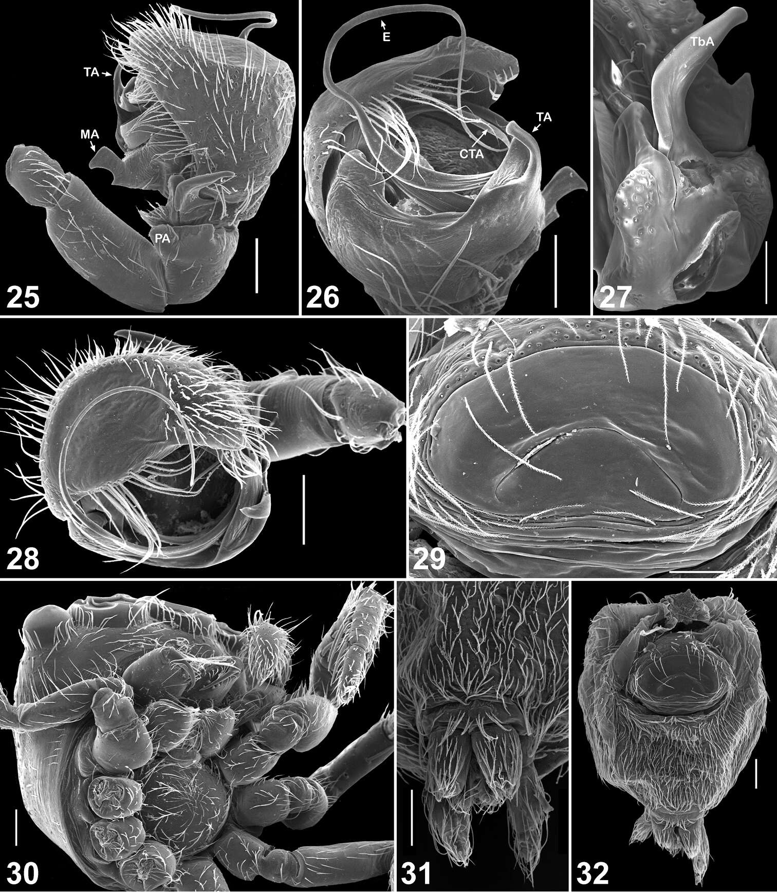
Figures 25–32.Copulatory organs and somatic characters of Eupoa lehtineni sp. n. 25 male palp, retrolateral view 26 ditto, median view 27 male tibial apophysis, retrolateral view 28 male palp, apical view 29 epigyne, ventral view 30 female carapace, ventral view 31 female spinnerets, ventral view 32 female abdomen, ventral view. Abbreviations as explained in ‘Material and methods’. Scale bars: 50 μm (27, 29), 0.1 mm (25–26, 28, 30–32).
-
Dmitri V. Logunov, Yuri M. Marusik
Zookeys
Figures 9–16.Somatic characters of Eupoa lehtineni sp. n. (9–11, 13–16) and Eupoa thailandica sp. n. (12). 9 female leg I, median view 10 female tarsus I, lateral view 11 female tarsus III, lateral view 12–13 tarsal organ on female tarsus I, dorsal view 14 trichobotrial base, female tarsus I, dorsal view 15–16 female palp and the claw at its tip (arrowed). Scale bars: 1 μm (13–14), 5 μm (12), 10 μm (11, 15), 50 μm (10, 16), 0.1 mm (9).
-
Dmitri V. Logunov, Yuri M. Marusik
Zookeys
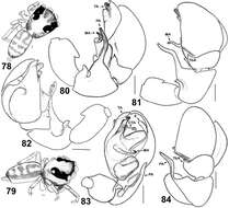
Figures 78–84.General appearance and copulatory organs of Eupoa prima (♂ paratype). 78 male body, dorsal view 79 ditto, lateral view 80 male palp, retrolateral view 81, 84 ditto, dorsal view 82 ditto, median view 83 ditto, ventral view. Abbreviations as explained in ‘Material and methods’. Scale bars: 0.1 mm.
-
Dmitri V. Logunov, Yuri M. Marusik
Zookeys
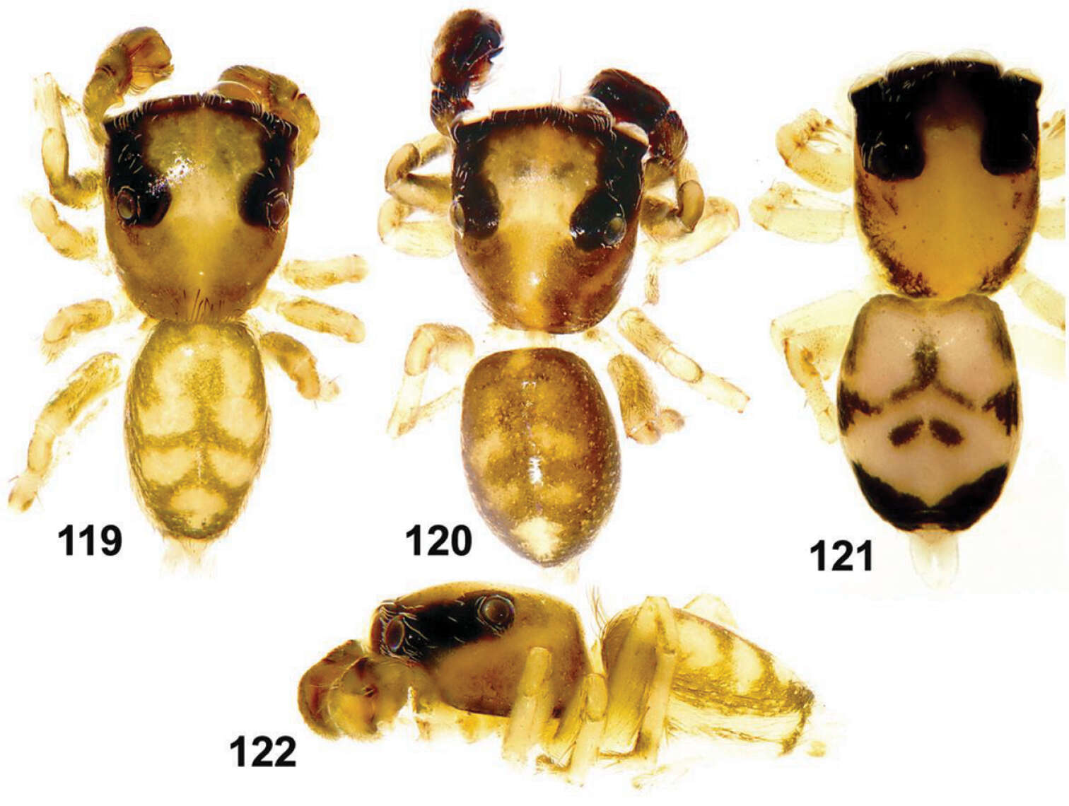
Figures 119–122.General appearance of Eupoa yunnanensis from Laos. 119, 120 male body, dorsal view 121 female body, dorsal view 122 male body, lateral view.
-
Dmitri V. Logunov, Yuri M. Marusik
Zookeys
Figures 123–128.Copulatory organs of Eupoa yunnanensis from Laos. 123 male palp, median view 124 ditto, dorsal view 125 ditto, ventral view 126 ditto, retrolateral view 127 epigyne, verntral view 128 ditto, dorsal view. Abbreviations as explained in ‘Material and Methods’. Scale bars: 0.1 mm.
-
Dmitri V. Logunov, Yuri M. Marusik
Zookeys
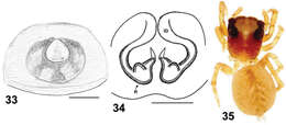
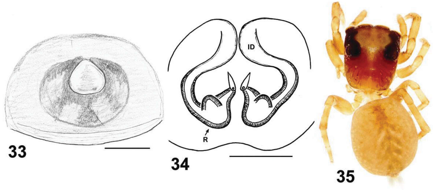
Figures 33–35.Copulatory organs and somatic characters of Eupoa daklak sp. n. 33 epigyne, ventral view 34 vulva, dorsal view 31 female body, dorsal view. Abbreviations as explained in ‘Material and methods’. Scale bars: 0.1 mm.
-
Dmitri V. Logunov, Yuri M. Marusik
Zookeys
Figures 1–8.Somatic characters of Eupoa lehtineni sp. n. 1–3 plumose scales on female carapace. 4 male carapace, frontal view 5 female carapace, frontal view 6 ditto, lateral view 7 female chelicerae, frontal view 8 female fang and cheliceral teeth. Scale bars: 10 μm (1–3), 50 μm (7–8), 0.1 mm (4–6).
-
Dmitri V. Logunov, Yuri M. Marusik
Zookeys
Figures 9–16.Somatic characters of Eupoa lehtineni sp. n. (9–11, 13–16) and Eupoa thailandica sp. n. (12). 9 female leg I, median view 10 female tarsus I, lateral view 11 female tarsus III, lateral view 12–13 tarsal organ on female tarsus I, dorsal view 14 trichobotrial base, female tarsus I, dorsal view 15–16 female palp and the claw at its tip (arrowed). Scale bars: 1 μm (13–14), 5 μm (12), 10 μm (11, 15), 50 μm (10, 16), 0.1 mm (9).
-
Dmitri V. Logunov, Yuri M. Marusik
Zookeys
Figures 17–24.Skin structures of Eupoa lehtineni sp. n. (17, 19–22) and Eupoa thailandica sp. n. (18, 23, 24). 17 male patella I, dorsal view 18, 20, 22 female carapace, lateral view 19, 21, 23–24 female patella I, dorsal view. Scale bars: 5 μm (21, 24), 10 μm (17–18, 20, 22), 50 μm (19, 23).