-
Shields, Wickham, and Kuris 1989
Nemertea
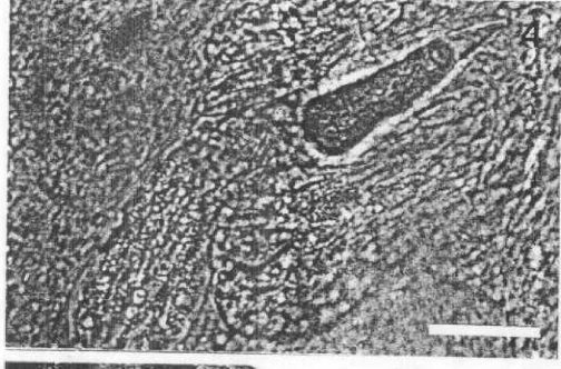
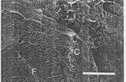
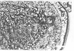
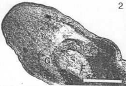
Frontal section of C. regicides showing cerebrum (C), foregut (F), and the absence of a distinct precerebral septum. Scale bar..Frontal section of C. regicides showing cerebrum (C), foregut (F), and the absence of a distinct precerebral septum. Scale bar = 50 μm.
-
Shields, Wickham, and Kuris 1989
Nemertea
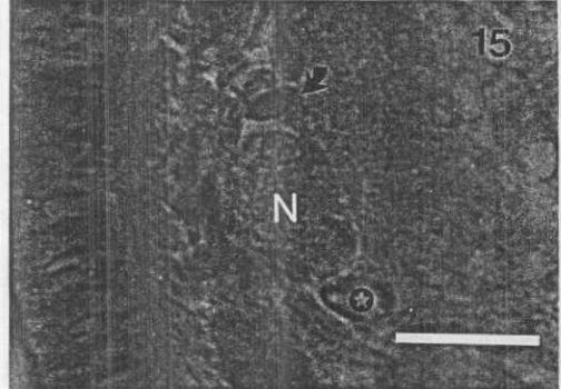
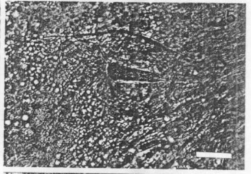
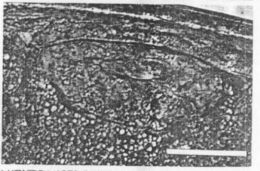
Cross section of C. regicides showing lateral nerve (N), submuscular gland (arrow), and ventral blood vessel (star). Scale bar..Cross section of C. regicides showing lateral nerve (N), submuscular gland (arrow), and ventral blood vessel (star). Scale bar = 25 μm
-
Shields, Wickham, and Kuris 1989
Nemertea
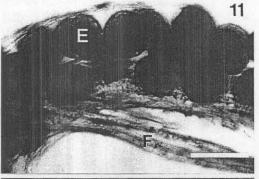
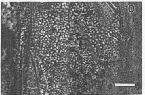
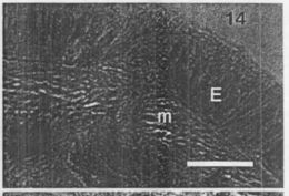
Ovarian pore in cross section; star, pore; m, muscle layer; E, epithelium. Scale bar = 25 μm
-
Shields, Wickham, and Kuris 1989
Nemertea
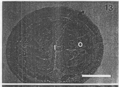
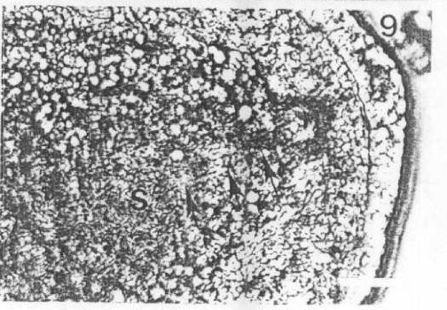
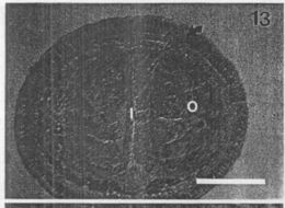
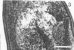
Female C. regicides in cross section; arrow, ovarian pore; 0, ovary; I, intestine. Scale bar = 100 μm
-
Shields, Wickham, and Kuris 1989
Nemertea
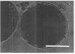
Female C. regicides in cross section; arrow, ovarian pore; 0, ovary; I, intestine. Scale bar = 100 μm
-
Shields, Wickham, and Kuris 1989
Nemertea
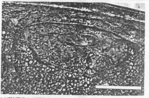
Larva of C. regicides (Van Cleave's hematoxylin). Note the epithelial layer surrounding the inner mass (arrow). Scale bar = 50 u
-
Shields, Wickham, and Kuris 1989
Nemertea
Egg string of C. regicides in situ on the funiculus of an egg of Paralithodes camtschatica; E, nemertean embryo; F, funiculus...Egg string of C. regicides in situ on the funiculus of an egg of Paralithodes camtschatica; E, nemertean embryo; F, funiculus. Scale bar = 100 um.
-
Shields, Wickham, and Kuris 1989
Nemertea
Detail of the mucous sheath of C. regicides . Note the grainy appearance and lack of lapilli. Scale bar = 200 μm
-
Shields, Wickham, and Kuris 1989
Nemertea
Detail of posterior aspect of male C. regicides; S, seminal vesicle; arrows, male gonoduct. Scale bar = 25 μm .
-
Shields, Wickham, and Kuris 1989
Nemertea
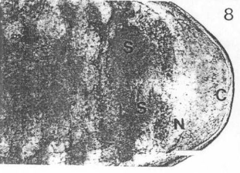
Posterior aspect of male C . regicides; S, seminal vesicle; N, posterior nerve ganglia; C, cloaca . Note the blunt posterior...Posterior aspect of male C . regicides; S, seminal vesicle; N, posterior nerve ganglia; C, cloaca. Note the blunt posterior form. Scale bar = 100 μm.
-
Shields, Wickham, and Kuris 1989
Nemertea
Detail of ovarian pore of C. regicides. Scale bar = 25 um.
-
Shields, Wickham, and Kuris 1989
Nemertea
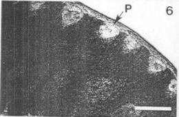
Lateral aspect of female C. regicides showing ovarian pore (P). Scale bar = 100 μm .
-
Shields, Wickham, and Kuris 1989
Nemertea
Lateral aspect of the stylet (stilleto-like) in the anterior proboscis chamber. Scale bar = 25 μm
-
Shields, Wickham, and Kuris 1989
Nemertea
Dorsoventral aspect of the stylet (dagger-like) in the anterior proboscis chamber of C. regicides. Scale bar = 25 μm.
-
Shields, Wickham, and Kuris 1989
Nemertea
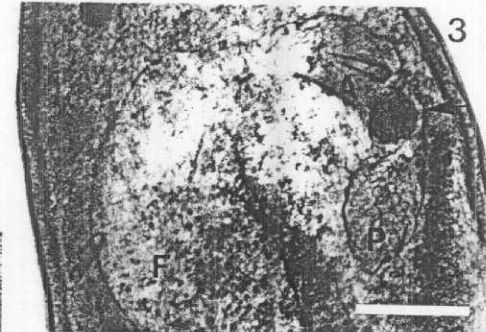
Detail of the feeding armature of C. regicides; F, posterior of foregut-esophagus; A, anterior proboscis chamber; arrow...Detail of the feeding armature of C. regicides ; F, posterior of foregut-esophagus; A, anterior proboscis chamber; arrow, middle proboscis chamber; P, posterior proboscis chamber. Scale bar = 100 μm .
-
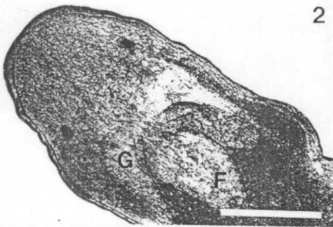
Anterior end of a female C. regicides with the foregut-esophagus retracted (F) . Note the prominent dorsal ganglia (G)...Anterior end of a female C. regicides with the foregut-esophagus retracted (F) . Note the prominent dorsal ganglia (G) . Scale bar = 100 μm.
-
Shields, Wickham, and Kuris 1989
Nemertea
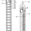
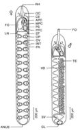
Composite drawing of female (left) and male (right) Carcinonemerles regicides from Paralithodes camtschatica...Composite drawing of female (left) and male (right) Carcinonemerles regicides from Paralithodes camtschatica . The foregut of the male is everted for comparison . Note that the reproductive system of the male is represented on the right, while the digestive system is figured on the left . APC, anterior proboscis chamber; CE, cerebrum; CL, cloaca; FO, foregut-esophagus; INT, intestine; LN, lateral nerve cord; MPC, middle proboscis chamber; OC, ocelli; OP, ovarian pore; OV, ovary; PA, parenchyma; PPC, posterior proboscis chamber; PS, proboscis sheath; RE, rhynchodaeum; ST, stomach; SV, seminal vesicle; TE, testis; VD, vas deferens.