-
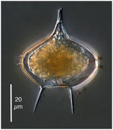
A heterotrophic dinoflagelate from the Amundsen Sea
-

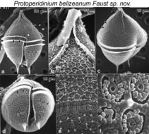
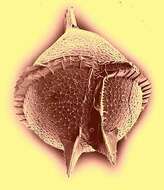
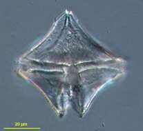
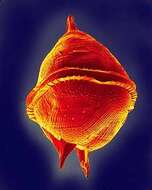
Figs. 1.SEM of Protoperidinium belizeanum sp. nov. (a) Ventral view. Cell is pyriform with an apical horn and two antapical spines. Apical plate 1' is meta. Cingulum is equatorial and ascending. Cingular wall reticulated (arrowheads). Postcingular plates narrow, qua¬drangular and wide (arrows), (b) Apical pore complex (APC) (X) includes an apical horn and a flange (arrows). Apical horn partially covered with biodetritus. (c) Epitheca excavated ventrally. Location of apical intercalary plate 3a (arrowheads), (d) Hypotheca is round. Cin¬gular list is prominent (arrowheads). Postcingular plates narrow (short arrows). Sulcus deep bordered by narrow list. Antapical spines are two, each with three fins, associated with antapical plates 1" and 2". (e) Thecal surface with three distinct ornamentation: 1) retic¬ulated pattern of ridged hexagonal depressions with a knob at network junctions (arrowheads), 2) depressions with a central rimmed pore (large arrows), and 3) vertical striation of paired particles above intercalary band (short arrows), (f) Depressions with a central rimmed pore (arrows) surrounded by small oblong particles Miniscule pore within the rimmed pore (arrowhead).
EMu: SEM NEGATIVE # 155141; SEM STUB # 155; FIELD # 631-93; ACCESSION # 407168; CATALOG # 1598; FIGURE # 3.
-
-
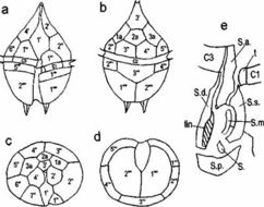
FIG. 2. Line drawing of Protoperidinium belizeanum sp. nov. Thecal plates are (a) ventral view, (b) dorsal view, (c) apical view, and (d) antapical view, (e) Details of the sulcal platelets.
EMu: SEM NEGATIVE # 155141; SEM STUB # 155; FIELD # 631-93; ACCESSION # 407168; CATALOG # 1598; FIGURE # 3.
-
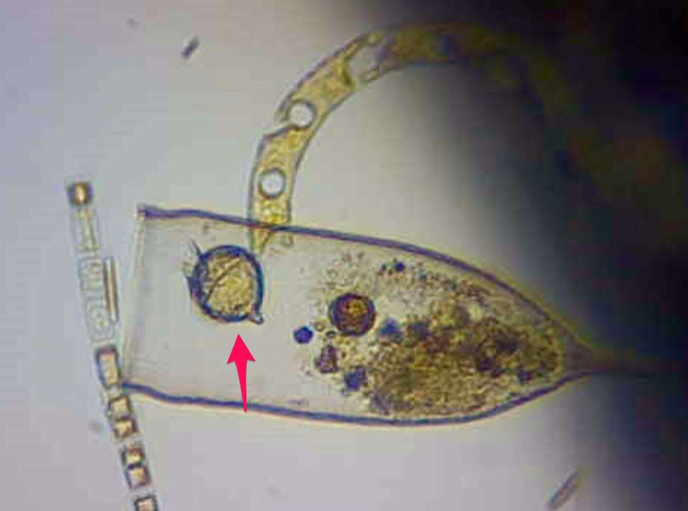
A heterotrophic dinoflagellate found with Phaeocystis on which it likely feeds.
-
-
-
Balea, Galicia, Spain
-
Balea, Galicia, Spain
-
O Grove, Galicia, Spain
-
Njar, Andalusia, Spain
-
Njar, Andalusia, Spain
-
Reboredo, Galicia, Spain
-
Reboredo, Galicia, Spain
-
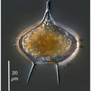
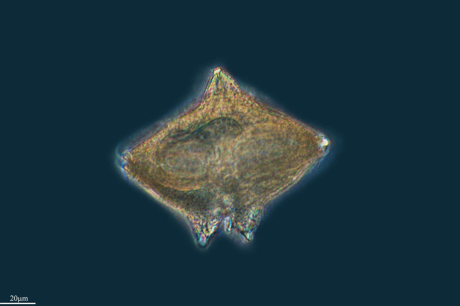
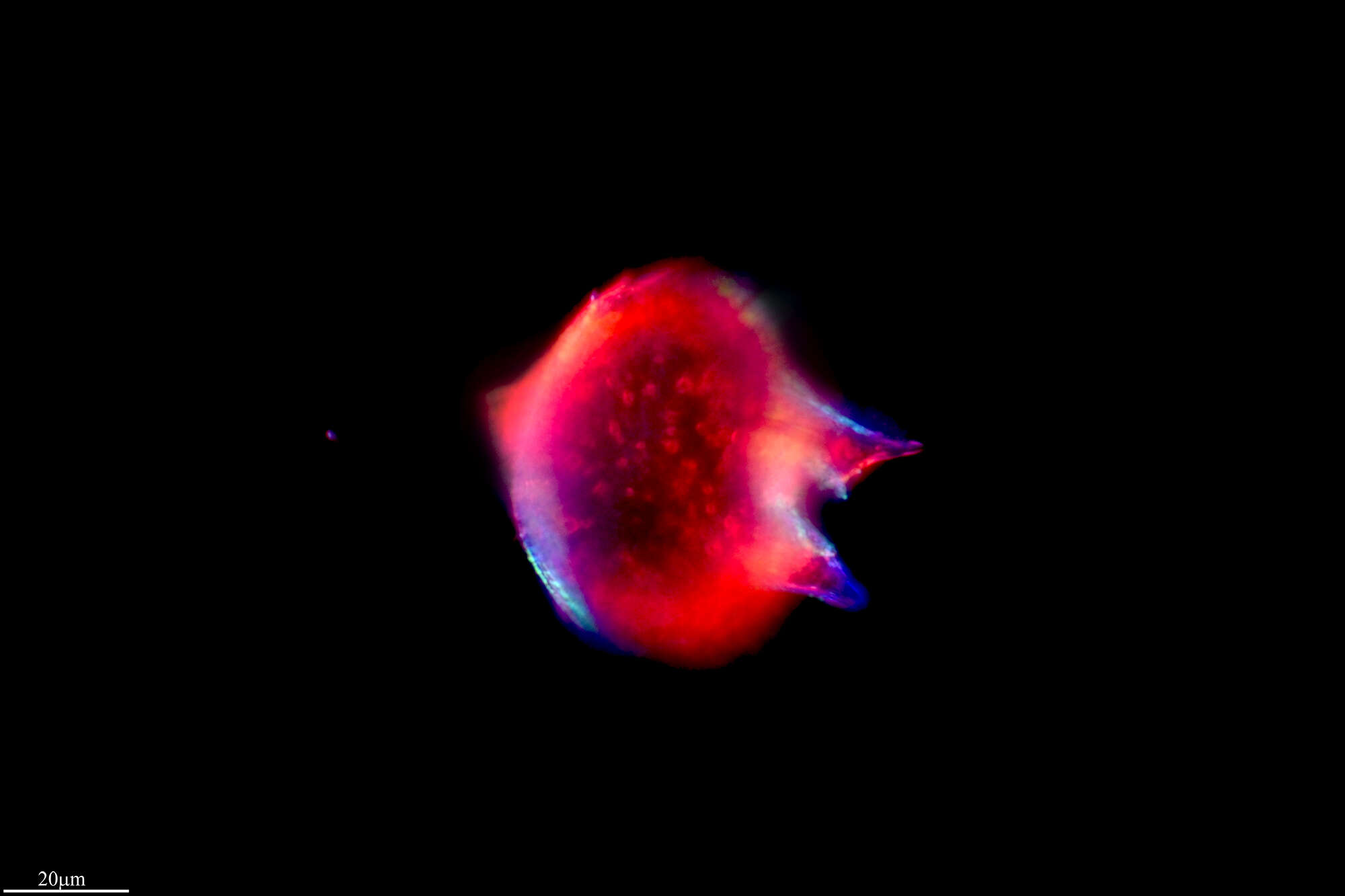
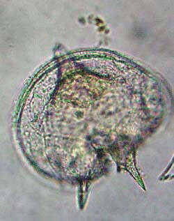
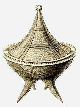
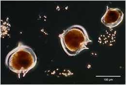
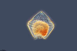
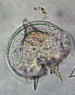
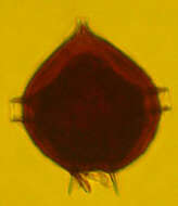
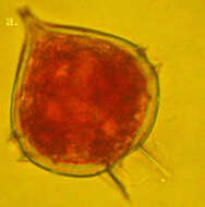
Protoperidinium (pro-toe-perry-din-ee-um) heterotrophic dinoflagellate. This is a cyst, but we can see that the margins of the circumferential groove have projecting margins, and there is an anterior point and two short posterior spines. Phase contrast.
-
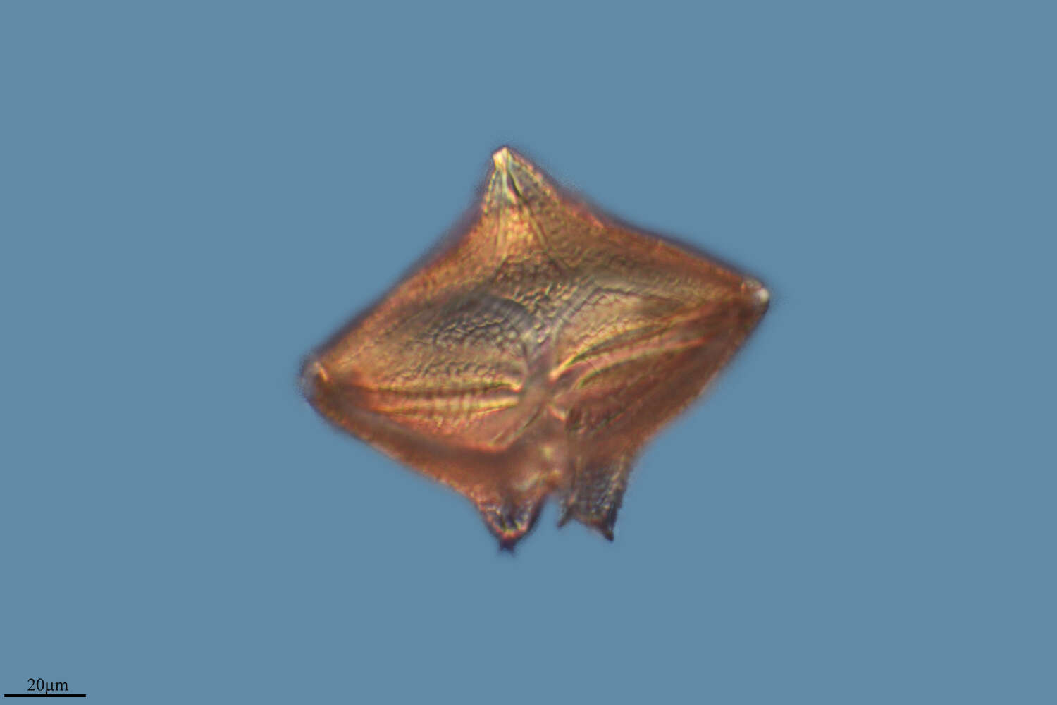
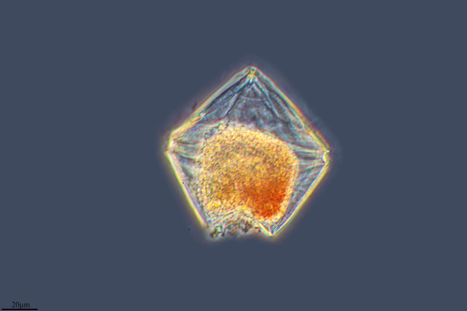
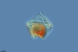
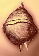
Ventral view showing girdle and sulcus. Armouring of the cell is also evident.
-
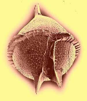
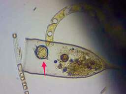
Theca remaining from a deceased cell showing the grooves in which the flagella lie, and, in profile, the lists or shelves that project around the girdle.
-
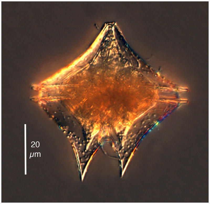
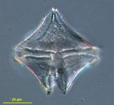
A large distinctive species. It has a long apical horn and long divergent antapical horns. P. depressum is a cosmopolitan species.
-
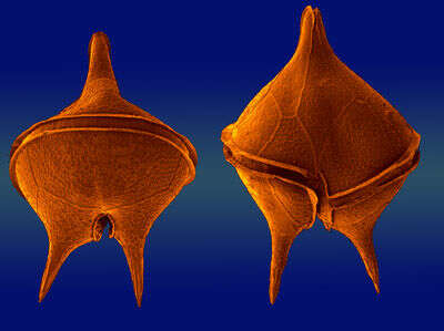
The two faces of ...
-
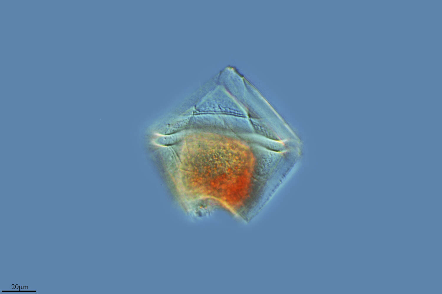
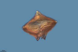
A medium-sized cell with short antapical horn and and winged antapical spines. The species can be confused with P. pallidum. A distinguishing feature is a prominent winged spines originating from a sulcal list.
-
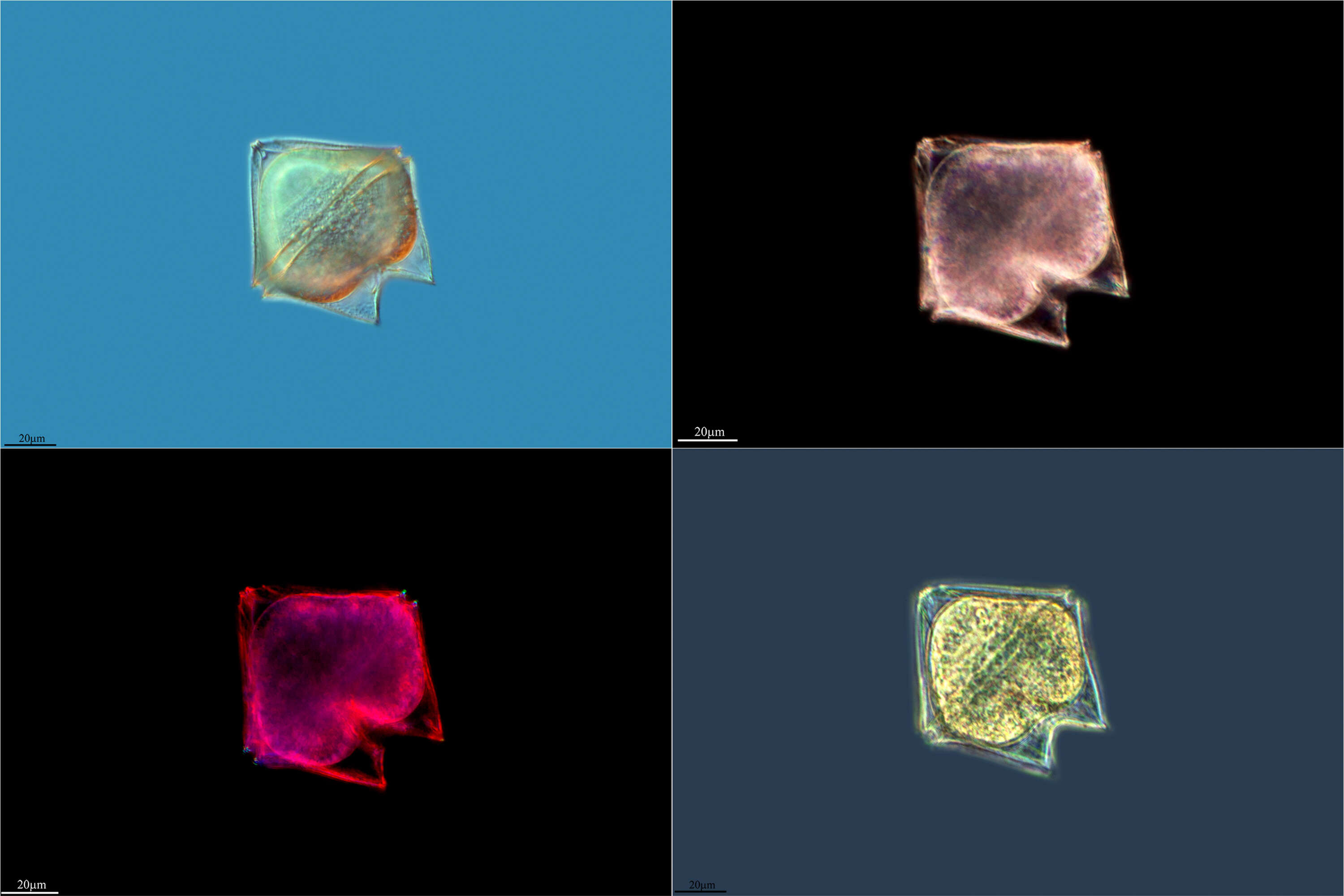
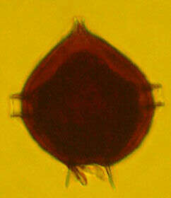
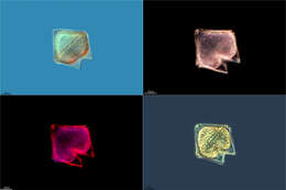
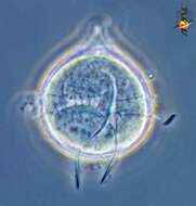
This micrograph emphasizes the armored character of the genus.
-
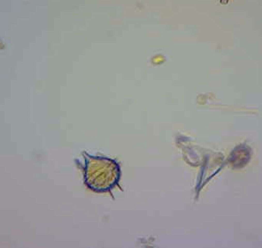
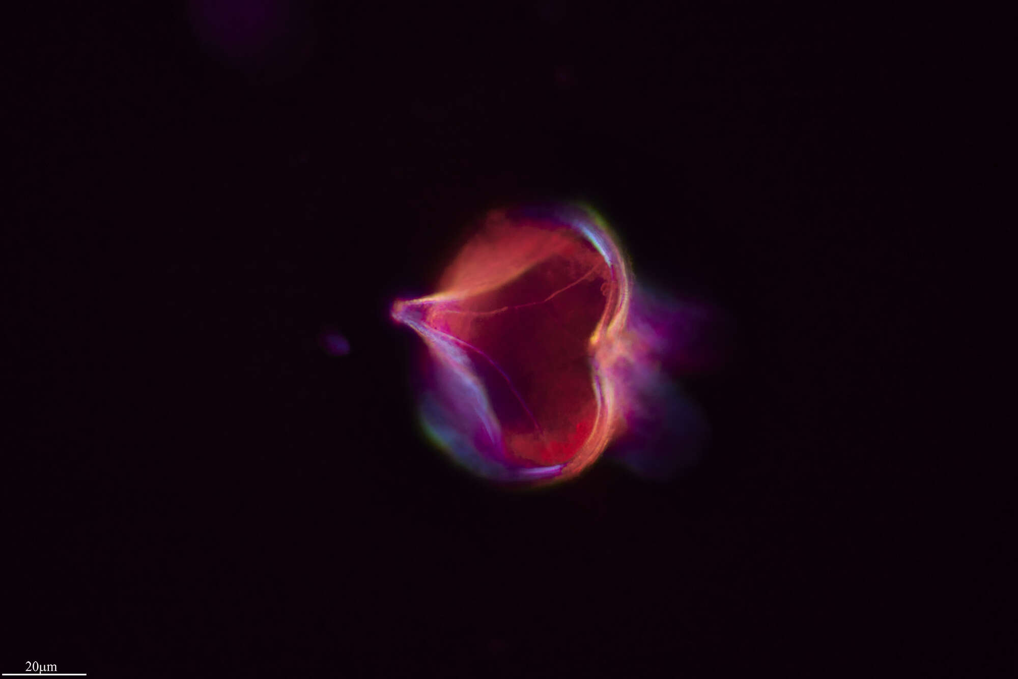
An armoured dinoflagellate.
-
The cells are pyriform with a long apical horn and a rounded hypotheca. There are two long, winged antapical spines. The girdle is right handed and bears distinct lists supported by spines.
-
Peridinium divergens.